

A short history
Before microscopes, the world of cells and microbes was a mystery, invisible to even the sharpest minds. But once scientists learned to bend light and magnify the unseen, biology was transformed forever. In this article, we’ll explore this journey from simple lenses to the cutting-edge technologies that reshaped science, medicine, and our understanding of the living world.
of microscopy
Imagine trying to understand how a city works by looking at it from outer space, with nothing but your naked eye. You’d probably be able to see general features and a broad layout, but many important details would be invisible: individual buildings, the roads connecting them, people living and working, and so on. This is the situation scientists faced before the invention of the microscope. Cells, not the speak of the crucial details of subcellular machinery, were far beyond view, seriously limiting our comprehension of biology and medicine.
Microscopy changed everything. Being able to see tiny details literally opened up a new dimension of science. Cells, their substructures, and how they work together could now be deciphered immediately, intuitively, directly, by observing them with one’s own eyes. The invisible became tangible.
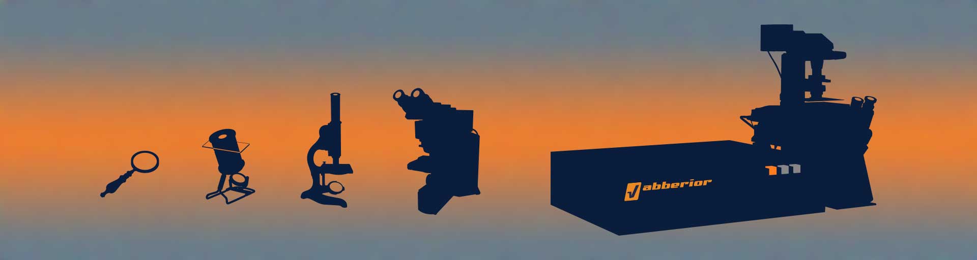
But this powerful tool didn’t appear overnight. It’s history and development spans several centuries, from simple lenses to sophisticated instruments capable of resolving single molecules. In this article, we’ll explore this journey and appreciate the cutting-edge technologies we are using today.
Ancient curiosity and early instruments
Long before real microscopes were invented, ancient civilizations already used “reading stones”, crystal quartz found as rocks in the nature, hewn into a round shape and polished. Yes, those were the first ever lenses.
Over time, mankind gained a better understanding of how these gadgets work. It was discovered that different curvatures of the glass surfaces produce different effects, the law of refraction came to be known, people learned to deliberately grind lenses with higher and higher precision, and finally, in the Netherlands of the 16th century, the first telescopes and microscopes appeared. These early instruments quickly were further improved by none other than Galilei and Newton and kick-started a revolution. Van Leeuwenhoek has been regarded as the father of microbiology since the late 1600s when he used hand-crafted single-lens microscopes to observe and describe bacteria, sperm cells, and protozoa.
A real turning point was the discovery that stacking lenses produced staggering effects. An optical train of multiple lenses could achieve greater magnification and correct for unwanted aberrations. Robert Hooke used these new instruments to produce the beautifully illustrated book Micrographia from 1665, showcasing his microscopic observations. Hooke discovered tiny compartments while examining cork bark and called them “cells”, from latin cella, meaning compartment, a name that endures to this day.
Modern times
In the 19th century, accelerating scientific progress had a profound effect on microscopy as well. Scientists began using fluorescent probes for staining tissues and pathogens, to increase observability and specificity. They learned to reliably design and produce compound microscopes consisting of many individual lenses. This led to larger fields of view with less distortion. It allowed the correction of color distortion, enabling clearer images. Oil immersion came into use; its high refractive index increases the numerical aperture of the objective lens, improving resolution. But then, in 1873, Ernst Abbe rigorously derived a natural boundary for the resolving power of a microscope, the diffraction limit. It states that one can only tell apart two points if and only if they are farther apart than half of the wavelength of the light used. That was a hard stop.
Of course, naturally, people looked for ways to break this barrier. Near-field optics is a way, at the expense of having to bring a probe very close (on the order of the wavelength) to the sample, thus essentially limiting it to surfaces. Atomic force microscopy has brought atomic resolution, but with similar limitations. Electron microscopes were invented in the early 1930s. These instruments use electron waves with much shorter wavelengths than visible light in order to achieve magnificent resolutions, without violating Abbe’s barrier. Much of the biological structural information we now have in the size range below 200 nanometers (e.g. viruses, mitochondria, nuclear pores) was first described using electron microscopes.
Yet, this didn’t stop people to think about ways to improve light microscopy, and rightly so. Because electron beams and the vacuum required by them are not exactly the best conditions for a healthy cell life. And looking at living cells is essential. Obviously, being able to observe our city as it buzzes with movement and interactions is much more informative than a mere still picture. This is the realm of light microscopy. So, phase-contrast and differential interference contrast were invented. Immunofluorescence labeling and fluorescent proteins were discovered, genetically encoded markers that could be linked to specific proteins within cells. In 1957, Marvin Minski conceived the confocal microscope, which greatly improves optical sectioning.
But still, the diffraction barrier stood firm. It was now considered a natural law, just like gravity or Heisenberg’s uncertainty relation (to which it is related, not incidentally).
Beyond the barrier
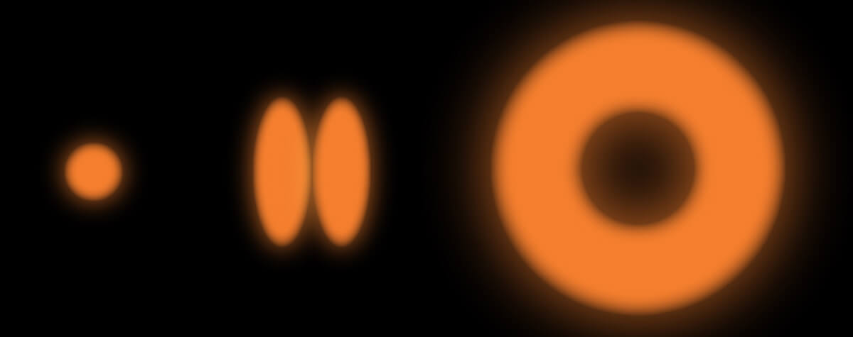
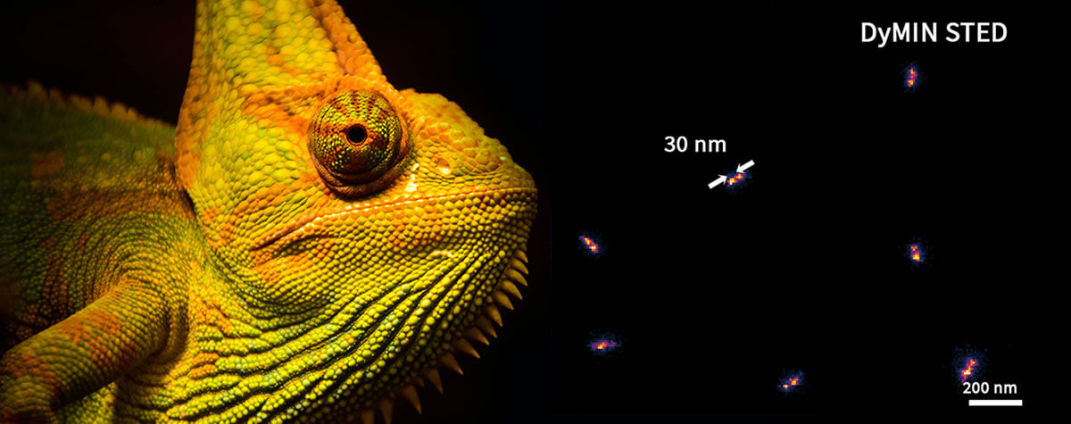
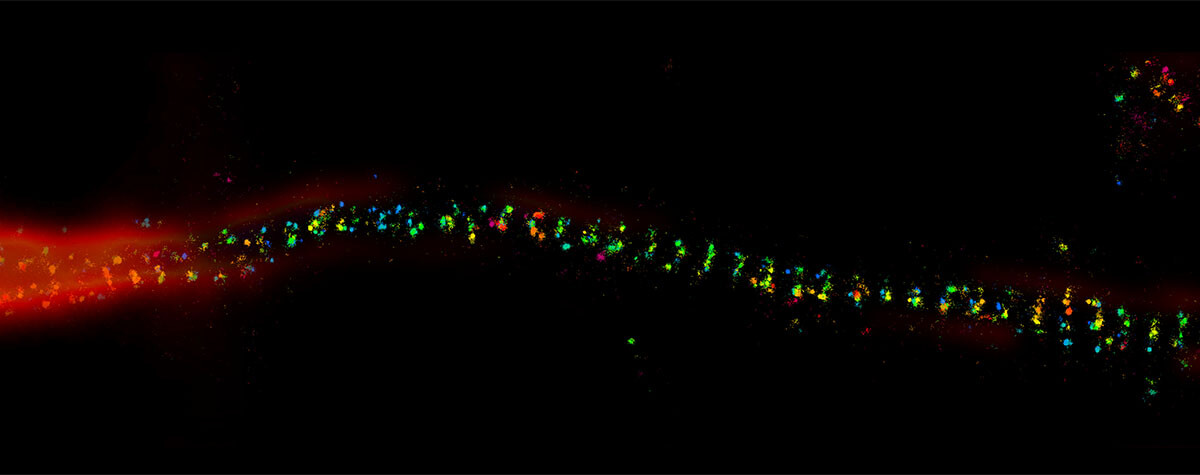
Until when in 1992, Stefan W. Hell figured out that if two points cannot be told apart when they are too close, we must try to look at them individually, that is, non-simultaneously. This led to several methods (STED, STORM) now collectively known as superresolution microscopy, which attain resolutions of about 20 nm, a tenfold improvement over the diffraction limit (read more in “How the donut changed the world”). They all work by ensuring that not every fluorescence molecule in the sample is emitting at the same time. This way, first one of them and then the other can be individually observed, rendering Abbe’s limitation inapplicable and thus irrelevant for this situation. In a sense it is a very clever way to circumvent it, which was rightly awarded with the Nobel Prize, essentially for realizing that the magic is not in the light, but in the dyes, and in making some of them temporarily invisible.
In the wake of these successes the resolution business gained traction. Soon, other (semi-)superresolution methods were introduced, such as SIM and pixel-reassignment, with intermediate resolution improvements of about a factor of two over the diffraction limit. Commercial instruments became available. Finally, in 2017, MINFLUX pushed the resolution to the scale of individual molecules.
This likely represents the ultimate limit. We can now routinely observe cellular cities and even their tiniest building blocks. At least for fluorescence microscopes, it seems obvious that a resolution finer than the size of the molecular markers used to label the sample does not make sense, but then again, who knows. The field of microscopy defied expectations for centuries, so we better brace for more breakthroughs!