


Application Snapshots
Take a look at our “Application Snapshots” – a small but steadily growing collection of concise, real-world examples of STED and MINFLUX superresolution microscopy. Be inspired by practical application examples, discover a wide range of possible uses, and learn how state-of-the-art microscopy techniques can advance your research.
Examples on superresolution microscopy
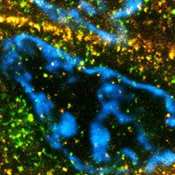
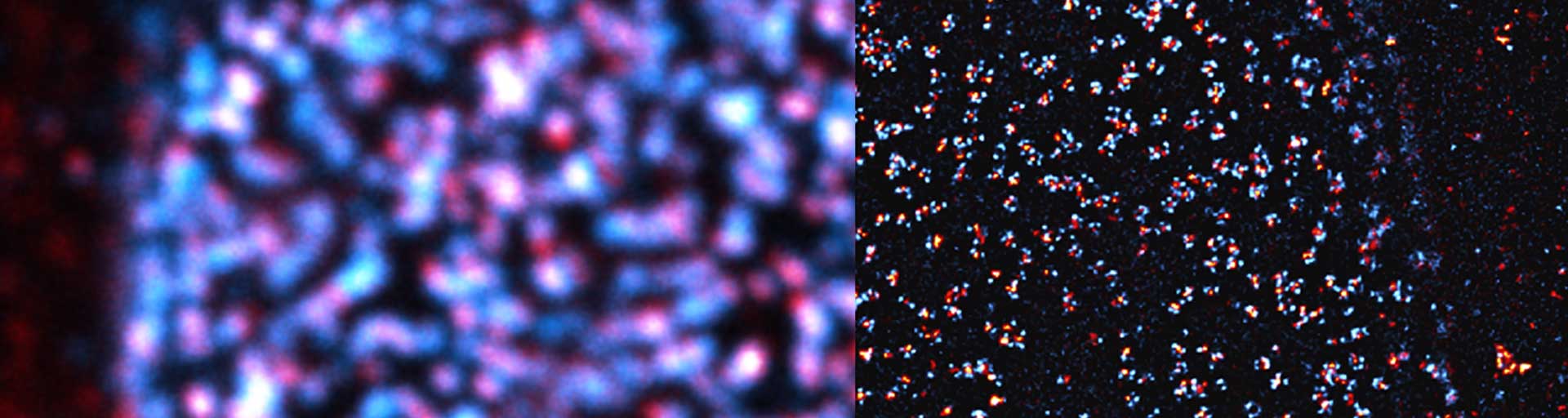
Image Quality
Learn more about the importance of labeling quality, antibody specificity, and expected localization to ensure reliable microscopy data.
Neuroscience
Read about how MINFLUX advance abberior's understanding of brain function and neurological disease at the molecular level.
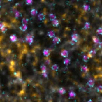
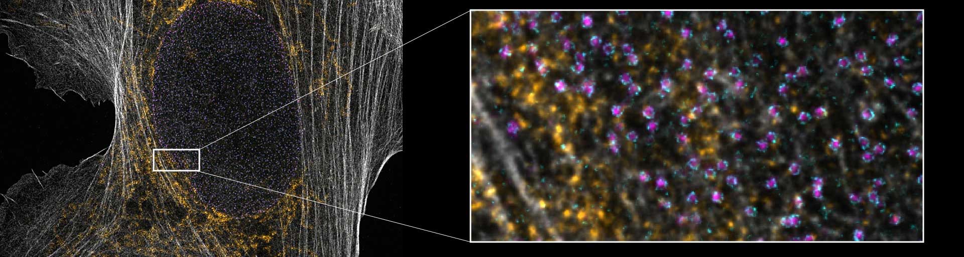
Multi-color imaging
Find out how abberior enables multi-color STED imaging of up to four channels using a single 775 nm depletion laser with minimal photobleaching.
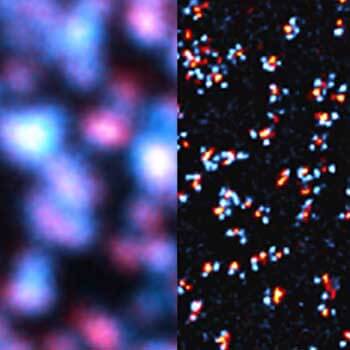
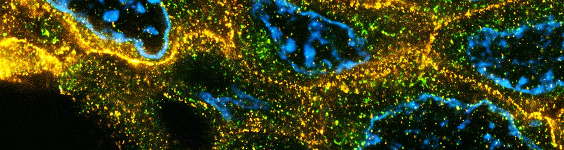
Celluar architecture
Learn how STED microscopy enabling accurate and quantitative colocalization analysis.