


Elevate your research,
We are excited to announce the next chapter in innovation! While the renowned FACILITY microscope has reached the end of its sales journey, we’re proud to introduce its extraordinary successor: please refer to our new MIRAVA POLYSCOPE for cutting-edge confocal, MATRIX, STED, and MINFLUX microscopy. Support for FACILITY will of course continue.
Stay at the forefront of progress by upgrading your FACILITY microscope with our EVERGREEN sustainability initiative that breathes new life into your deserving equipment – boosting user-friendliness and power, all while driving a more sustainable future.
go EVERGREEN and upgrade now!
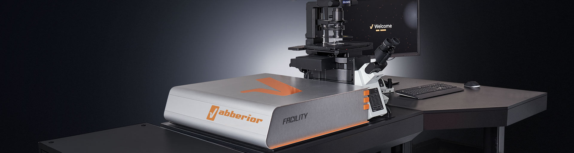
FACILITY
Our FACILITY platform offers you a workhorse instrument that boasts top-of-the-line superresolution STED and confocal imaging. It combines cutting-edge microscopy with unprecedented ease-of-use. Our software allows beginners to intuitively arrive at a top-notch image within three clicks, while also giving experts full control over the instrument. Microscopy has never felt better.
when every detail matters

Description
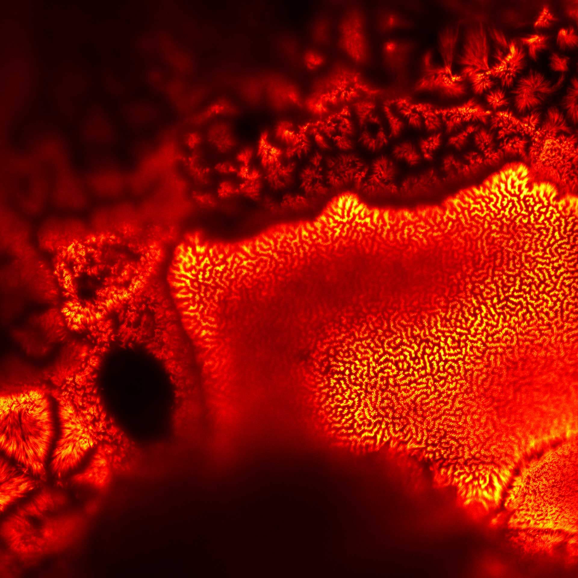
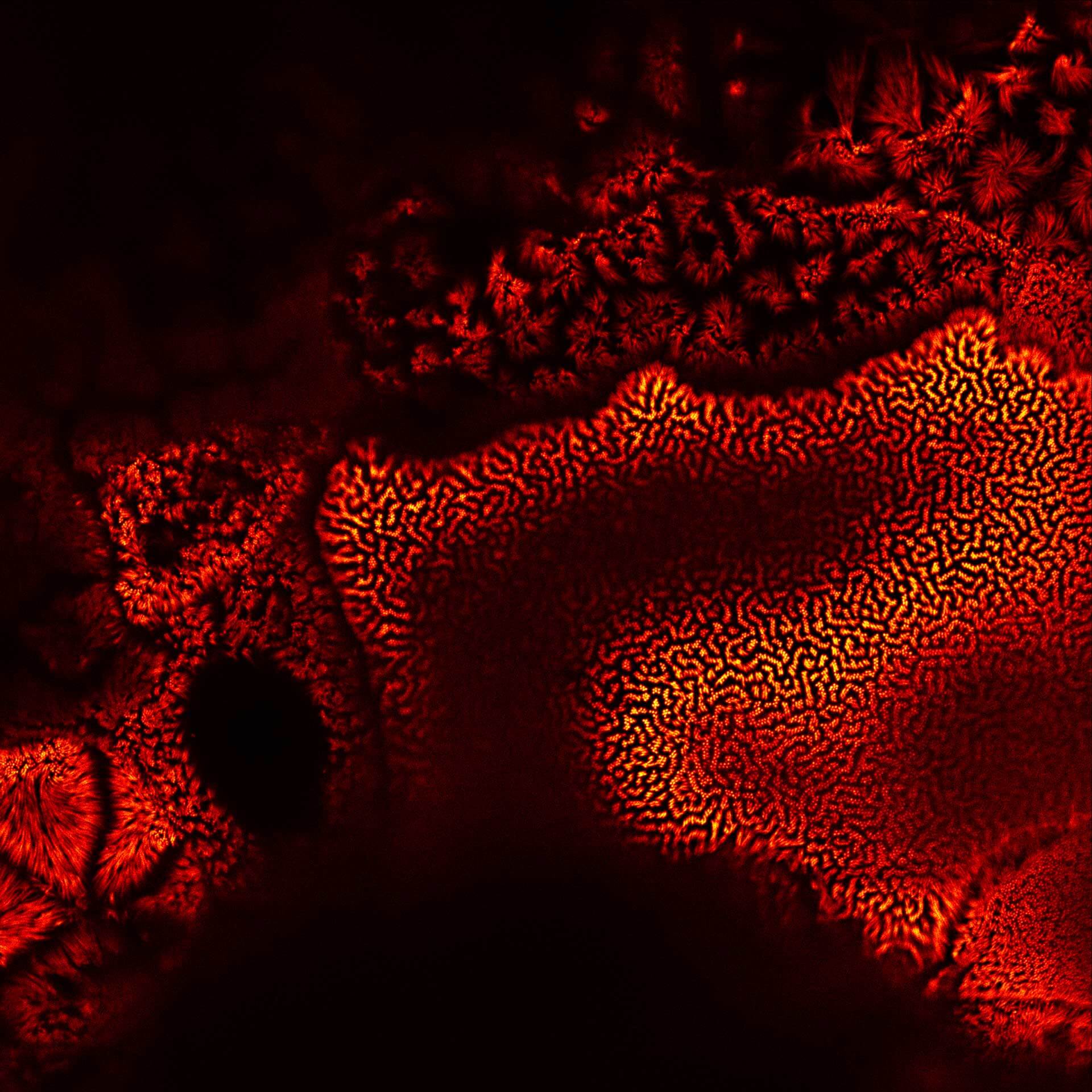
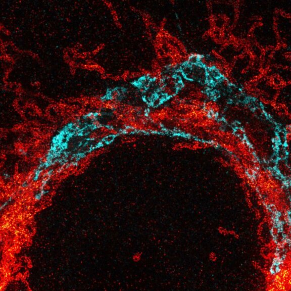
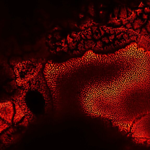
Actin in microvilli of Caco2 cells. Fixed cells were stained with abberior STAR RED phalloidin. MATRIX detection removes background signal and dramatically improves optical sectioning for 2D-STED.
Samples by Prof. Dorothee Günzel & Roman Mannweiler, Institut für Klinische Physiologie, Charité – Universitätsmedizin Berlin.
Modules:
Description
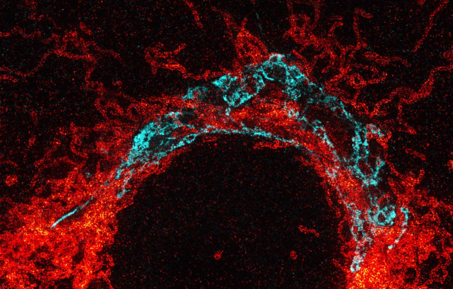
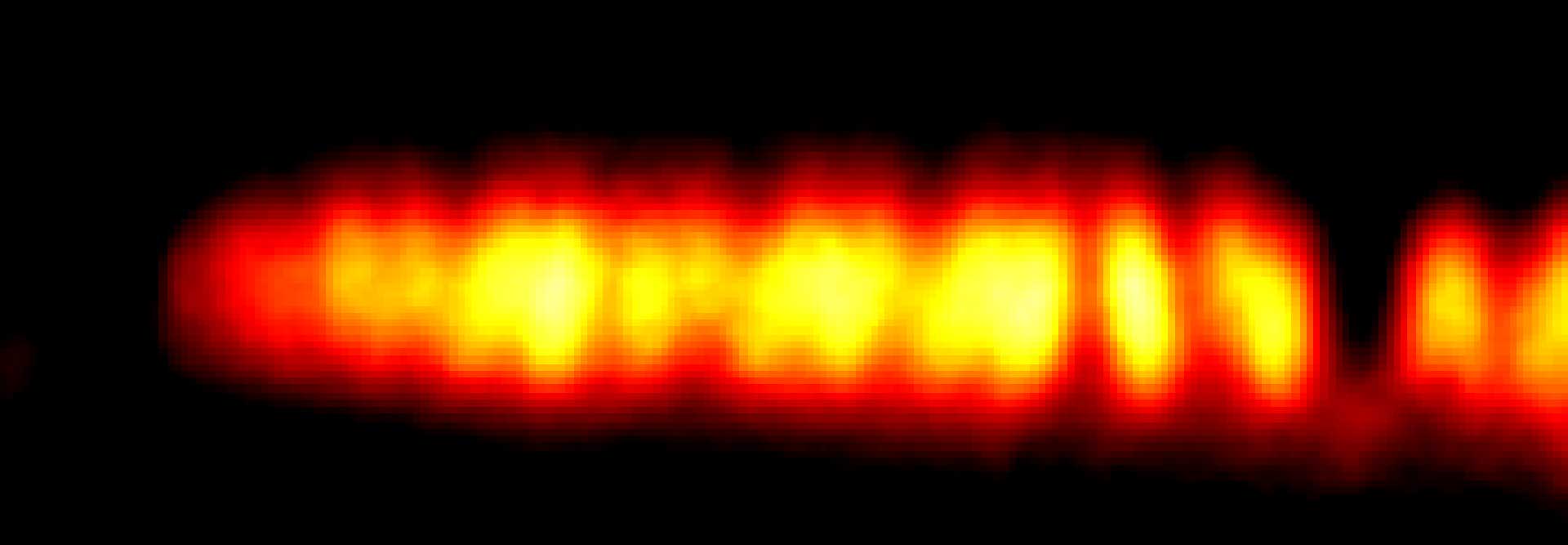
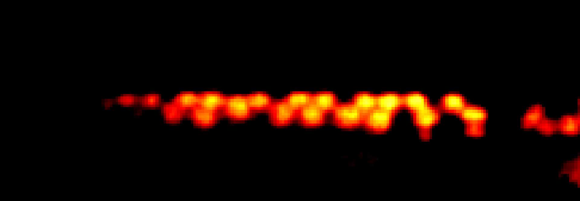
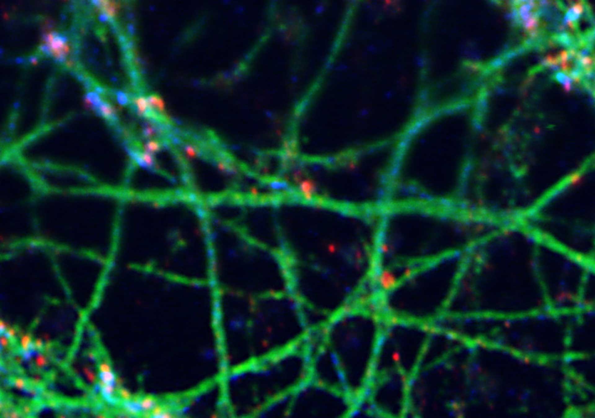
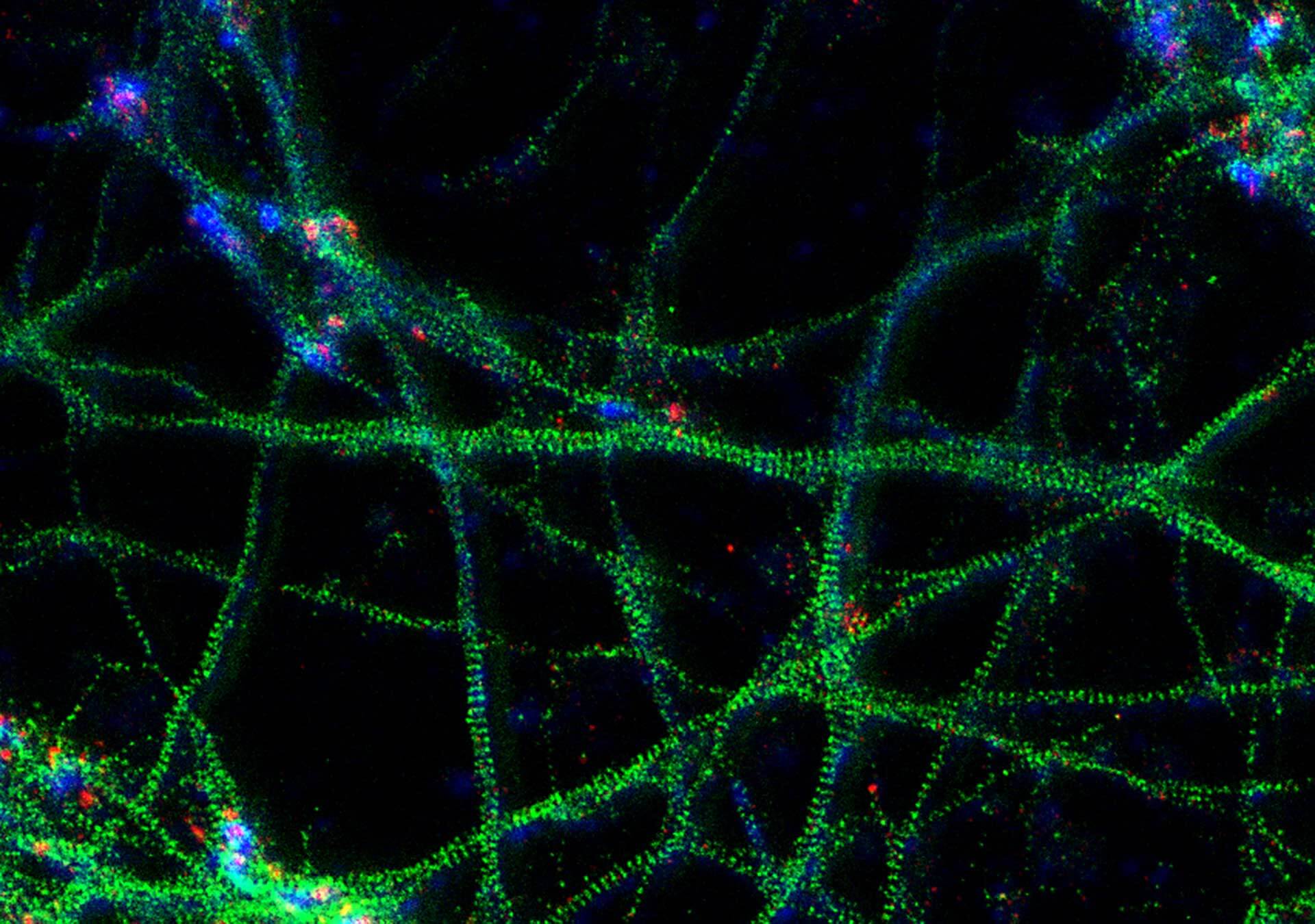
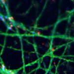
3-color STED image of primary hippocampal neurons. Please note the characteristic ~190 nm beta II spectrin periodicity along distal axons (green) which is only visible in the STED image. Labelled structures: beta II spectrin (green, abberior STAR 635P), Bassoon (red, abberior STAR 580), Actin cytoskeleton (blue, phalloidin, Oregon Green 488). Imaged with abberior Expert Line with 595nm and 775nm STED laser. Sample was prepared by Elisa D’Este @ MPIBPC, Göttingen.
Modules:


FACILITY
best for imaging
Our FACILTY system is a truly cutting-edge micro- and nanoscope that is incredibly easy to use. It combines advanced features for high-end confocal and superresolution imaging with foolproof operation. FLEXPOSURE adaptive illumination (DYMIN, RESCUE, MINFIELD), RAYSHAPE dynamic aberration correction, EASY3D STED, confocal & STED autofocus, spectral RAINBOW detection, and Full Autoalignment.
Software
sensational
Our LiGHTBOX software was designed from scratch to furnish a user interface that allows beginners to intuitively arrive at a top-notch STED image within three clicks, while it triggers a steep learning curve and allows expert users full control over the instrument. The unprecedented workflow outshines everything you have seen before.
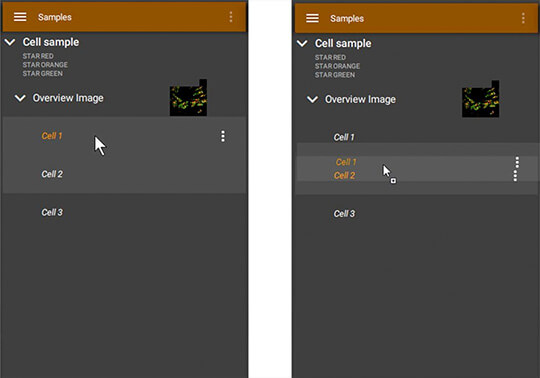
All images are grouped hierarchically in TreeView, where context between measurements is kept from the sample level over large overviews down to final images. This way, STED and confocal measurements can always be spatially related to the greater environment of the sample, and you can switch between different modes at the push of a button. There is no need to zoom out to find a good area, followed by zooming in, adjusting settings such as pixel size and line frequency, taking an image, zooming out again, re-adjusting pixel size… all this is one click in LiGHTBOX. On top, TreeView supports drag and drop (right) to transfer settings to other images. Once you have found good settings for your sample, they can be applied lightning fast to all images, in this case from a measured “Cell 1” to a new region of interest called “Cell 2”
The LiGHTBOX supports new and advanced users alike. After entering the dyes that are in your sample, the machine knows everything about excitation wavelengths, detection bands, STED settings, etc. From there, you simply select a region you want to image and the first scan will give you a good superresolution measurement. After this, it’s a breeze to optimize settings.
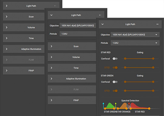
All functions are grouped in convenient tabs that can be escalated to adapt to different user levels. In the example above, Light path controls are unfolded, first to show basic objective and pinhole controls, while a more advanced user can move one escalation higher to manipulate gating and detection settings. This way, you only see what you need in the moment, cluttered screens with hundreds of buttons are avoided, and new users don’t feel overwhelmed, while advanced users are in full control.
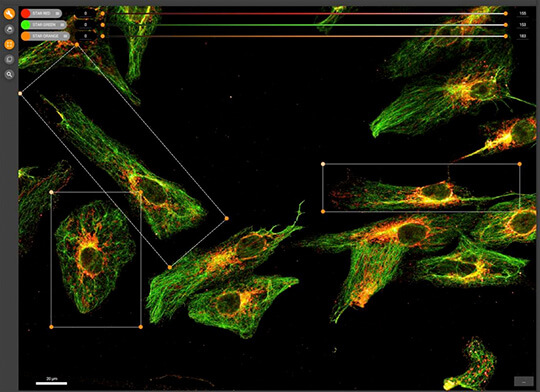
The LiGHTBOX allows tiled scans, and in every scan you can select an arbitrary number of regions of interests (ROIs, corresponding to items in TreeView, see above). Every ROI itself can serve as an overview again, meaning that you can easily climb down from sample level overviews to detailed superresolution images, while spatial context is saved at all times. Of course, the FACILITY supports arbitrarily rotated ROIs, even in STED mode!
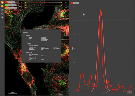
After you’ve acquired your images, our software offers powerful analysis features, such as measurements, plus, images can be sent to Huygens SVI for stitching, deconvolution, and rendering.
Visit the LiGHTBOX software page to explore even more features.
What others say
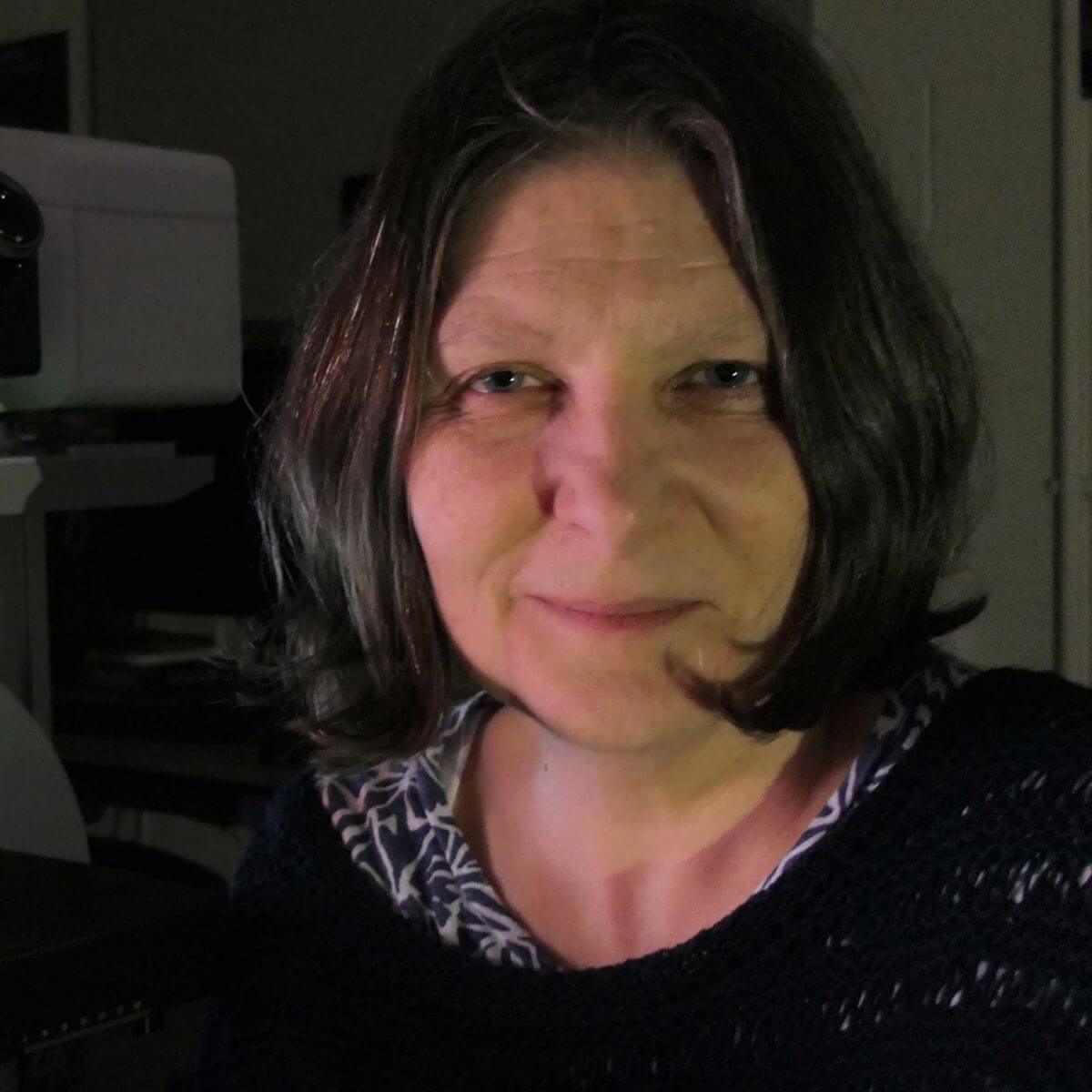
Prof. Michelle Peckham
University of Leeds
Leeds, United Kingdom
“Like the STEDYCON, the FACILITY line is very easy to use and it will work very well in an imaging facility.”
Dr. Antonio Virgilio Failla
Head of the UKE Microscopy Imaging Facility
Hamburg, Germany
“The FACILITY line is the best way to produce world class scientific results. It is driven by a fantastic user interface and I would recommend it to every imaging facility.”


Dr. Herlinde de Keersmaeker,
Manager of the Centre for Advanced Light Microscopy
Ghent University, Belgium
“After the installation of our FACILITY line, we are very pleased with the excellent and very fast after sales service and support that abberior instruments offers. This is highly appreciated and necessary to operate as imaging Core Facility.”


Prof. Christian Eggeling
Friedrich-Schiller-Universität Jena, Germany, and
University of Oxford, United Kingdom
“The FACILITY line is a highly flexible turn-key instrument that combines extremely powerful superresolution imaging, including unique features such as adaptive optics, and intellingent scanning schemes, with a fantastic user interface.”
Dr. Hans-Ulrich Fried
Head of the Light Microscope Facility, DZNE
Bonn, Germany
“The FACILITY line is an impressive superresolution and confocal microscope combining ease of use with cutting edge technology. The usage of intelligent illumination schemes allows live-cell recordings with high-resolution STED at an extraordinary low light level.”
- Advanced confocal and superresolution features (EASY3D, tiling/stitching, multi-ROI recordings, adaptive illumination, full autoalignment, …)
- Unprecedented workflow
Powerful performance combined with sublime useability

Why do we usually recommend APDs in our microscopes and why aren’t we worried about the supposedly lower dynamic range?
Having too many photons is never a problem. Therefore, detectors with the highest quantum efficiency are always the best choice, such as in a MATRIX array.
MATRIX Detector
Many eyes see more than one. The MATRIX detector drastically improves signal-to-background ratio, resolution, and dynamic range.
Modules
MODULES expand your microscope and turn it into a powerful instrument ready to take up any imaging challenge.