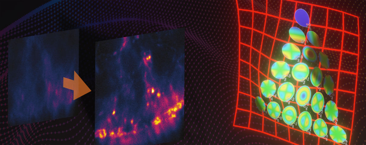
Confocal and multi-photon microscopy are used for deep tissue imaging, but misconceptions about their utility have led to their misuse. We’ll plunge into tissue depths to reveal a gap in obtaining sharp images that RAYSHAPE – a solution for dynamic aberration correction – fills with clarity and brightness. Details >
Knowledge Base
Have you ever wondered how superresolution microscopy works? What’s the difference between STED, STORM, and MINFLUX? What is “resolution” and what is a “PSF”? What is so special about the STEDYCON? Read on to find out.
If you have any suggestions, questions or ideas for our knowledge base, we would be very happy to hear from you.
ContactEverything about microscopes, dyes, and superresolution
Expansion microscopy turns the attention to the specimen. It achieves high-resolution images via a chemical rather than optical approach. Preserved specimens are physically enlarged within a swellable hydrogel to allow 3D nano-imaging using conventional microscopes. Tuning the sample may sound tempting, but it comes with some relevant drawbacks. Details >
How does STED work?

You have heard of STED but don’t have a clear idea how it overcomes the diffraction-limited resolution of confocal microscopes? You maybe even think it to be somewhat complicated? In fact, it isn’t. It’s just physics, smartly applied. Details >
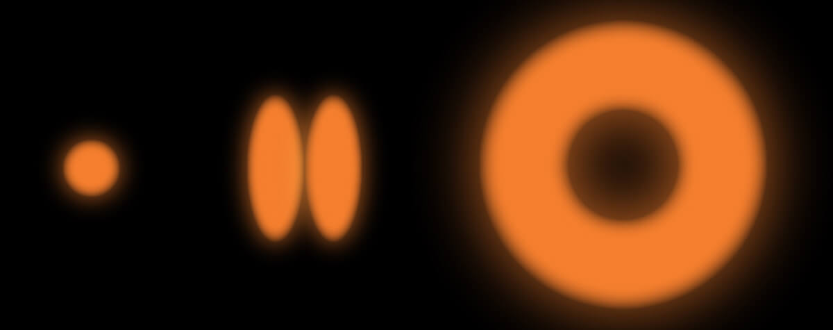
The donut-shaped de-excitation beam is one of the most important practical ingredients for superresolution STED microscopy. But how do you put a hole into a beam of light? Surprisingly, it’s not that difficult if you know how to do it, but it’s very difficult to get it right in practice. Details >
What has to be inside a STED microscope to achieve superresolution? How does its hardware differ from a confocal setup? (Hint: Not very much.) And what does that mean for the user? (Many good things.) Is handling a STED system any more complicated than using a confocal? (Not really.) Important questions – here are some in-depth answers. Details >
Since the 1990s, confocal microscopes have been a staple in labs visualizing biological or material specimens. The development of STED microscopy prompted the question: how does the established confocal microscope compare to the (now not so) “new kid on the block”? Details >
How the donut changed the world

For over a century, we stood at the edge of microscope resolution and cursed the inexorable blur of diffracted light. Instruments improved, but the fog never lifted. Then, one man stopped trying to control how light behaves. Armed with a donut-shaped laser beam, he instead commanded where it shines and untethered resolution forever. Details >
PALM and STORM are often used as synonyms, and in fact they have a lot in common. But there are slight differences that can be important for your application. And then there are other superresolution techniques, too – like STED and MINFLUX. Details >
Structured illumination microscopy offers some advantages over confocal, most notably increased resolution. Comparing it to STED, however, reveals its limitations. Details >
Which microscope has the best resolution?

The elctron microscope achieves the highest magnification and resolution. But does "highest" always equal "best"? Well, that depends on what you want to do with the resolution. Details >
Confocal microscopy offers superior optical sectioning. But what is that exactly? And what about other ways to get rid of the background, such as array-based detectors like the MATRIX? Details >
Today’s research microscopes are increasingly powerful, modular, and combinatorial. There’s a lot of options out there. While the price is unquestionably a deal-breaker for purchase, a more helpful criterion is value. Details >
For centuries, conventional light microscopy was and continues to be the workhorse of labs to visualize cells and cellular details. But the advent of electron microscopy brought about a new level of detail. Let's take a closer look at the two techniques. Details >
Deep and clear: where confocal beats out wide-field microscopy

Confocal microscopes were designed to get rid of background signal. How do they work? And when do you know it’s time to use one? The answer is in the pinhole. Details >
Fluorescent labeling strategies have become more and more sophisticated and offer ever-new options to improve microscopic imaging. Among the latest are exchangeable HaloTag ligands that put an end to photobleaching for good. Details >
Today’s high-end fluorescence microscopy is unthinkable without lasers. Reason enough to take a closer look at these sophisticated light sources. Details >
How to correct for aberrations in light microscopy

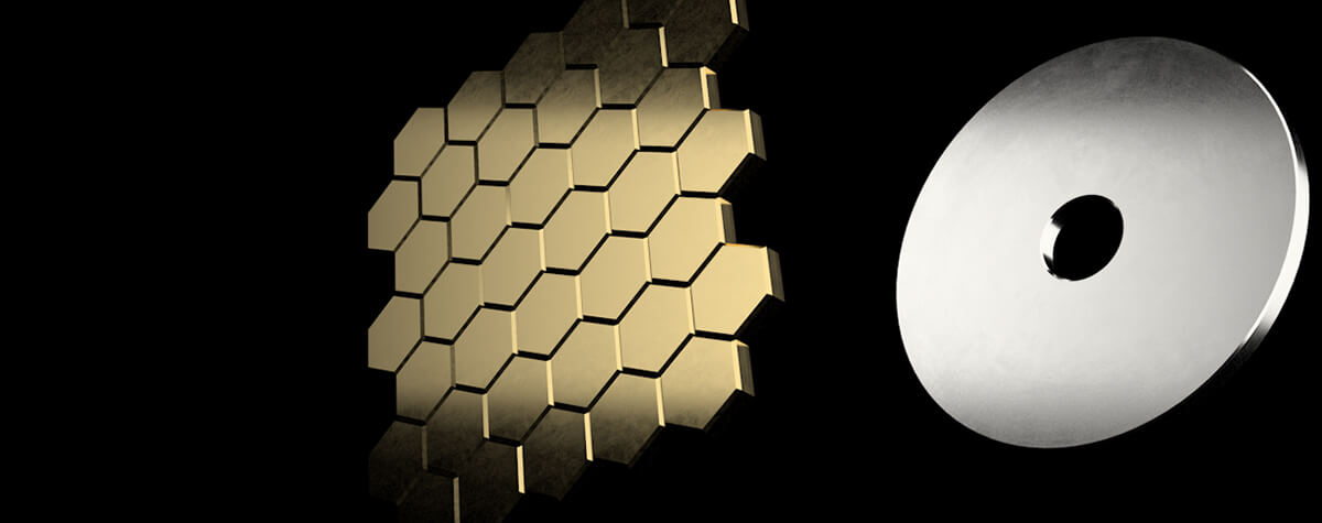
Aberrations can give microscopists a hard time. They belong to microscopy like pathogens belong to life. There are ways to bring diverted rays back on track, but some are better than others. The question is: deformable mirror or correction collar? Details >
Every technique that allows to observe cells is more or less invasive and fluorescence microscopy is no exception. Many imaging situations profit from a reduction in light dose as provided by FLEXPOSURE adaptive illumination. Details >
MATRIX STED is the next level of STED microscopy – combining superior resolution with outstanding signal quality and clarity. Details >
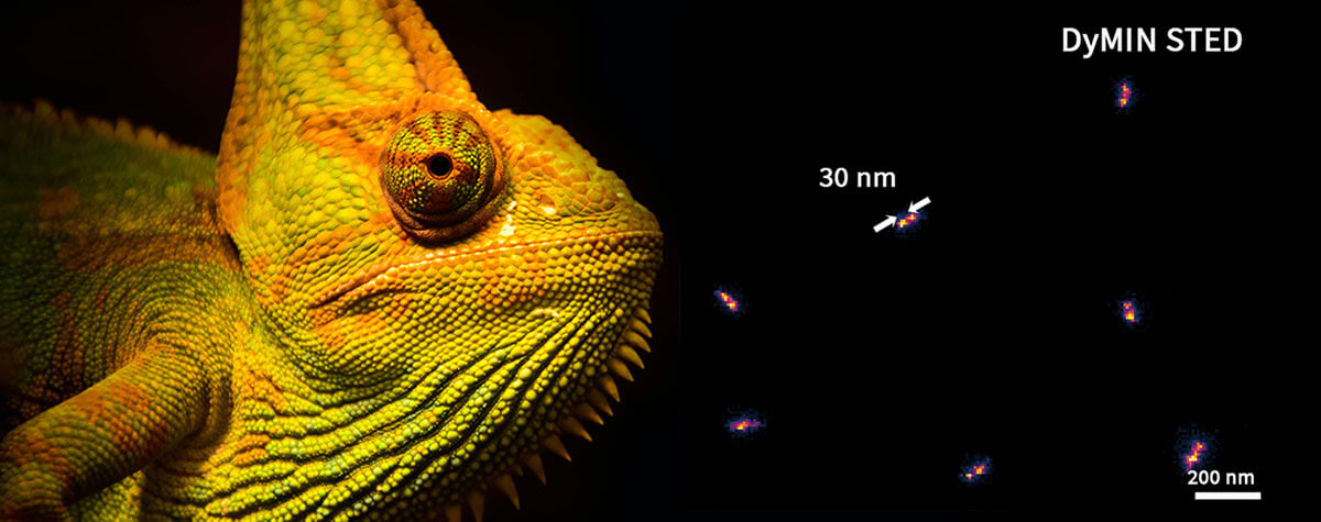
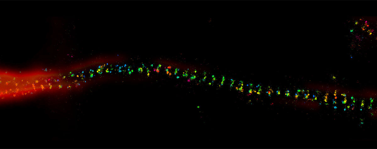
MINFLUX reaches unprecedented spatio-temporal resolution in light microscopy and provides 2D and 3D localization precisions in the single-digit nanometer range. Details >
Ideal imaging conditions are often compromised by imperfections in the optical path. These can severely compromise a microscope’s performance, unless they are eliminated by RAYSHAPE's deformable mirror. Details >
Why do superresolution microscopists love alpacas?

It is a very simple yet very important fact: the localization precision of any superresolution microscope can only be as good as the size of the fluorescent staining allows. In other words, when your fluorescent dye is too big or too far away from the protein you want to label, you will never be able to reach a resolution that is higher than this offset. The good news is: there are ways to reduce the offset between target protein and fluorescent label. And one of these are nanobodies. Details >
Let the cells shine with immunofluorescence labeling

The most versatile and therefore most common strategy to bring the dye to the sample is immunofluorescence. In case you always wanted to know how immunofluorescence works and which properties of antibodies make it so powerful and at the same time define its limits! Details >
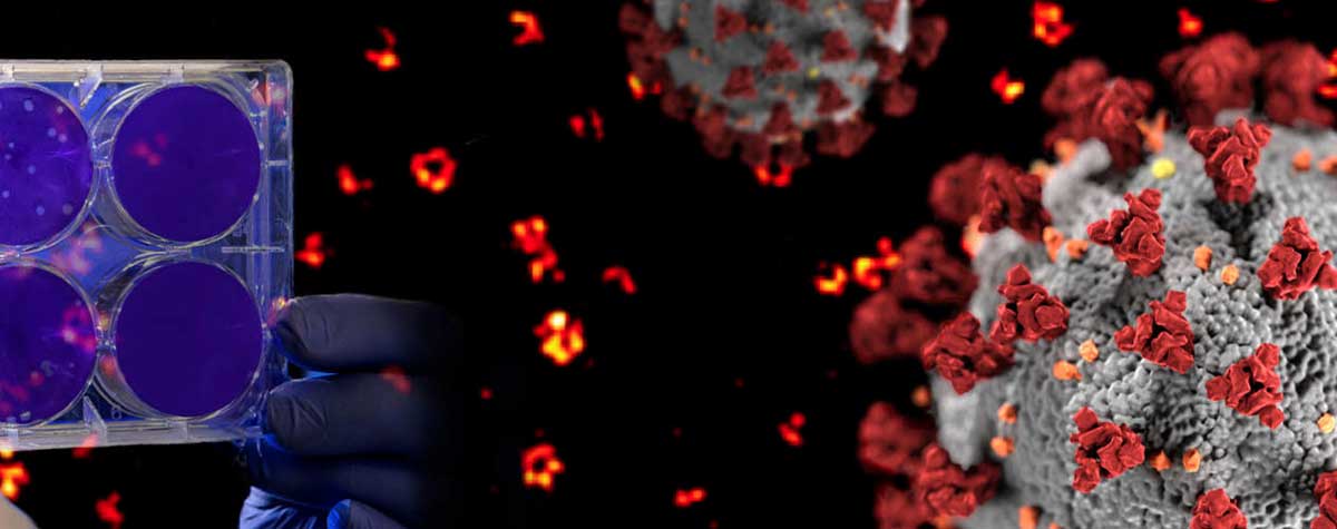
A little insight into the advances in virus research made possible by STED microscopy and a hint to were the journey might go. Details >
The combination of STED microscopy and PAINT circumvents the physical limitations of current labeling technology. Details >
For STED microscopy, similar sample preparation techniques may be utilized as for conventional microscopy. However, the increase in special resolution requires additional precautions to ensure the structural preservation of the specimen. Details >
Superresolution for biology: when size, time, and context matter

The spatial resolution achievable with today’s light microscopes has unveiled life at the scale of individual molecules. Size is no longer a barrier to seeing biology at the most fundamental level. But life is not static. It emerges from movement and change. How do superresolution technologies hold up to the challenges of documenting dynamic biological mechanisms? Details >
Photon numbers from the emitting fluorophore. Width of the PSF. How do they impact the resolution of a microscope? Here’s a simple graphic that lays out those effects. Details >
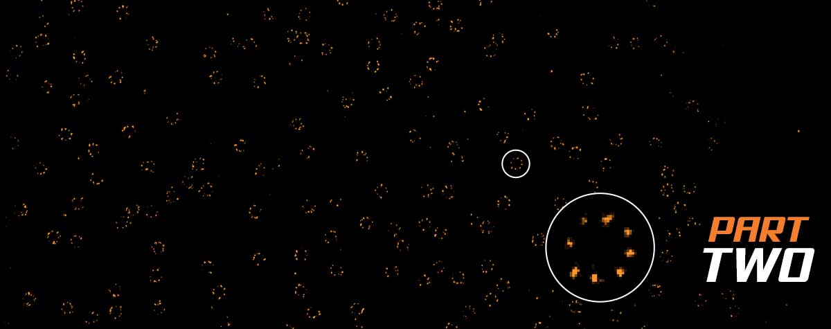
For all the talk about criteria and definitions, measuring the resolution of a microscope is more nuanced than you’d think. The scales at which microscopes operate today are subject to noise and background that obscure and distort signals. What you use for the measurement can make a big difference. The second article in our "Resolution" series. Details >
STEDYCON: ease-of-use in a shoebox

A sleek, black-and-orange box transforms your widefield microscope into a confocal and a superresolution STED instrument and your exploration of subcellular structures into a seamless, discovery-rich experience. Carefully designed with masterly engineering, STEDYCON breaks the stereotype of the finicky, hard-to-use scope. It opens new possibilities at the press of a button for any user and almost any location. How does it do it? The secret’s in the box. Details >


