


MINFLUX 3D
The MINFLUX platform offers an unprecedented array of imaging possibilities and allows you to resolve structures as small as a molecule, along all three dimensions.
This unmatched resolution capability combined with unprecedented speeds reveals sample details never seen before. The MINFLUX is the world’s most powerful fluorescence microscope.
unrivaled resolution and speed

Description
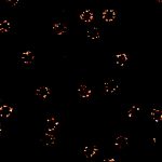
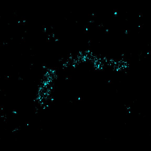
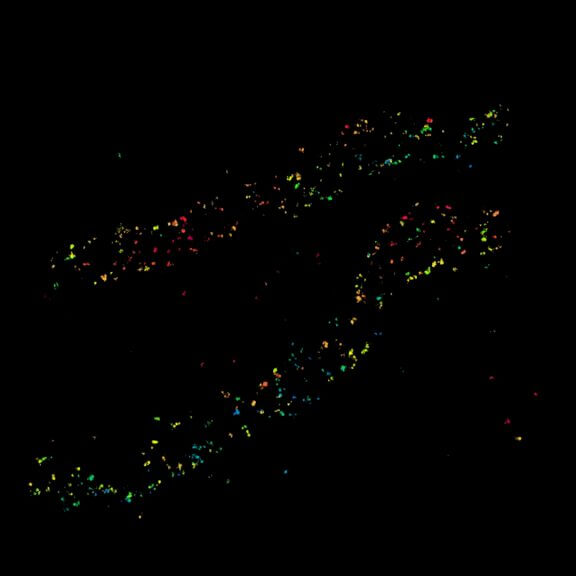
2D MINFLUX nanoscopy of Y. enterocolitica sorting platform protein Halo-YscL.
Sample from Alexander Carsten, Prof. Martin Aepfelbacher, Institute of Medical Microbiology, Virology and Hygiene, University Medical Center Hamburg Eppendorf, Germany
Description
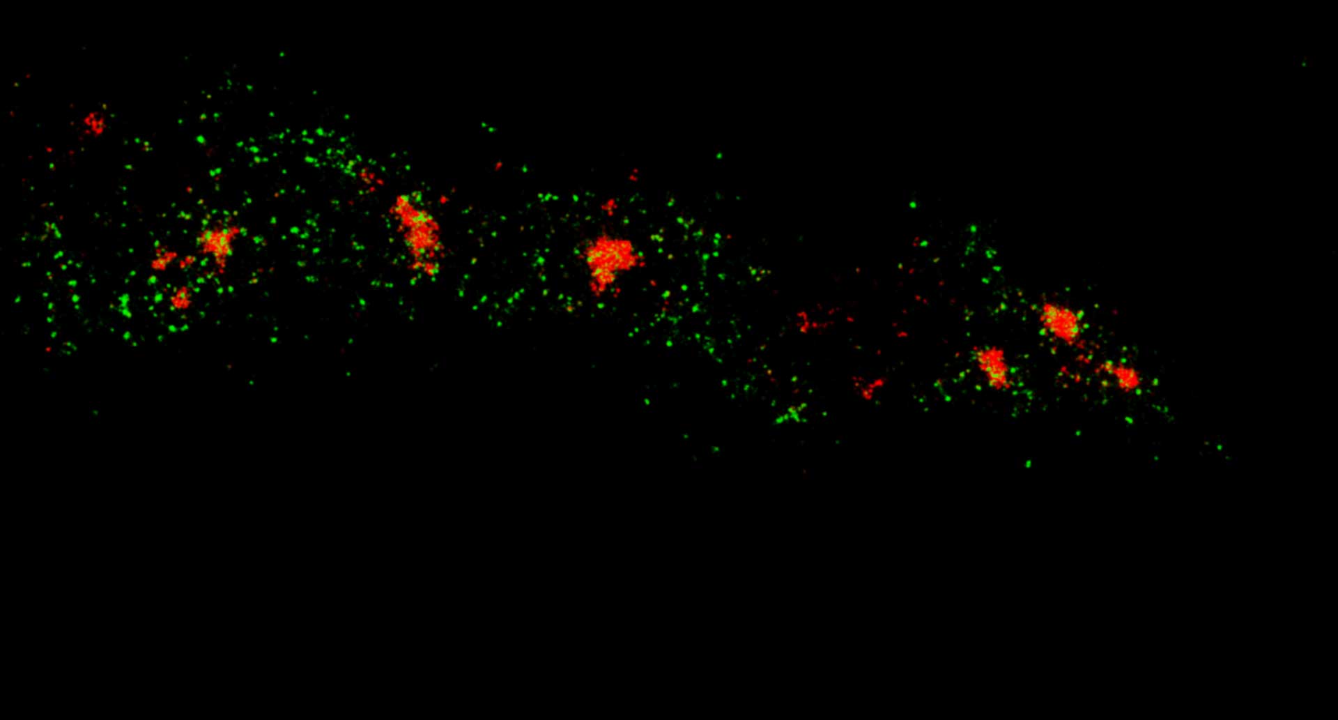
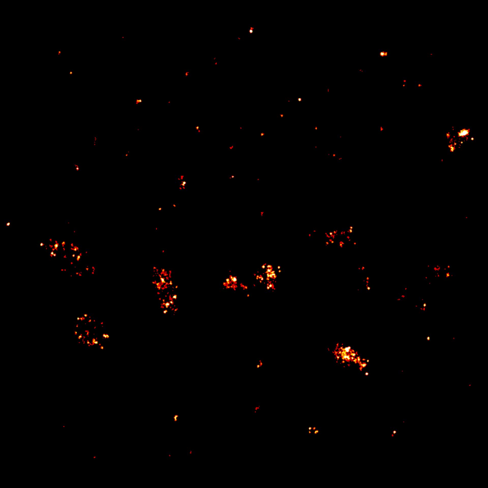
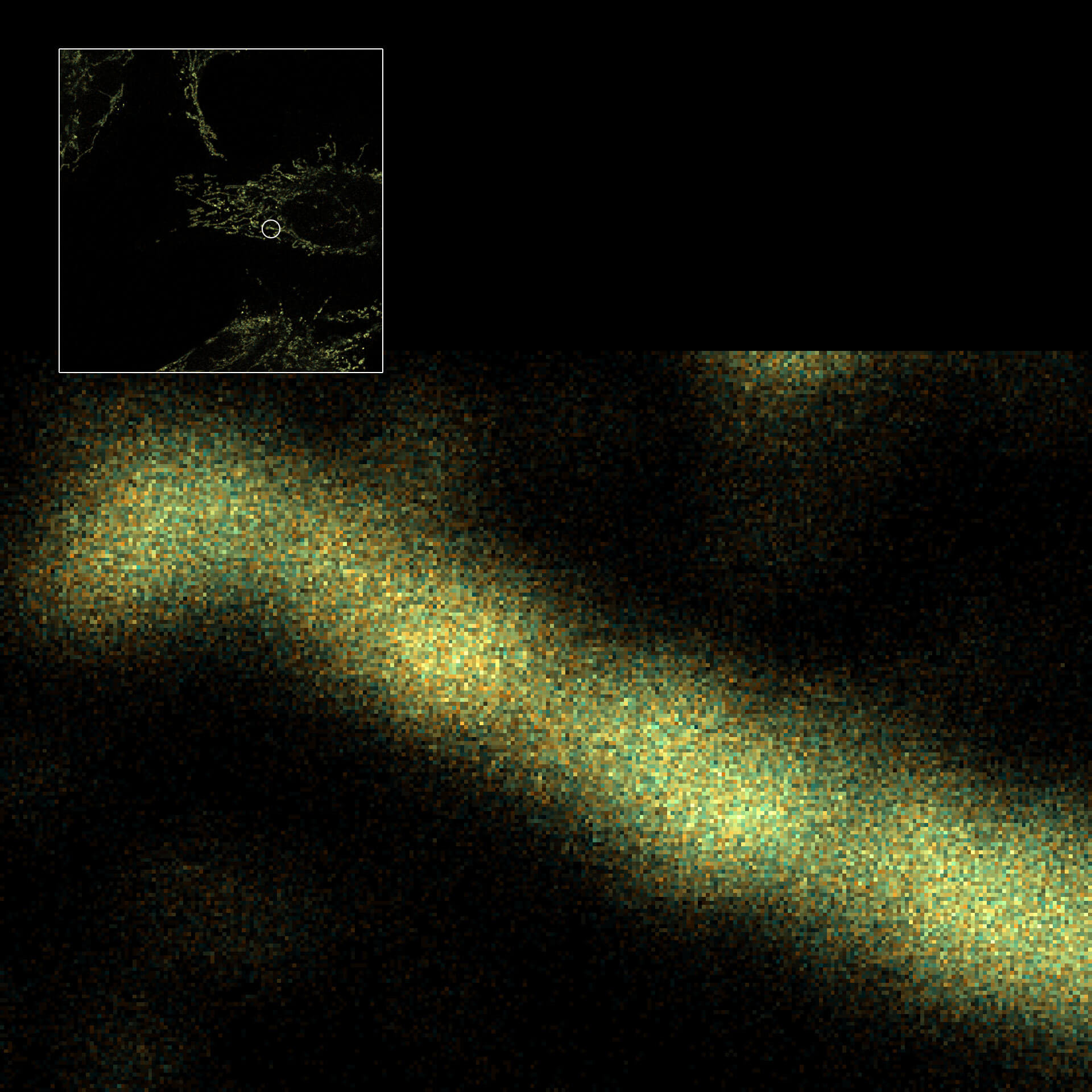
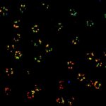
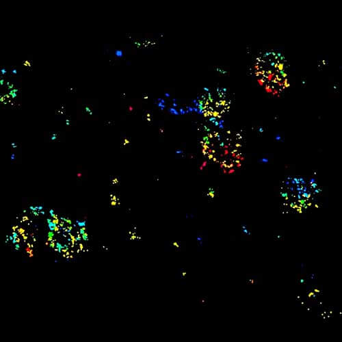
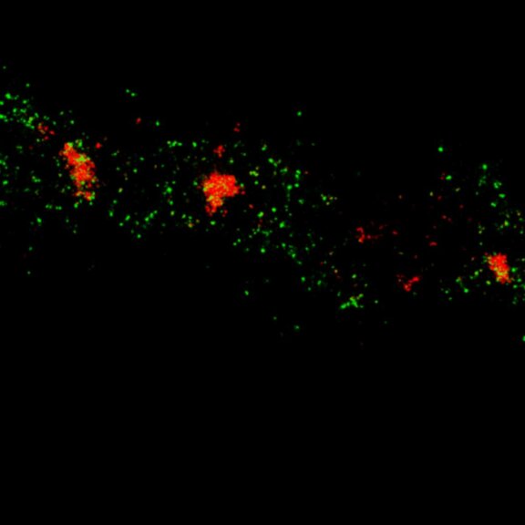
Two-color MINFLUX on mitochondria samples. The mitochondrial proteins TOM20 (green) and mtDNA (red) were labeled in mammalian cells with indirect immunofluorescence using secondary antibodies coupled to sCy5 and CF680. Two-color confocal (A) and MINFLUX (B) was performed using a ratiometric detection strategy. Please note that the labeling density of both structures is highly dissimilar. For TOM20 single proteins are labeled in the mitochondrial membrane, whereas numerous binding sites are decorated in the mtDNA. MINFLUX enables the visualization and separation of both structures.
Description
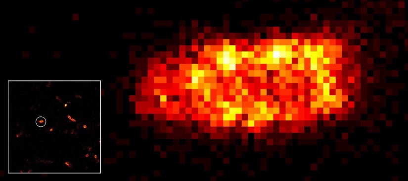
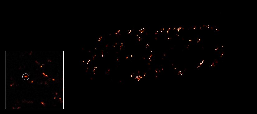
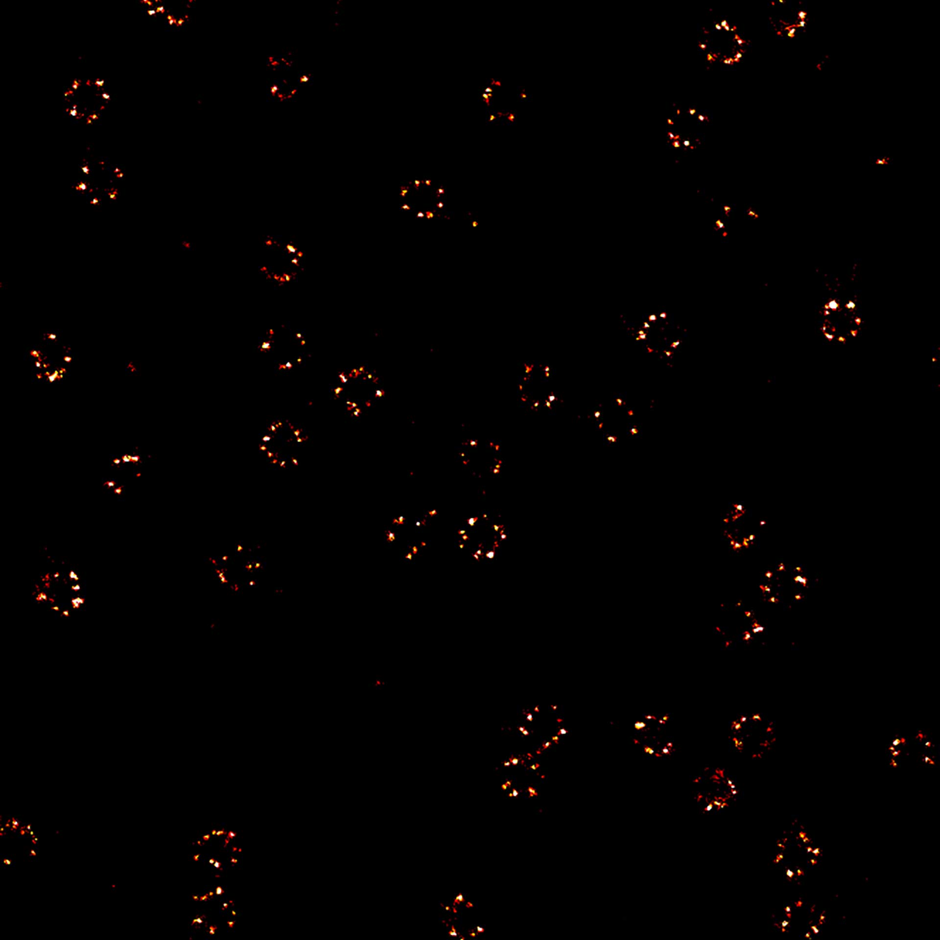
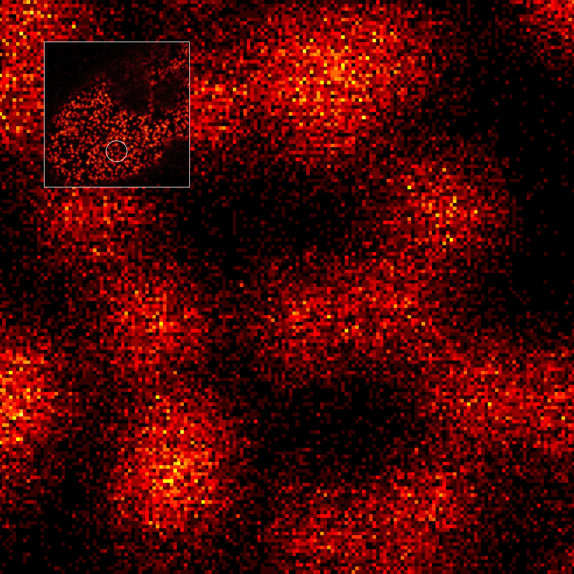
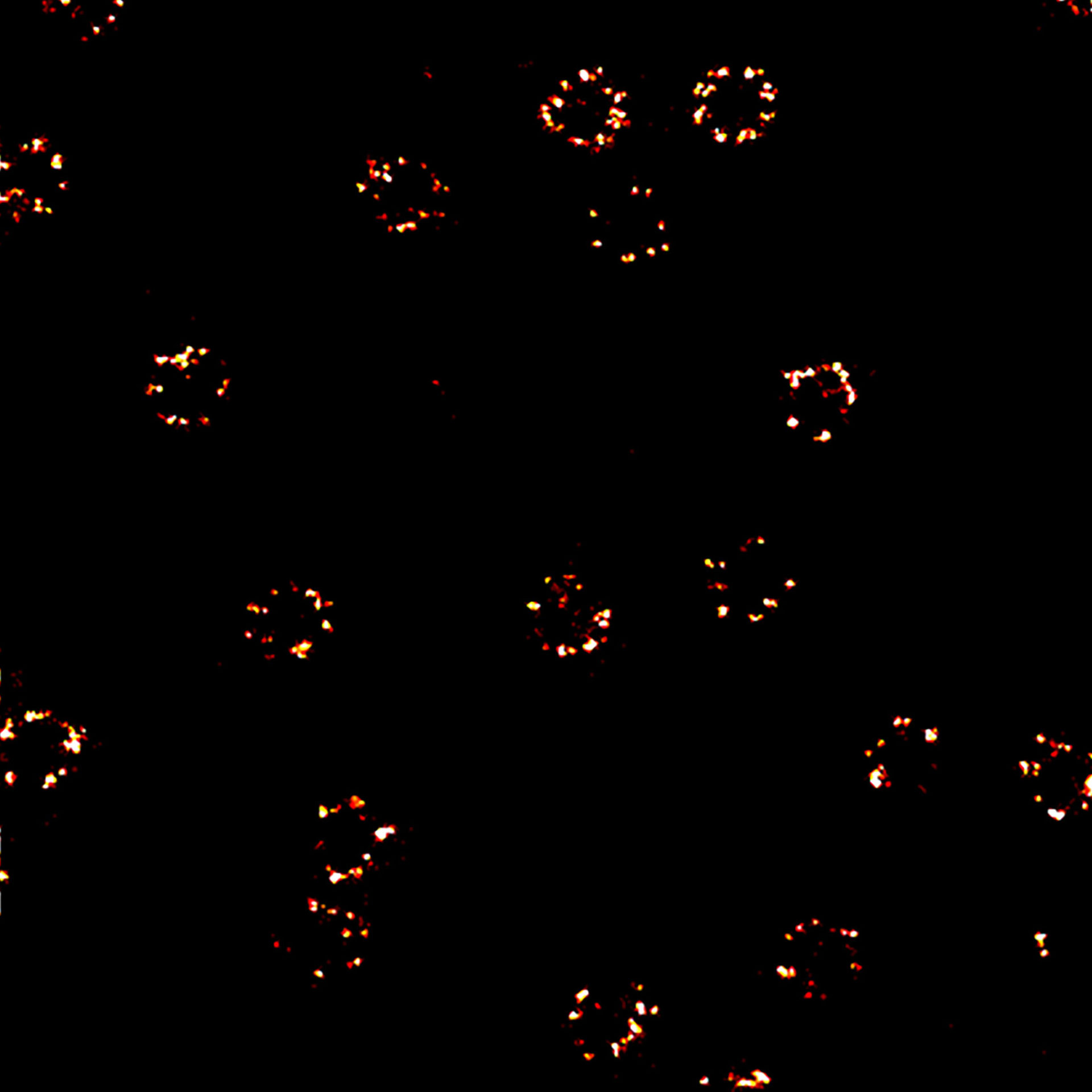
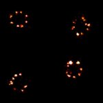
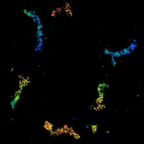
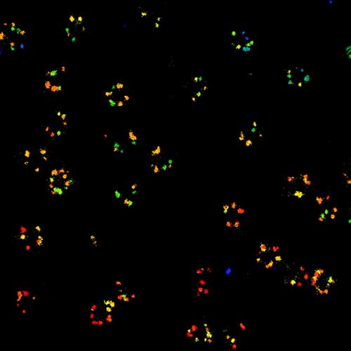
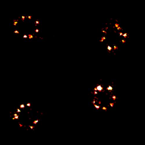
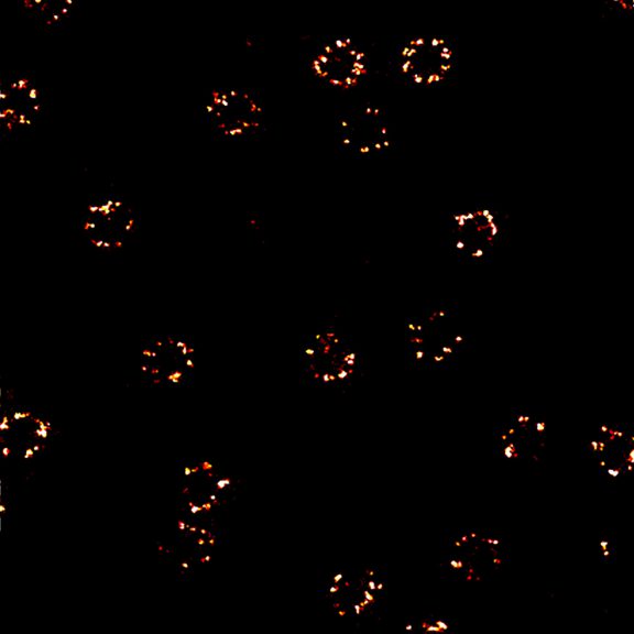
2D MINFLUX nanoscopy of the nuclear pore complex subunits. NUP96-SNAP/SNAP-Alexa Fluor 647 lend themselves as benchmark structures to test superresolution light microscopes. In contrast to confocal microscopy, 2D MINFLUX allows to visualize the shape and arrangement of individual subunits of the nuclear pore complex. Here, we reach localization precisions of ~2 nm in raw localization data.

Description
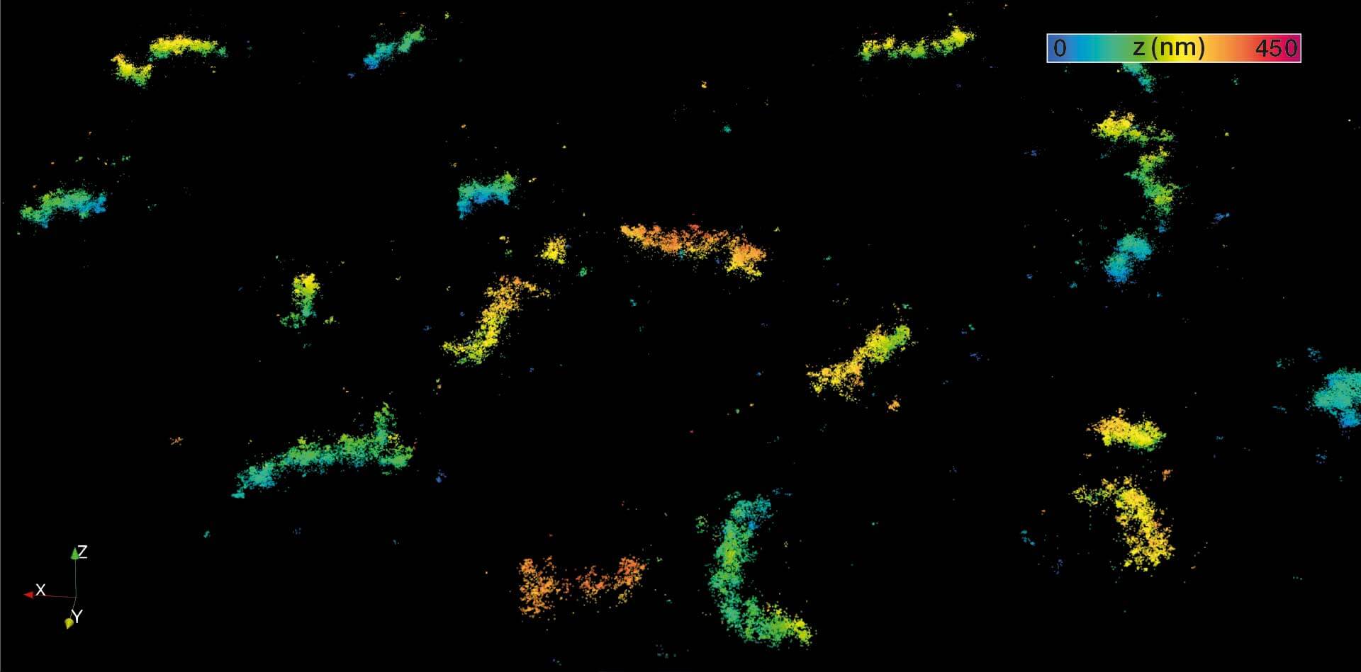
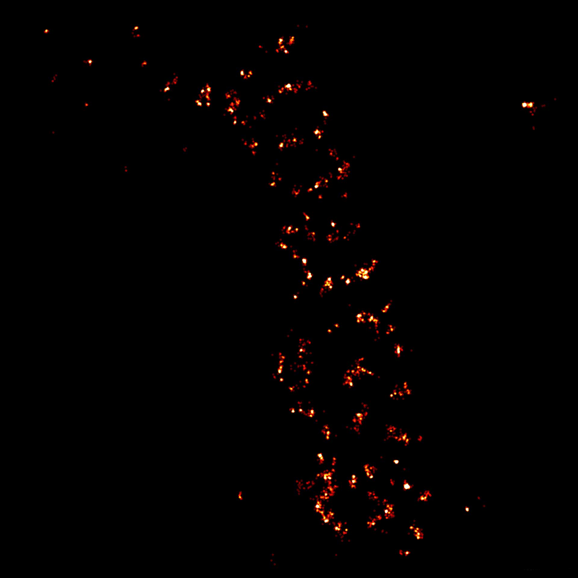
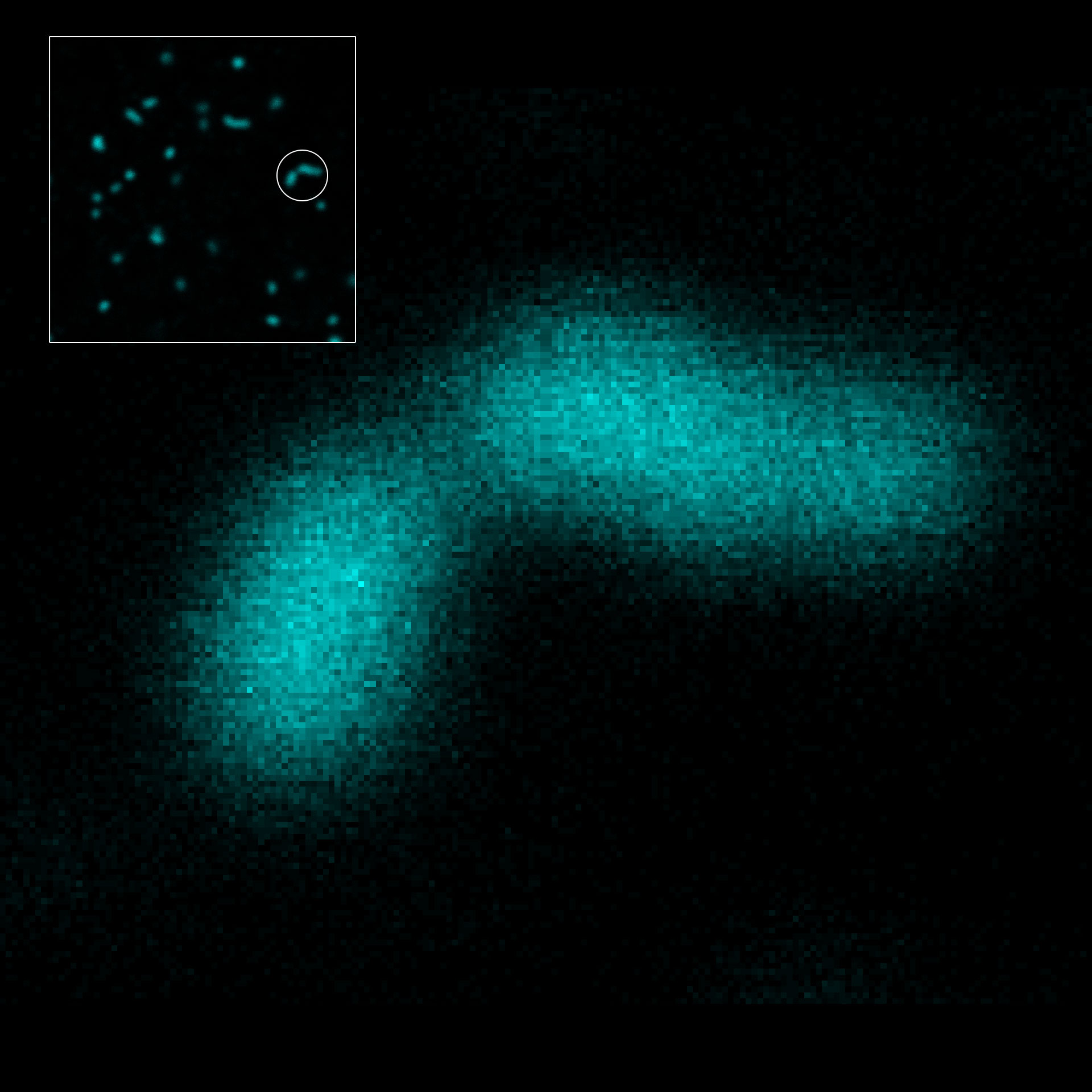
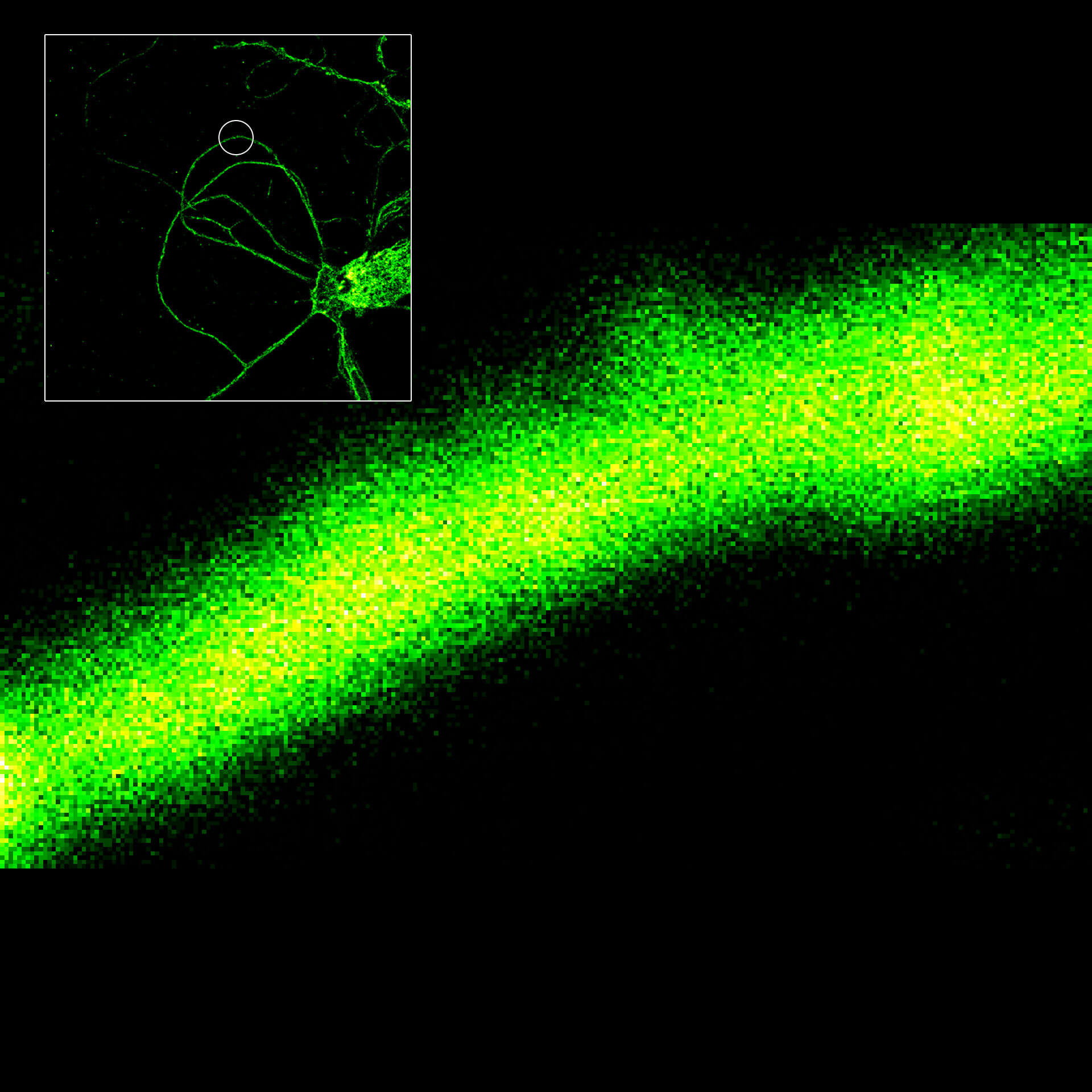
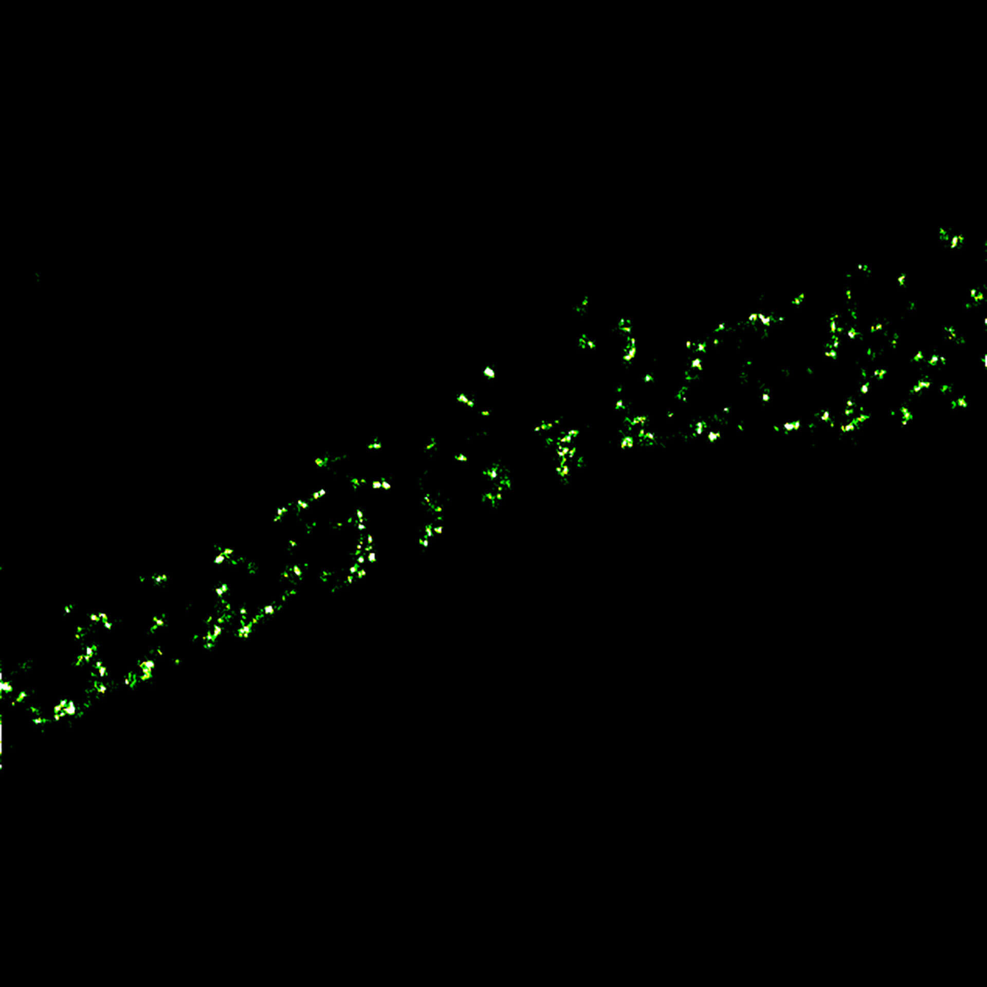
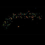
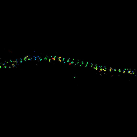
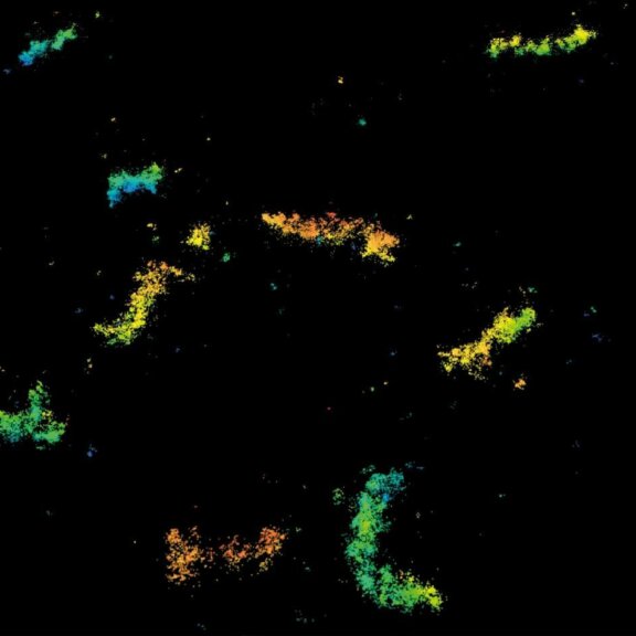
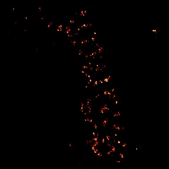
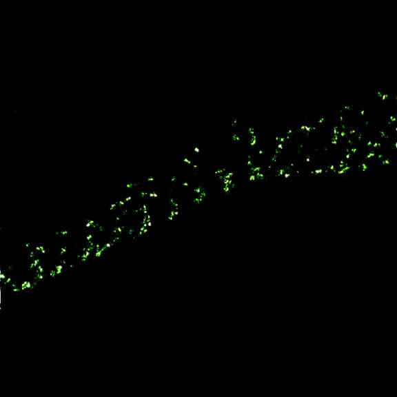
MINFLUX image of axonal bII spectrin in primary hippocampal neurons with < 2 nm resolution. Note the periodic arrangement of spectrin along the axon, and the absence of any details in the confocal counterpart image.
Description
2D MINFLUX imaging of the peroxisomal membrane protein PMP70 labeled with Alexa Fluor 647 in fixed mammalian cells using indirect immunofluorescence
Description
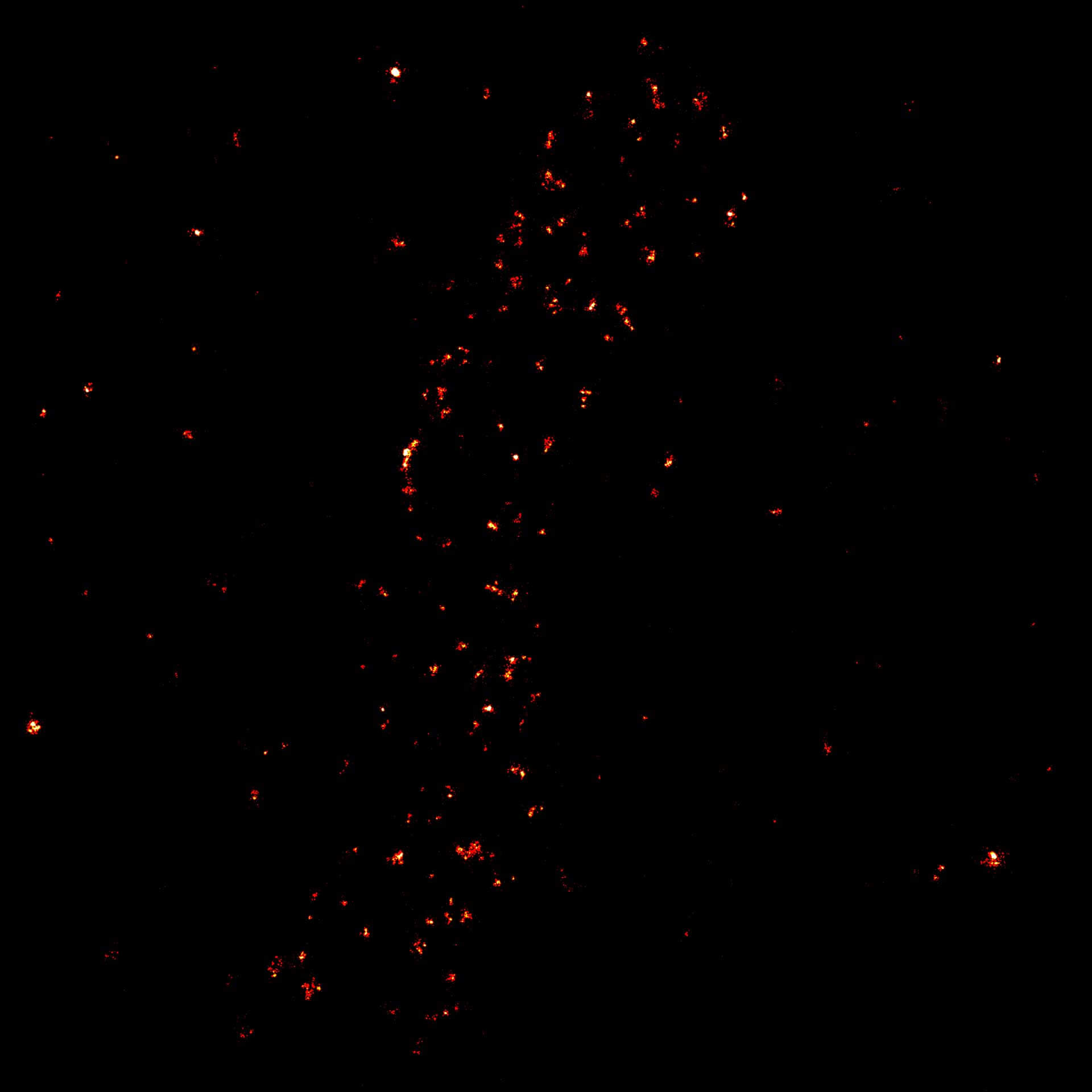
2D MINFLUX image of the mitochondrial import receptor Tom20 labeled with Alexa Fluor 647 in fixed mammalian cells using indirect immunolabeling.

Description
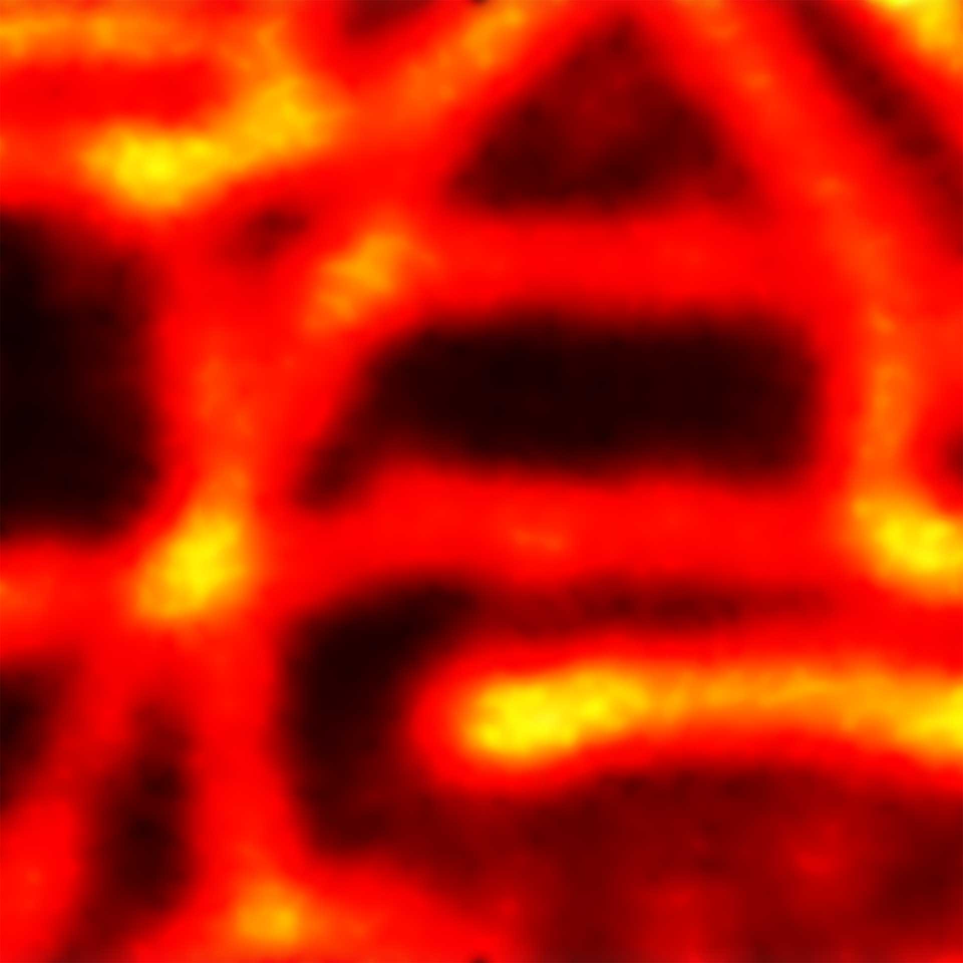
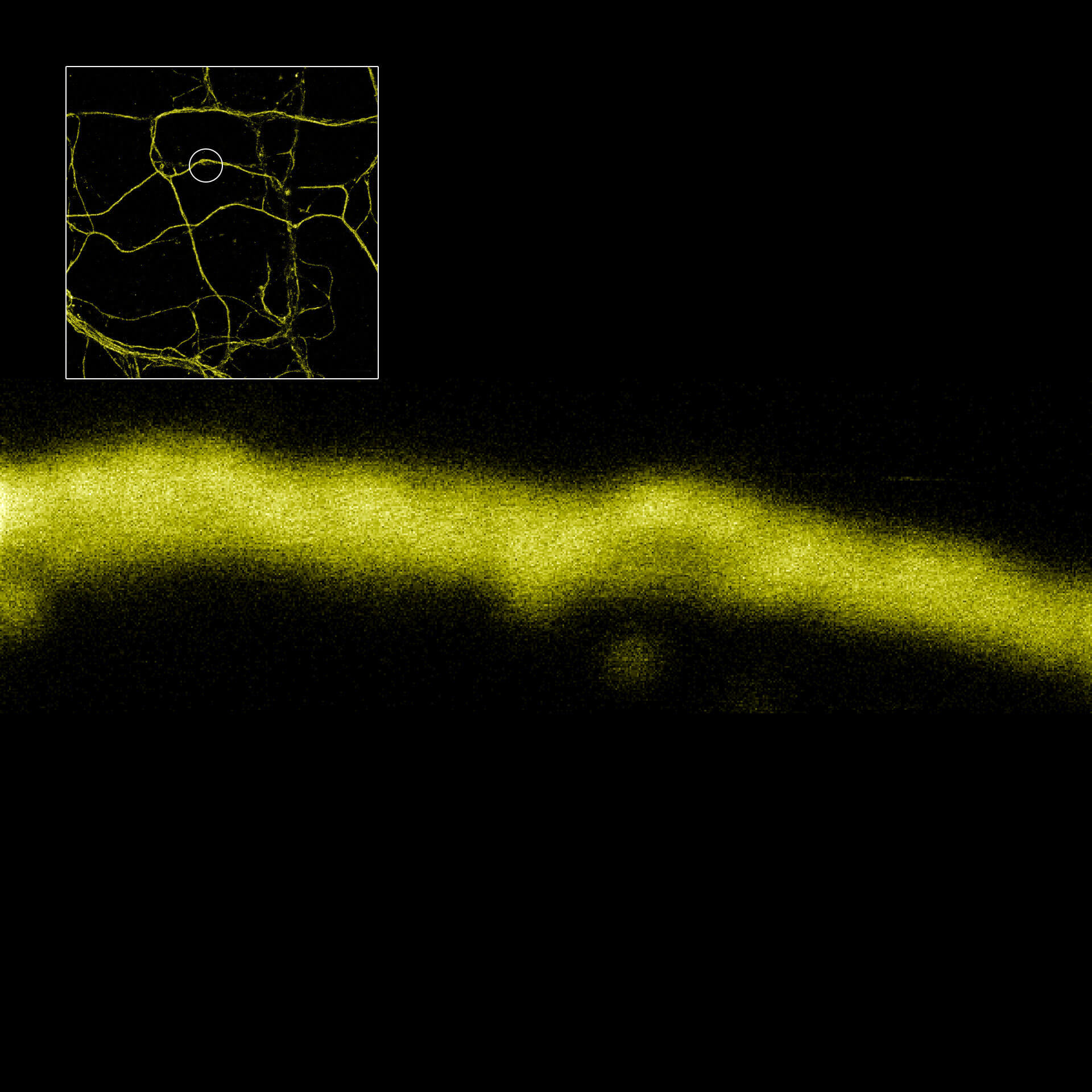
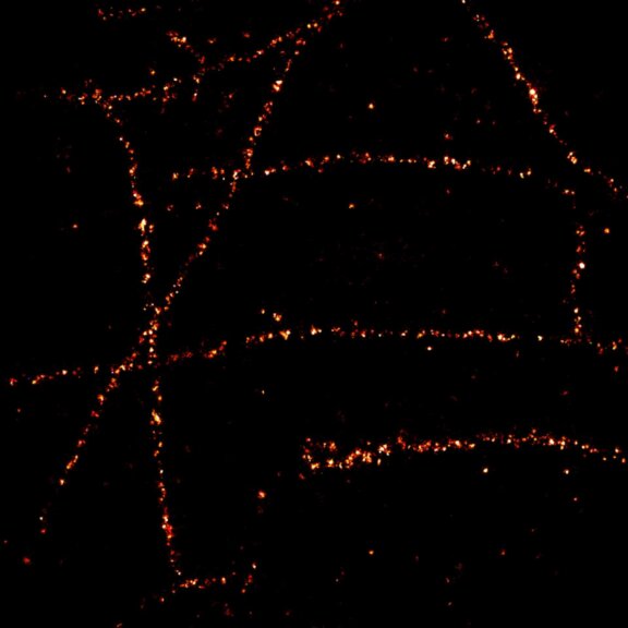
2D MINFLUX imaging of the cytoskeletal protein vimentin. Vimentin was labeled with Alexa Fluor 647 in fixed mammalian cells using indirect immunofluorescence. Note the individual filaments at intersections are invisible in the confocal image.
Description
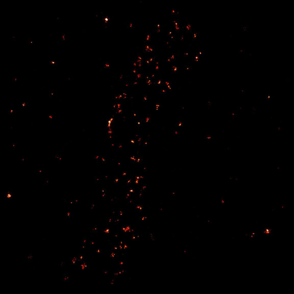
2D tracking of a lipid-coupled Atto 647N molecule embedded in a supported lipid bilayer. The movement of the labeled lipid was tracked with a frequency of up to 10 kHz.

Description
2D MINFLUX imaging of the peroxisomal membrane protein PMP70 labeled with abberior FLUX 640 in fixed mammalian cells using indirect immunofluorescence.
Description
2D MINFLUX nanoscopy of the nuclear pore complex subunits. Fixed mammalian cells expressing SNAP-tag® NUP96 were labeled with abberior FLUX 647 SNAP. In contrast to confocal microscopy, 2D MINFLUX allows to visualize the shape and arrangement of individual subunits of the nuclear pore complex.
Description
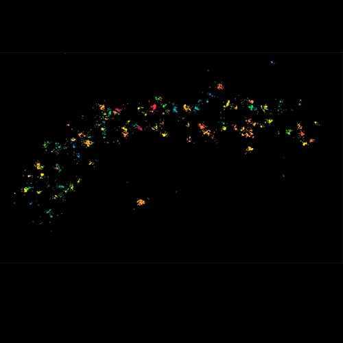
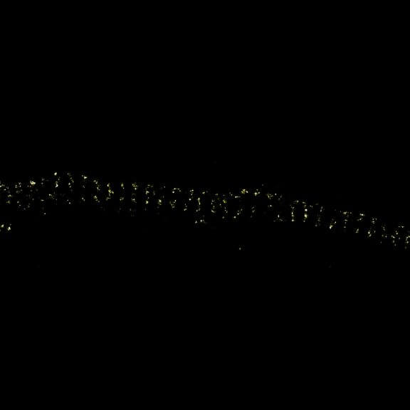
MINFLUX image of axonal βII spectrin labeled with abberior FLUX 660 in primary hippocampal neurons. Note the periodic arrangement of spectrin along the axon, and the absence of any details in the confocal counterpart image.
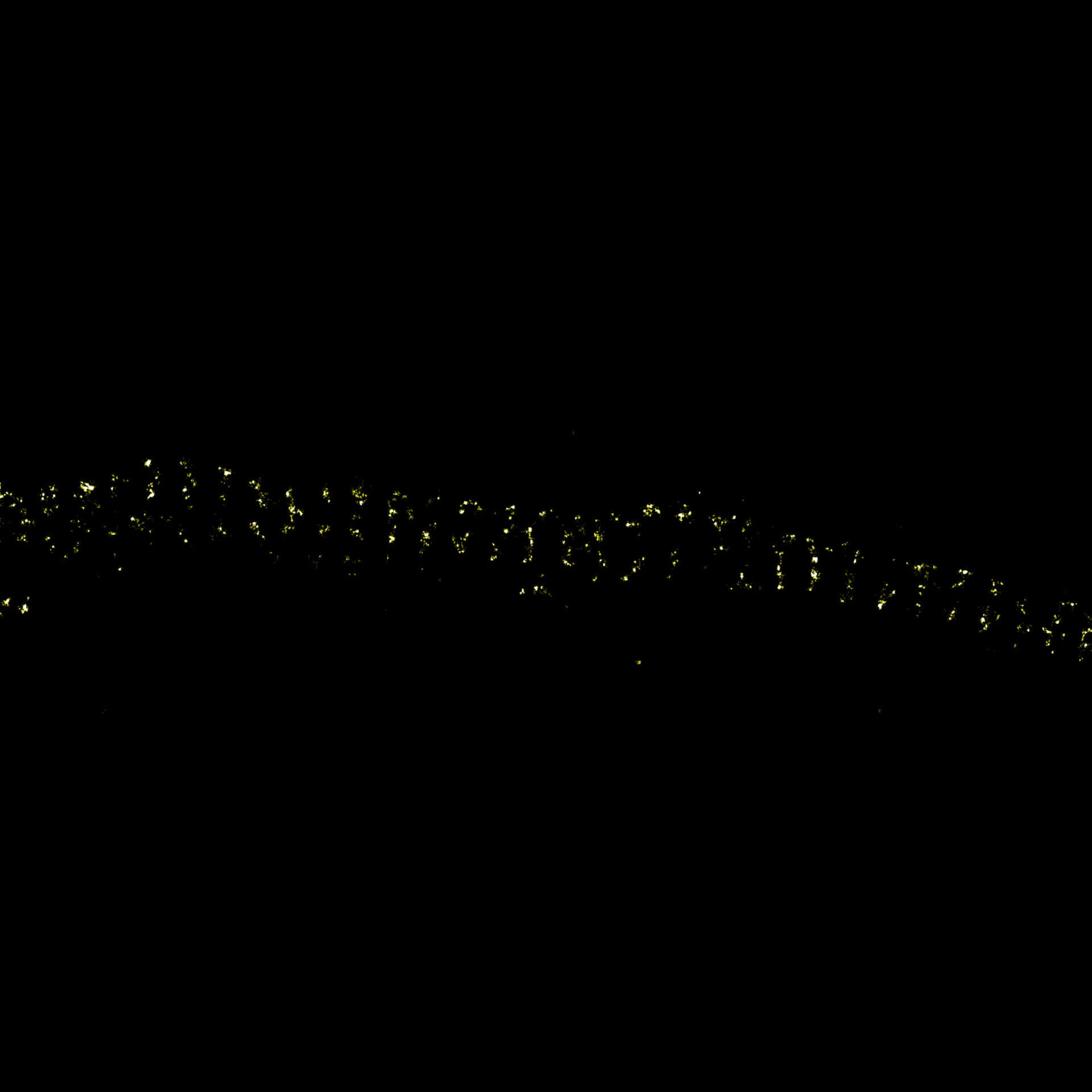
Description
MINFLUX imaging of βII spectrin in a primary hippocampal neuron labeled with abberior FLUX 680 by indirect immunofluorescence. Please note the periodic arrangement of spectrin along the axon.
Description
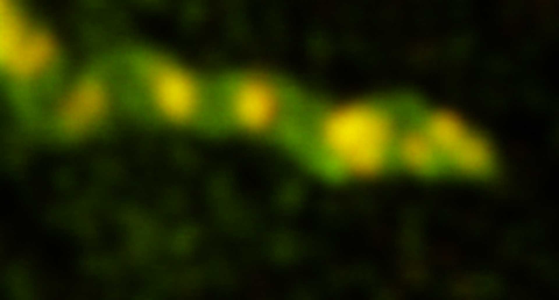
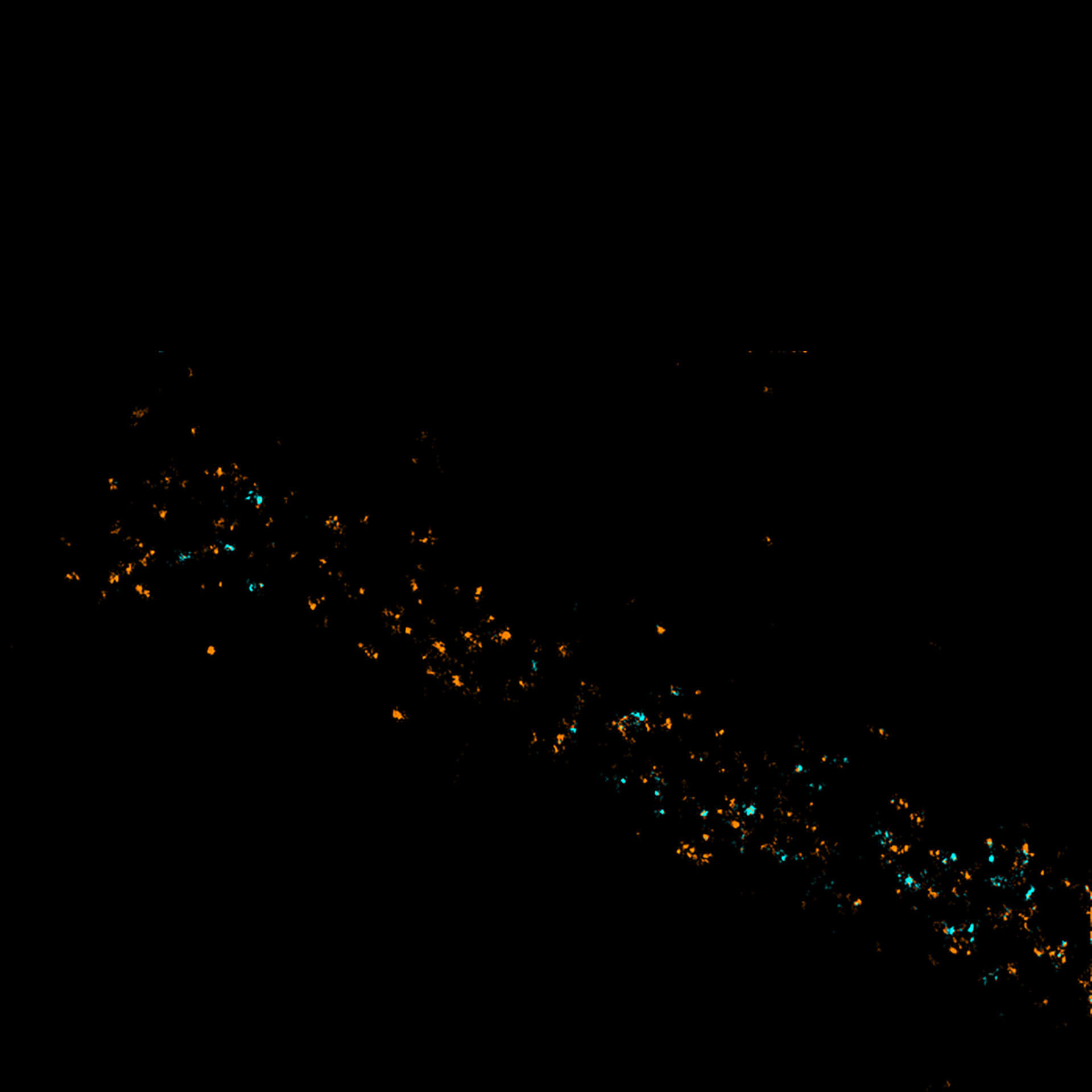
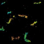
Two-color MINFLUX revealing an inner and outer mitochondrial membrane marker. Cultured mammalian cell labeled with indirect immunofluorescence using secondary antibodies coupled to abberior FLUX 640 (orange) and FLUX 680 (cyan). MINFLUX enables the visualization and separation of both structures.


100x sharper
than a confocal microscope
Although impactful, previous superresolution methods have failed to achieve resolutions on the length scales of the fluorescent molecules. Harnessing an entirely new and revolutionary localization principle, MINFLUX has finally accomplished this feat. Expect a resolution leap of 10-fold compared to other superresolution methods and 100-fold compared to confocal fluorescence imaging.
MINFLUX is the most precise and most photon-efficient way of localizing fluorescent molecules. With its new MINFLUX system, abberior has produced the first commercial fluorescence microscope that can achieve localization precisions of 1-3 nm (3D) in biological samples on large fields of view (10 x 15 μm). MINFLUX imaging means getting the maximal localization information out of your sample.
100x faster tracking
than a camera
MINFLUX tracks molecular movements at frequencies up to 10 kHz, resolving molecular motion every 100 μs. That’s 100 times faster than conventional camera-based methods. Of course, tracking works in all three dimensions, i.e. along x, y, and z. Due to the low number of photons required for each localization, single molecules can be monitored with unprecedented spatio-temporal resolution (e.g. 28.000 localizations each at 20 nm resolution).
MINFLUX is the fastest way to localize fluorescent molecules. With its revolutionary MINFLUX microscope, abberior is raising the bar for molecular tracking with world-record temporal resolution, opening new doors for life-scientists across all disciplines.
Our MINFLUX microscope runs an iMSPECTOR software version tailored to the needs of the platform’s unique abilities. Go ahead and take control of single-digit nanometer measurements and millisecond tracking.
Choose between MINFLUX molecular imaging and tracking at the push of a button!

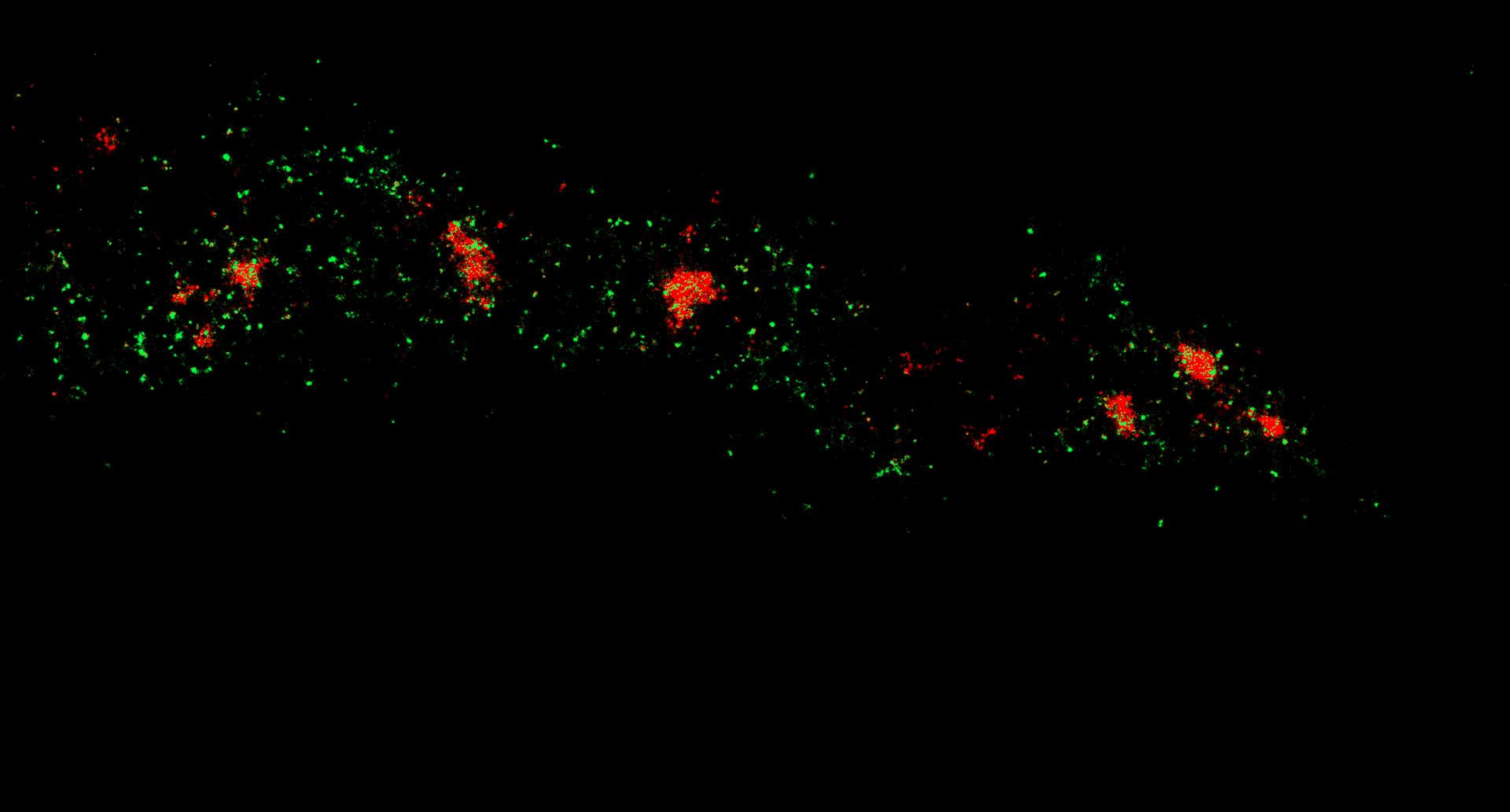
Two-color confocal and MINFLUX images of Tom20 (green) and mitochondrial DNA (red) stained with sCy5 and CF680 in mammalian cells using indirect immunolabeling. The two fluorophores were distinguished by ratiometric detection strategy. Note the dissimilar labeling density of the two imaged structures.


Maximize resolution
with minimal emission
MINFLUX defines an entirely new class of superresolution methods that uses the best of STED microscopy and the single molecule localization family: 1) Emitters are activated one-at-a-time to obtain the best molecule separation possible, 2) Localization is performed with a light distribution for fluorescence excitation that has a central intensity zero instead of a maximum. This fundamentally reduces the number of emitted photons required for ultimate localization precision. The central intensity-zero of the excitation beam searches for the emitting molecule by performing a clever sequence of sub-nm sized probing steps. The closer the intensity-zero is to the molecule, the lower the resulting fluorescence. By optimizing for low emission rates, the MINFLUX microscope zooms in on the molecule, concomitantly increasing the precision with which the molecular position is revealed.
MINimizing fluorescence FLUXes by matching the dark center of the excitation beam with the molecule‘s position localizes molecules reliably and with 1-3 nanometer precision in 3D! By relying on emission minimization rather than maximization, MINFLUX localization is: i) inherently fast, ii) does not discard weakly emitting molecules, iii) minimizes bleaching, and iv) is less drift dependent. Inaccuracies due to unknown molecular orientations and tumbling that severely compromise camera-based spot-centroid localization are ruled out.

Standard body
work as usual
The scientists and developers at abberior understand the importance of smooth and simple operation when performing biological research. That’s why our MINFLUX system is built around a standard microscope body that provides a variety of options ranging from widefield fluorescence, DIC, phase contrast over confocal and STED, all the way up to MINFLUX. To maximize extendability, we built our MINFLUX system with reliable and time-tested elements assembled on a robust optical breadboard. We use rock-solid opto-mechanical building blocks that have been proven to function seamlessly in hundreds of abberior microscopes all around the world.
Moreover, in line with our design philosophy for top-of-the-line instruments, our MINFLUX systems are future-proof: they are designed to allow adaptations with the latest technologies available: performance in perpetuity.
Almost no drift
solid as a rock
When performing experiments with nanometer resolution, miniscule sample drifts and movements can compromise performance. That’s why our MINFLUX comes with active sub-nanometer stabilization technology. When MINFLUX imaging is performed, a fully automated stabilization system based on laser-illuminated fiducial markers keeps the sample perfectly still, with residual fluctuations < 1 nm in 3D.
MINFLUX technology, system and module
changing the game
Our MINFLUX technology allows 3-dimensional localization precision of 2 to 3 nm in biological samples on fields of view of several tens of microns. With MINFLUX, molecular movements can be tracked with frequencies of up to 10 kHz, pinpointing the location of the tracked molecule every 100 µs.
Our MINFLUX system offers unprecedented spatial and temporal resolution in 3D, with two colors. It is 100x sharper than a confocal microscope and 100x faster than a camera.
Our MINFLUX module, seamlessly integrated into MIRAVA POLYSCOPE, allows single-color imaging with resolutions down to 3 nm in 2D and is 100 % as easy to use as any fluorescent microscope.
iMSPECTOR
for MINFLUX
Our MINFLUX microscope runs an iMSPECTOR version tailored to the needs of the platform’s unique abilities. Go ahead and take control of single-digit nanometer measurements and millisecond tracking.
- more than 100 kHz line frequency: scan 100 times faster than any other method
- 1 – 3 nm localization precision in 3D: resolve 100 times better than confocal microscopy
- 100 µs time per localization: track fluorophores 100 times faster than with a camera
MINFLUX – a factor of hundred

Why is MINFLUX a game changer?
Nobel laureate Stefan W. Hell explains that MINFLUX is the first concept to reliably reach molecular resolution of a few nanometers in 3D.

New partnership for next generation superresolution microscopy
EMBL Imaging Centre will provide access to MINFLUX nanoscope from abberior