abberior LIVE dyes
With our abberior LIVE dye collection, we offer exceptional labeling options. All the advantages of organic dyes, but in living cells – unraveling biological functions is now a breeze!
live-cell imaging with organic dyes
Description
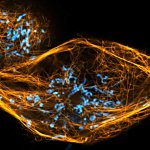
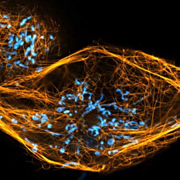
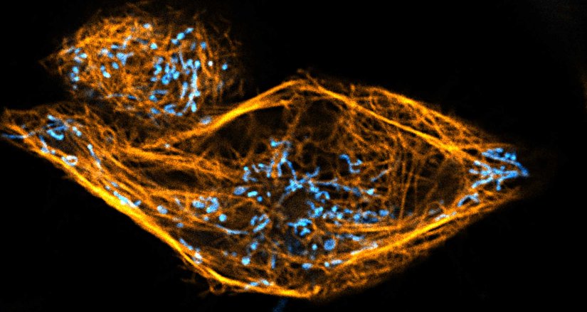
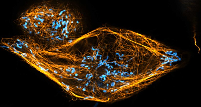
Two color live-cell confocal and STED image of a mammalian cell directly labeled with abberior LIVE 510 mito (cyan), and LIVE RED tubulin. This image was acquired with a STEDYCON microscope.
Description
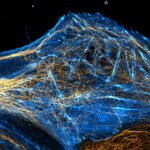
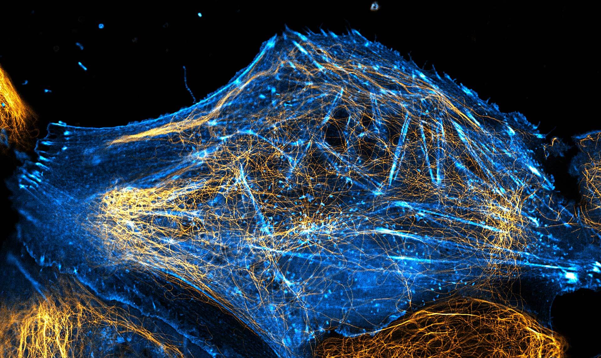
2-color STED image of a living cell expressing actinK118TAG incorporating TCO*A labeled with abberior LIVE 550 click (cyan). Tubulin filaments were stained with our direct probe abberior LIVE 610 tubulin (yellow).
Description
Description
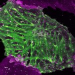
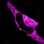
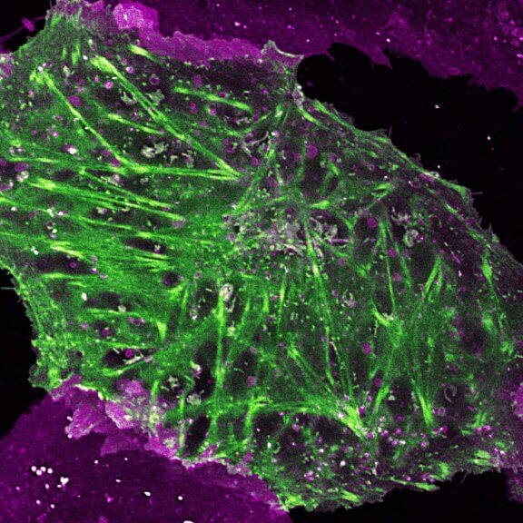
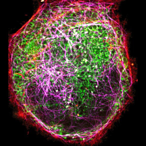
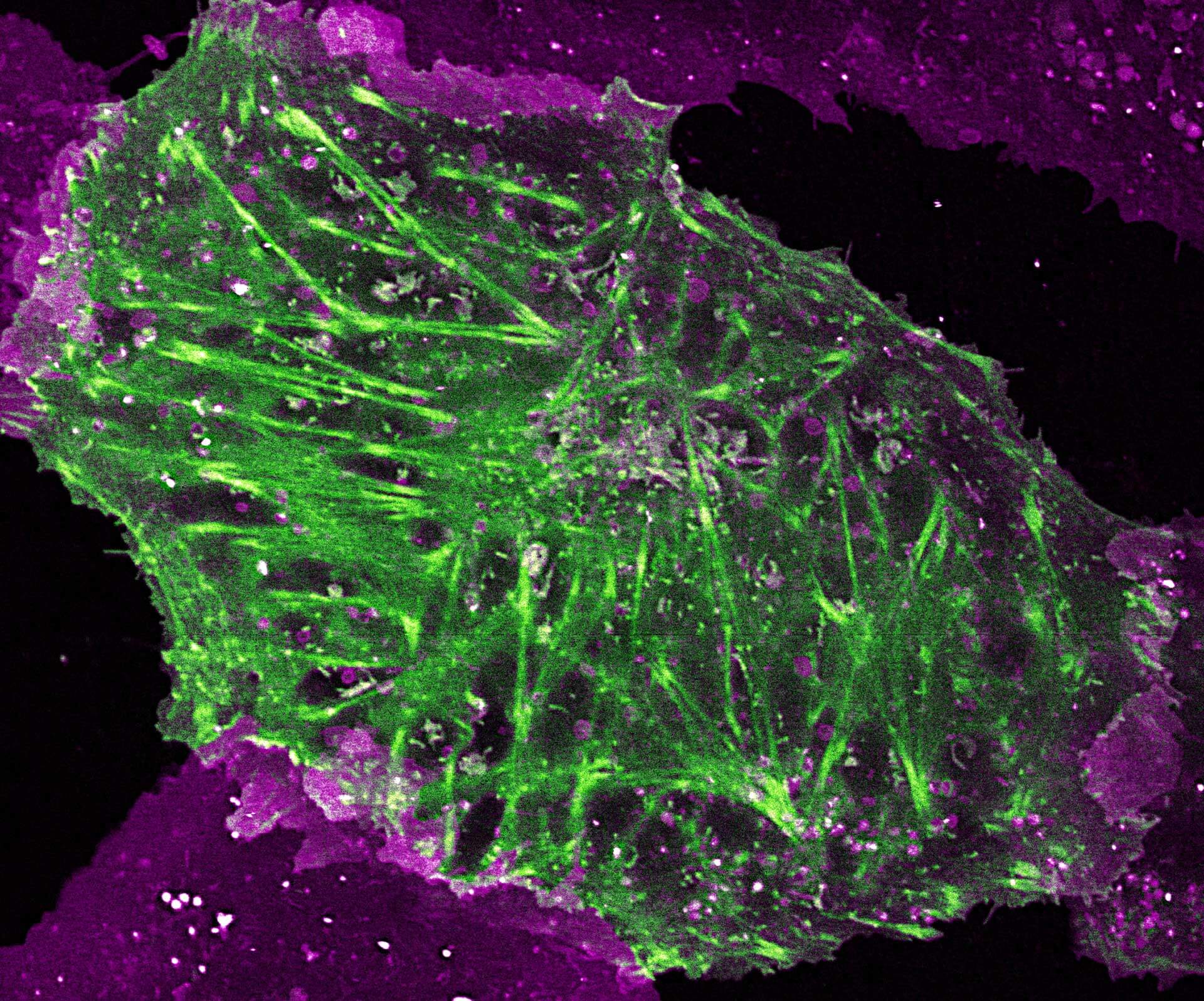
2-color STED image of a living cell expressing actin K118TAG incorporating TCO*A labeled with abberior LIVE 590 click (green). Plasma membrane is highlighted with abberior STAR RED membrane (magenta).


Description
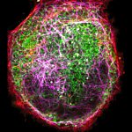
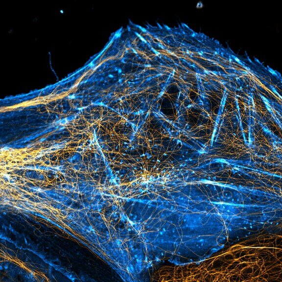
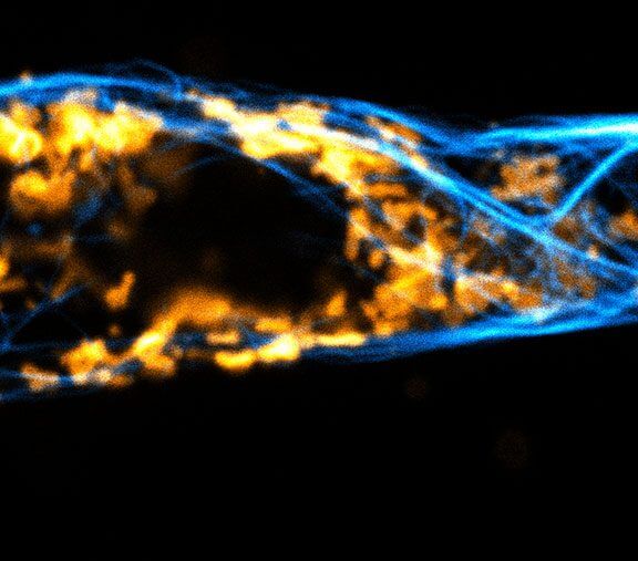
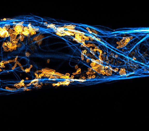
Two color live-cell confocal and STED image of a mammalian cell expressing a SNAP-tag® OMP25 fusion protein decoration the outer membrane of mitochondria. OMP25 is visualized by our new abberior LIVE 610 SNAP ligand (orange). Tubulin filaments are highlighted with abberior LIVE 550 tubulin (cyan).
The SNAP-OMP25 plasmid was a gift from Francesca Bottanelli, FU Berlin.
Description
We are happy to introduce the first commercial long Stokes-shift dye for live-cell imaging applications. The NEW abberior LIVE 460L makes 3-color live-cell STED at 775 nm possible!
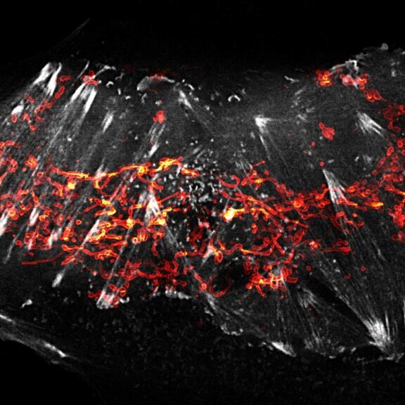
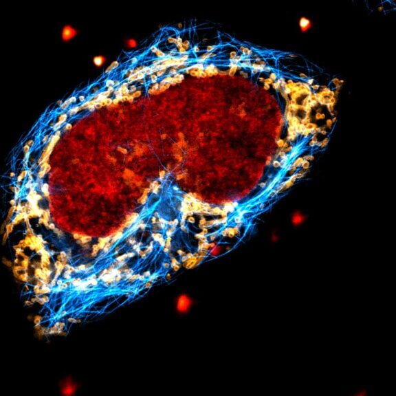
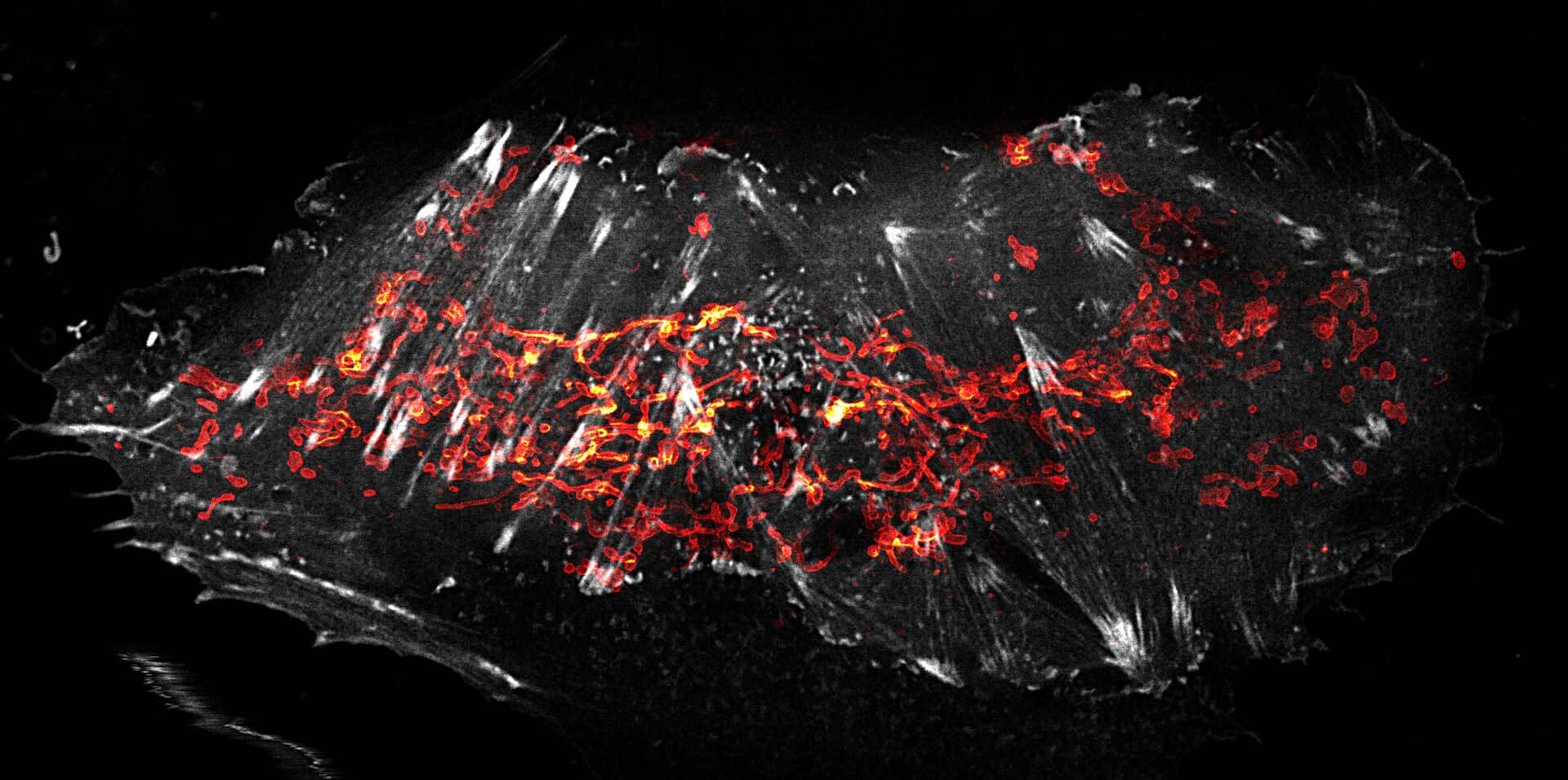
Live-cell STED image of a cell expressing a SNAP-tag® in an outer mitochondrial membrane protein visualized by our abberior LIVE 460L SNAP (orange). Tubulin filaments are highlighted with LIVE 610 (cyan) and the DNA with LIVE 560 DNA (red).
Description
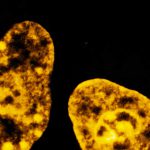
Live cell time lapse of abberior LIVE 590 DNA probe in cultured mammalian cells.
The use of very low concentrations of our abberior LIVE dyes reduces toxicity and allows long-term imaging of dynamic processes.
Image was acquired with the FACILITY microscope.
Description
Confocal and STED time lapse of a living cell expressing SNAP-tag® fusion protein fusion protein in the endoplasmic reticulum lumen with SNAP and C-terminal tetrapeptide KDEL. SNAP-KDEL was labelled with abberior LIVE 610 SNAP ligand (benzylguanine).
The SNAP-KDEL plasmid was a gift from Francesca Bottanelli, FU Berlin.
Description
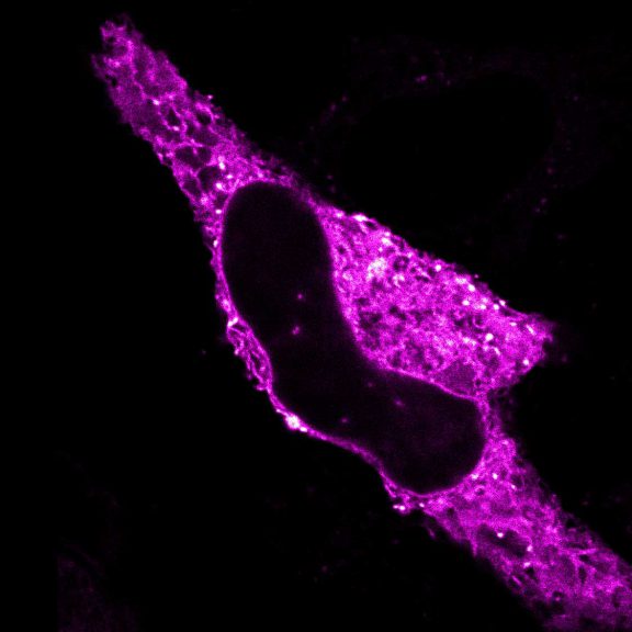
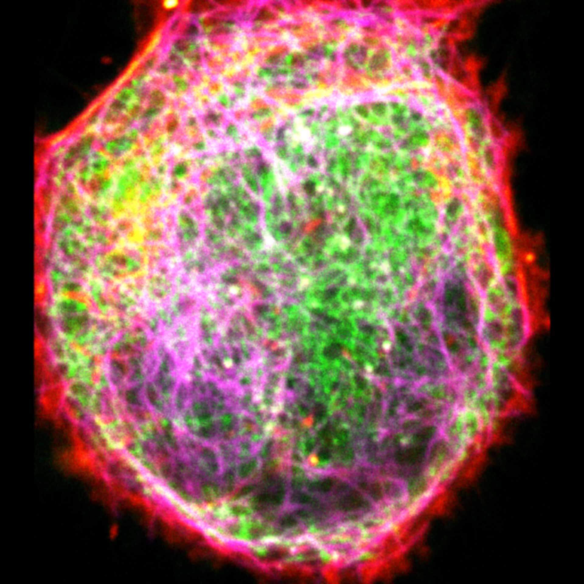
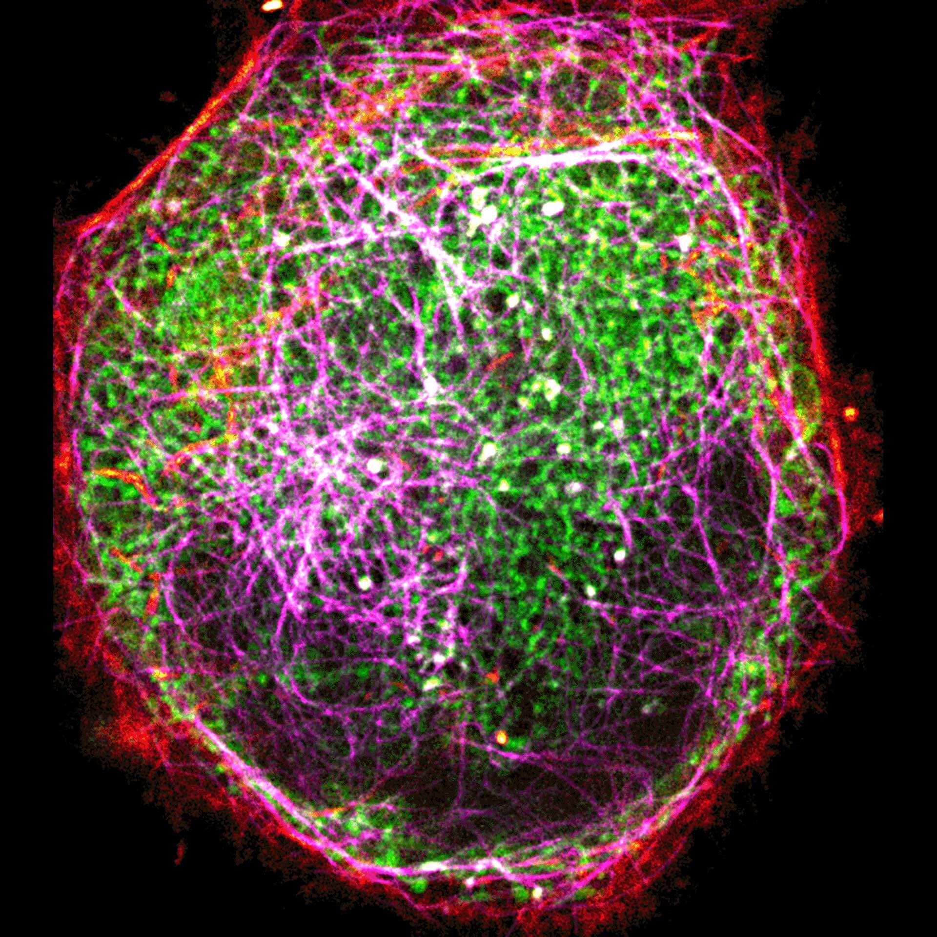
Three color live-cell STED at 775 nm: living cell labelled with abberior LIVE 460L (ER, green), LIVE 560 tubulin (magenta) and LIVE 610 actin (red).
Description
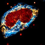
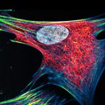
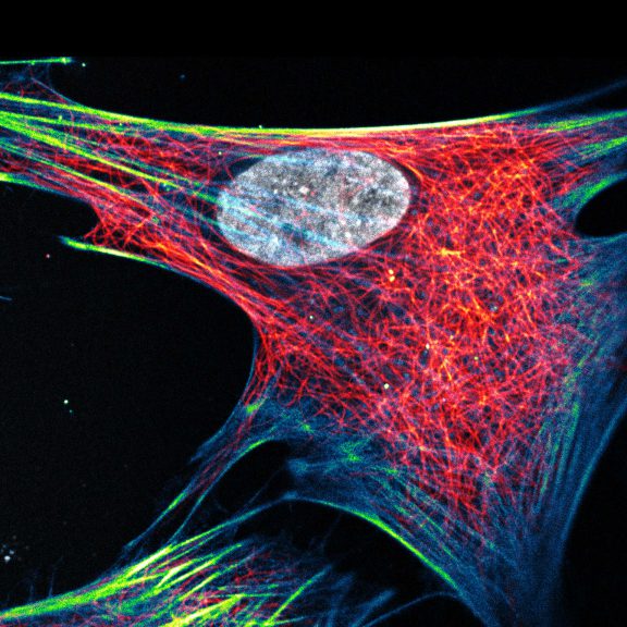
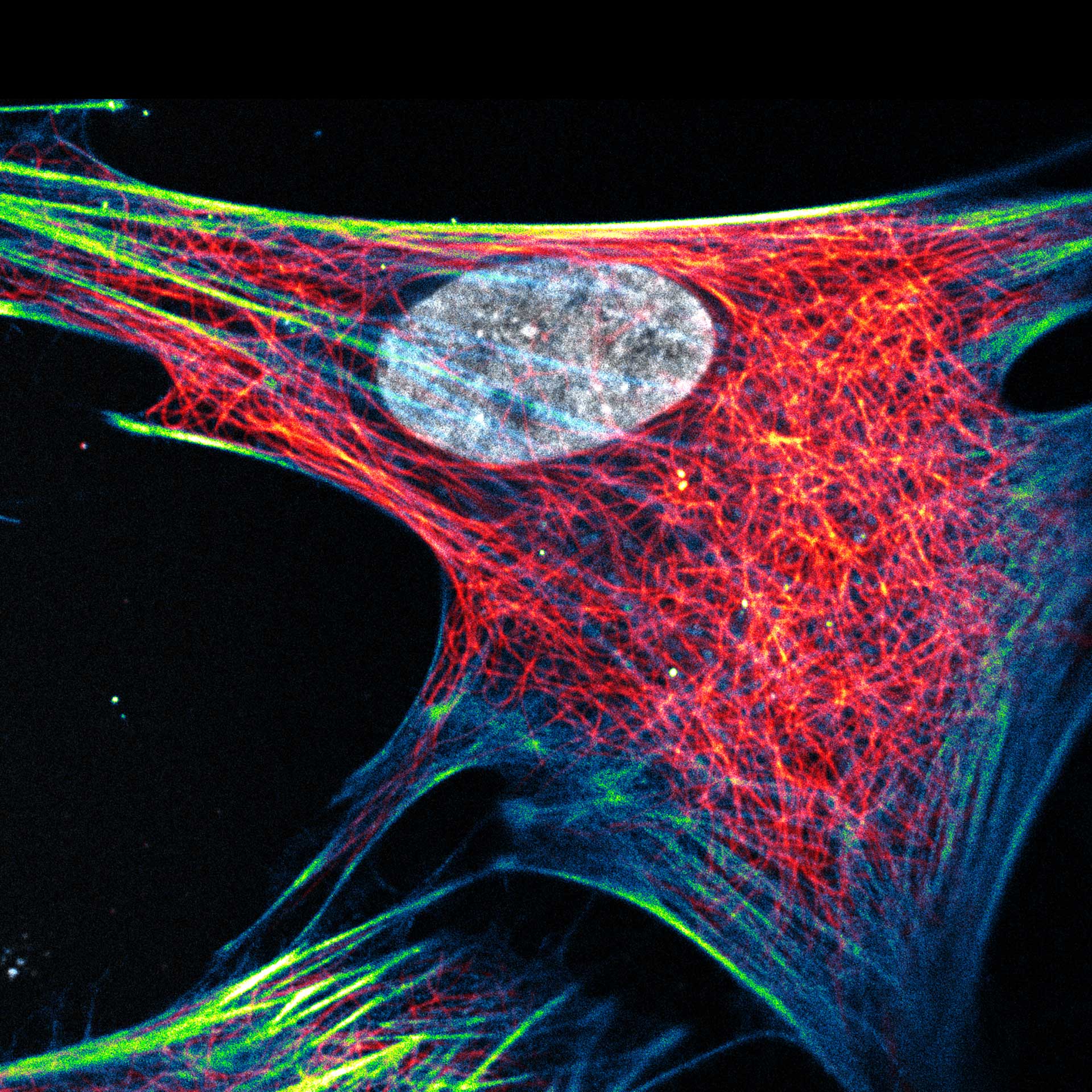
Composite of three color live-cell image of an adherent mammalian cell. This living cell was directly labelled with abberior LIVE 510 actin (blue/green), LIVE 560 DNA (gray) and LIVE 610 tubulin. This image was acquired with a STEDYCON microscope.
Description
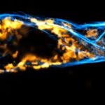
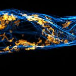
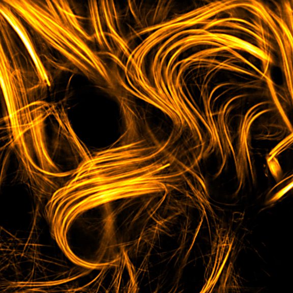
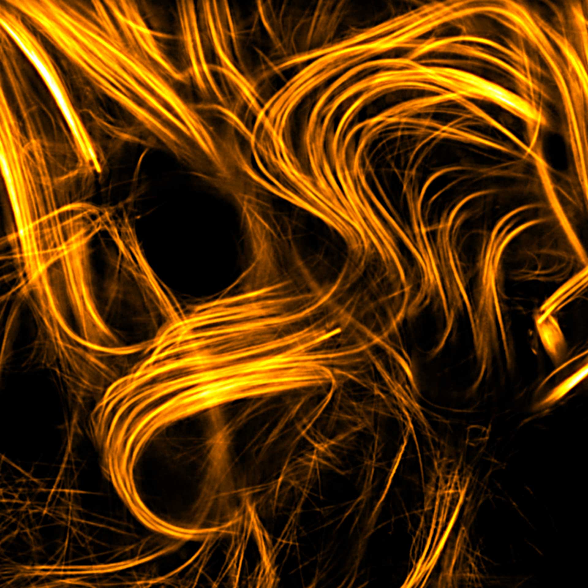
Drosophila spermatid tails were stained with abberior LIVE 610 tubulin. Testis were dissected from adult male fruit flies. Live cell imaging experiment was performed on a INFINITY microscope.
Description
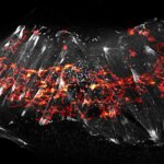
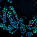
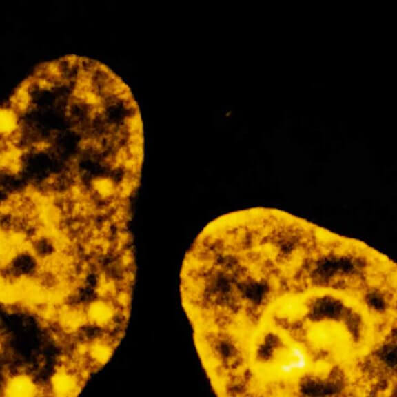
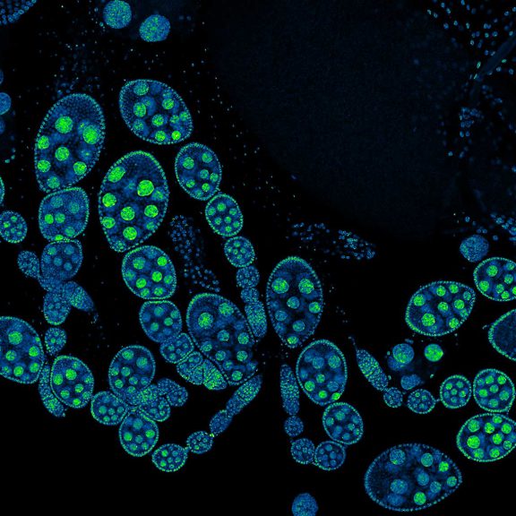
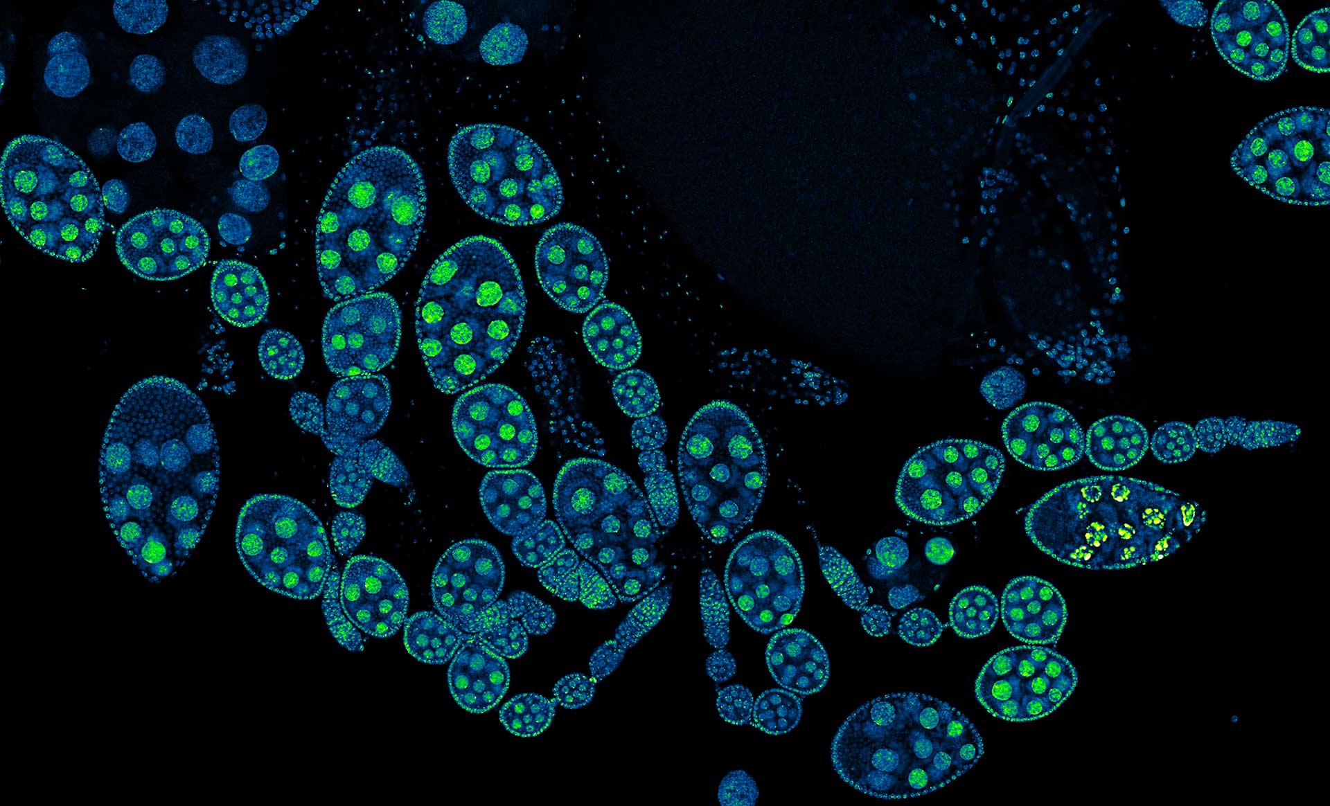
Drosophila ovariole stained with abberior LIVE 560 DNA showing nuclei in different cell types of the egg chamber. Ovaries were dissected from adult female fruit flies and were fixed prior to staining.
Image was acquired with the STEDYCON tiling feature and assembled with the SVI Huygens Stitcher.
Description
abberior – dyes that make you smile!
Description
Two color live-cell confocal and STED image of a mammalian cell directly labeled with abberior LIVE 510 mito (cyan), and LIVE RED tubulin. This image was acquired with a STEDYCON microscope.
Description
2-color STED image of a living cell expressing actinK118TAG incorporating TCO*A labeled with abberior LIVE 550 click (cyan). Tubulin filaments were stained with our direct probe abberior LIVE 610 tubulin (yellow).
Description
Description
2-color STED image of a living cell expressing actin K118TAG incorporating TCO*A labeled with abberior LIVE 590 click (green). Plasma membrane is highlighted with abberior STAR RED membrane (magenta).
Description
Two color live-cell confocal and STED image of a mammalian cell expressing a SNAP-tag® OMP25 fusion protein decoration the outer membrane of mitochondria. OMP25 is visualized by our new abberior LIVE 610 SNAP ligand (orange). Tubulin filaments are highlighted with abberior LIVE 550 tubulin (cyan).
The SNAP-OMP25 plasmid was a gift from Francesca Bottanelli, FU Berlin.
Description
We are happy to introduce the first commercial long Stokes-shift dye for live-cell imaging applications. The NEW abberior LIVE 460L makes 3-color live-cell STED at 775 nm possible!
Live-cell STED image of a cell expressing a SNAP-tag® in an outer mitochondrial membrane protein visualized by our abberior LIVE 460L SNAP (orange). Tubulin filaments are highlighted with LIVE 610 (cyan) and the DNA with LIVE 560 DNA (red).
Description
Live cell time lapse of abberior LIVE 590 DNA probe in cultured mammalian cells.
The use of very low concentrations of our abberior LIVE dyes reduces toxicity and allows long-term imaging of dynamic processes.
Image was acquired with the FACILITY microscope.
Description
Confocal and STED time lapse of a living cell expressing SNAP-tag® fusion protein fusion protein in the endoplasmic reticulum lumen with SNAP and C-terminal tetrapeptide KDEL. SNAP-KDEL was labelled with abberior LIVE 610 SNAP ligand (benzylguanine).
The SNAP-KDEL plasmid was a gift from Francesca Bottanelli, FU Berlin.
Description
Three color live-cell STED at 775 nm: living cell labelled with abberior LIVE 460L (ER, green), LIVE 560 tubulin (magenta) and LIVE 610 actin (red).
Description
Composite of three color live-cell image of an adherent mammalian cell. This living cell was directly labelled with abberior LIVE 510 actin (blue/green), LIVE 560 DNA (gray) and LIVE 610 tubulin. This image was acquired with a STEDYCON microscope.
Description
Drosophila spermatid tails were stained with abberior LIVE 610 tubulin. Testis were dissected from adult male fruit flies. Live cell imaging experiment was performed on a INFINITY microscope.
Description
Drosophila ovariole stained with abberior LIVE 560 DNA showing nuclei in different cell types of the egg chamber. Ovaries were dissected from adult female fruit flies and were fixed prior to staining.
Image was acquired with the STEDYCON tiling feature and assembled with the SVI Huygens Stitcher.
Description
abberior – dyes that make you smile!
abberior LIVE dyes
easy labeling
abberior LIVE dyes are designed for STED and confocal microscopy in living cells. These unique dyes combine highest brightness and photostability with effortless live-cell labeling.
We offer ready-to-use probes for DNA, tubulin, actin, mitochondria, click, and SNAP labeling. In addition, custom-made abberior LIVE dye conjugates can be provided.
All abberior LIVE dyes work remarkably well for labeling with ligands like Halo®-Tag fusion proteins in living cells. The required conjugates can be prepared easily from abberior LIVE carboxylic acid and commercially available Halo®-Tag amines.
- Get the performance of organic dyes with live-cell labeling
- Image at exceptional signal and resolutions combined with high biological relevance


