


abberior STAR dyes
Our abberior STAR dyes deliver the best performance in fluorescence light microscopy. They feature exceptional photostability and brightness. There is simply no reason for not using our STAR dyes for your STED or confocal imaging application.
best for STED and confocal
Description
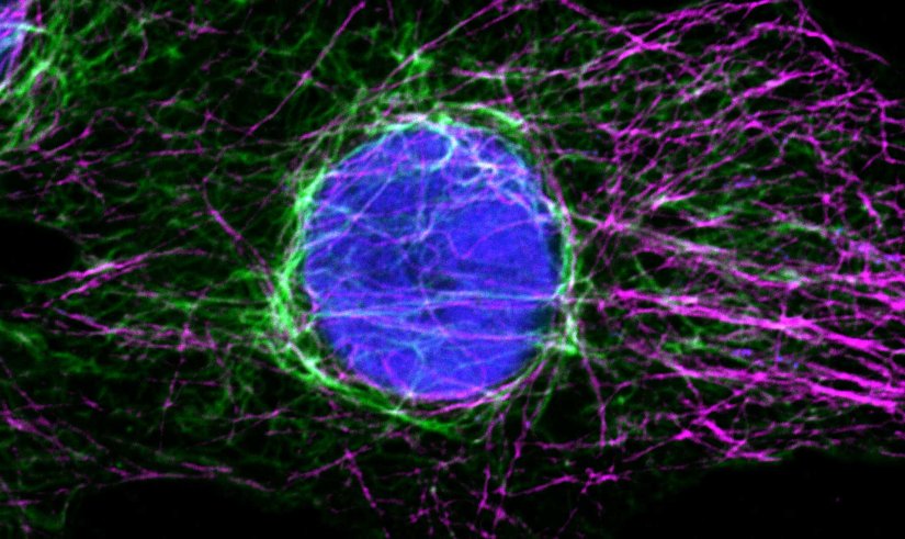
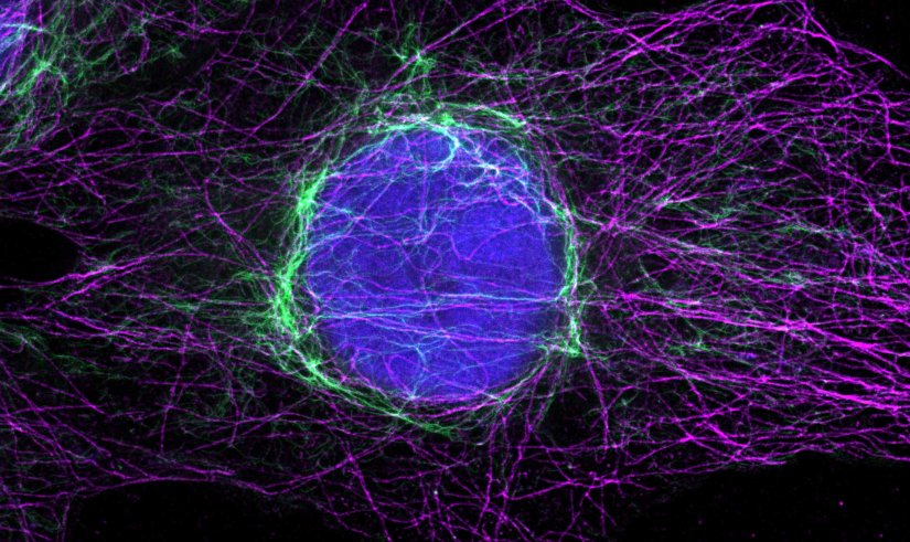
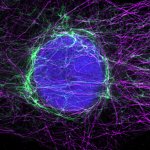
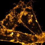
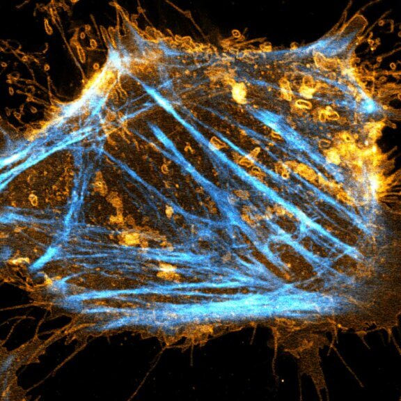
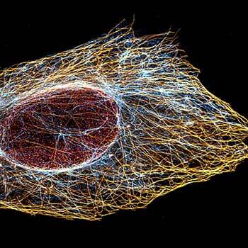
Indirect immunofluorescence staining with abberior STAR dyes coupled to polyclonal anti-mouse secondary nanobodies (alpaca VHH single domain antibodies) from Jackson ImmunoResearch shows excellent results in 3-color STED imaging. Vimentin (abberior STAR RED, green), tubulin (abberior STAR 580, magenta) and dsDNA (abberior STAR 460L, blue) were labeled in fixed mammalian cells and visualized with the STEDYCON.

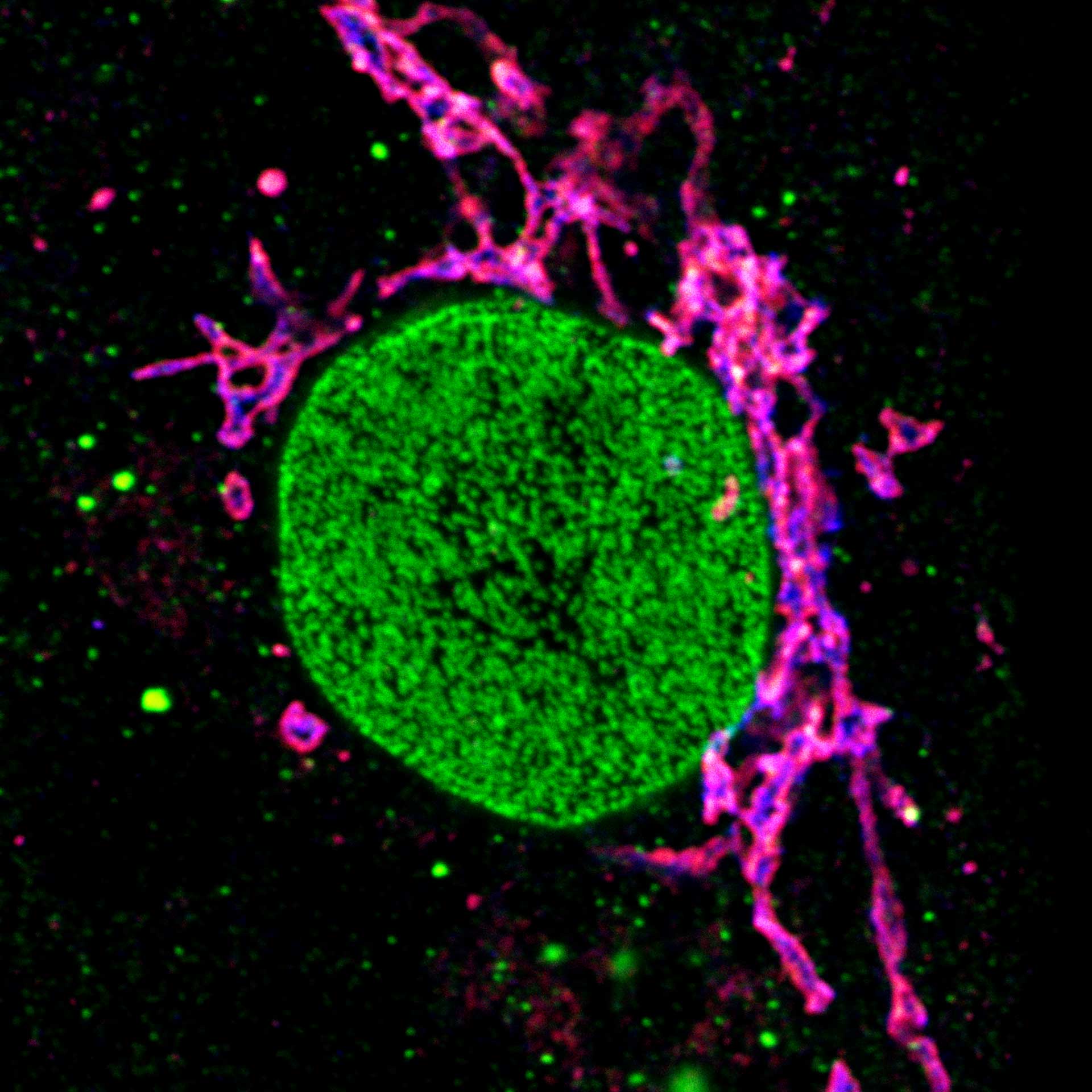
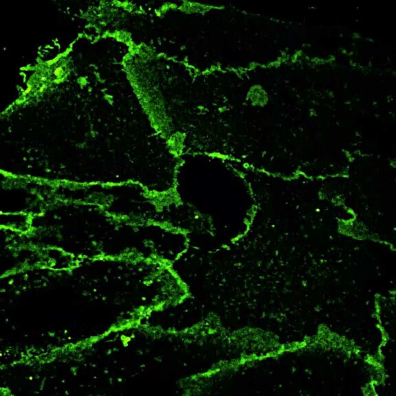
Description
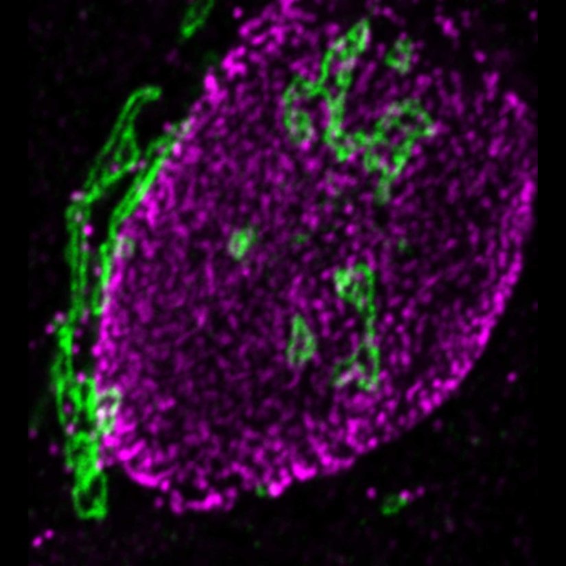
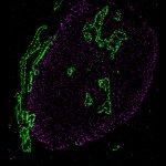
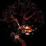
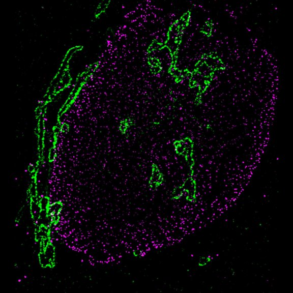
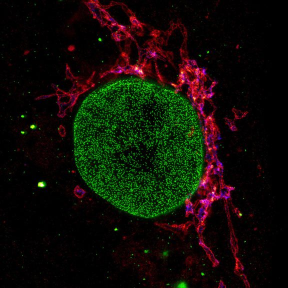
Proteins of the nuclear pore complex and the golgi apparatus were stained by indirect immunofluorescence using abberior STAR RED (nuclear pore complex, magenta) and abberior STAR 580 (golgi, green) coupled to polyclonal secondary nanobodies (alpaca VHH single domain antibodies) from Jackson ImmunoResearch. Images of the fixed mammalian cells were acquired with the STEDYCON.
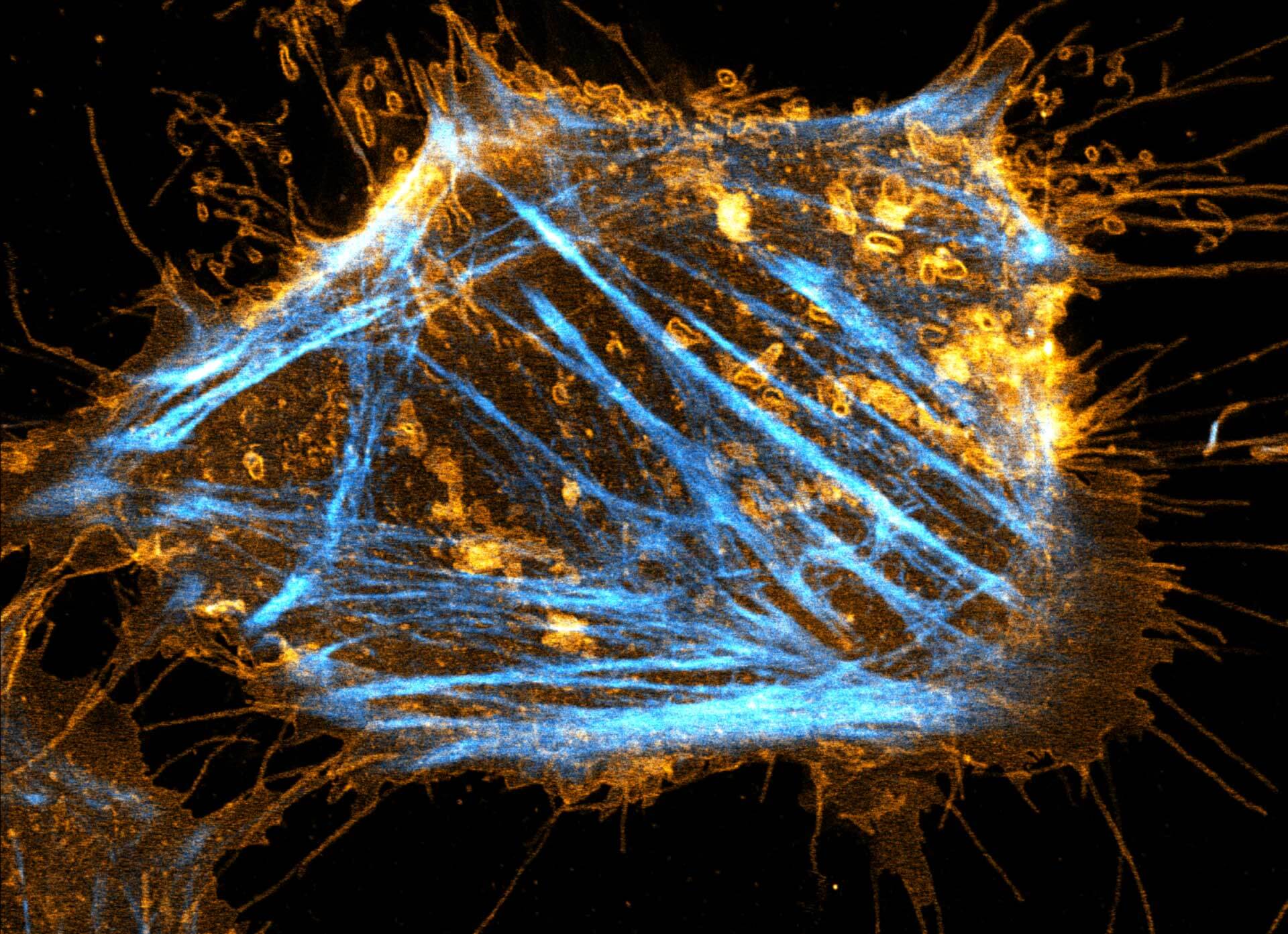
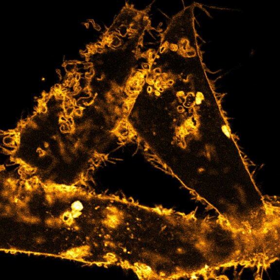
Description
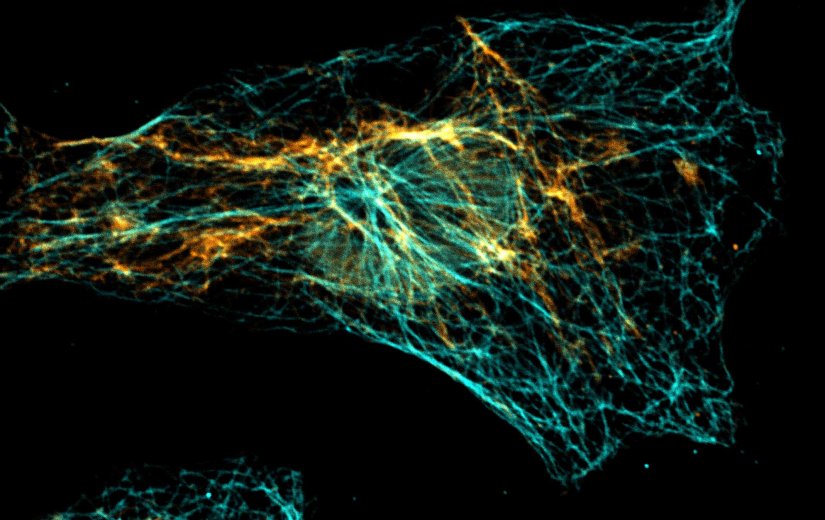
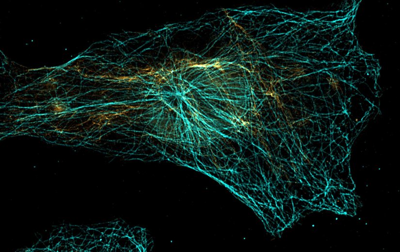
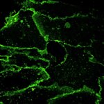
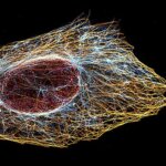
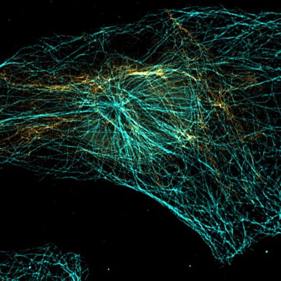
Vimentin and tubulin were stained by indirect immunofluorescence using abberior STAR RED (vimentin, orange) and abberior STAR 580 (tubulin, cyan) coupled to polyclonal secondary nanobodies (alpaca VHH single domain antibodies) from Jackson ImmunoResearch. Images of the fixed mammalian cells were acquired with the STEDYCON.
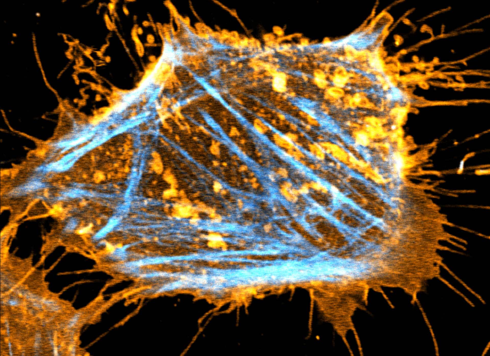
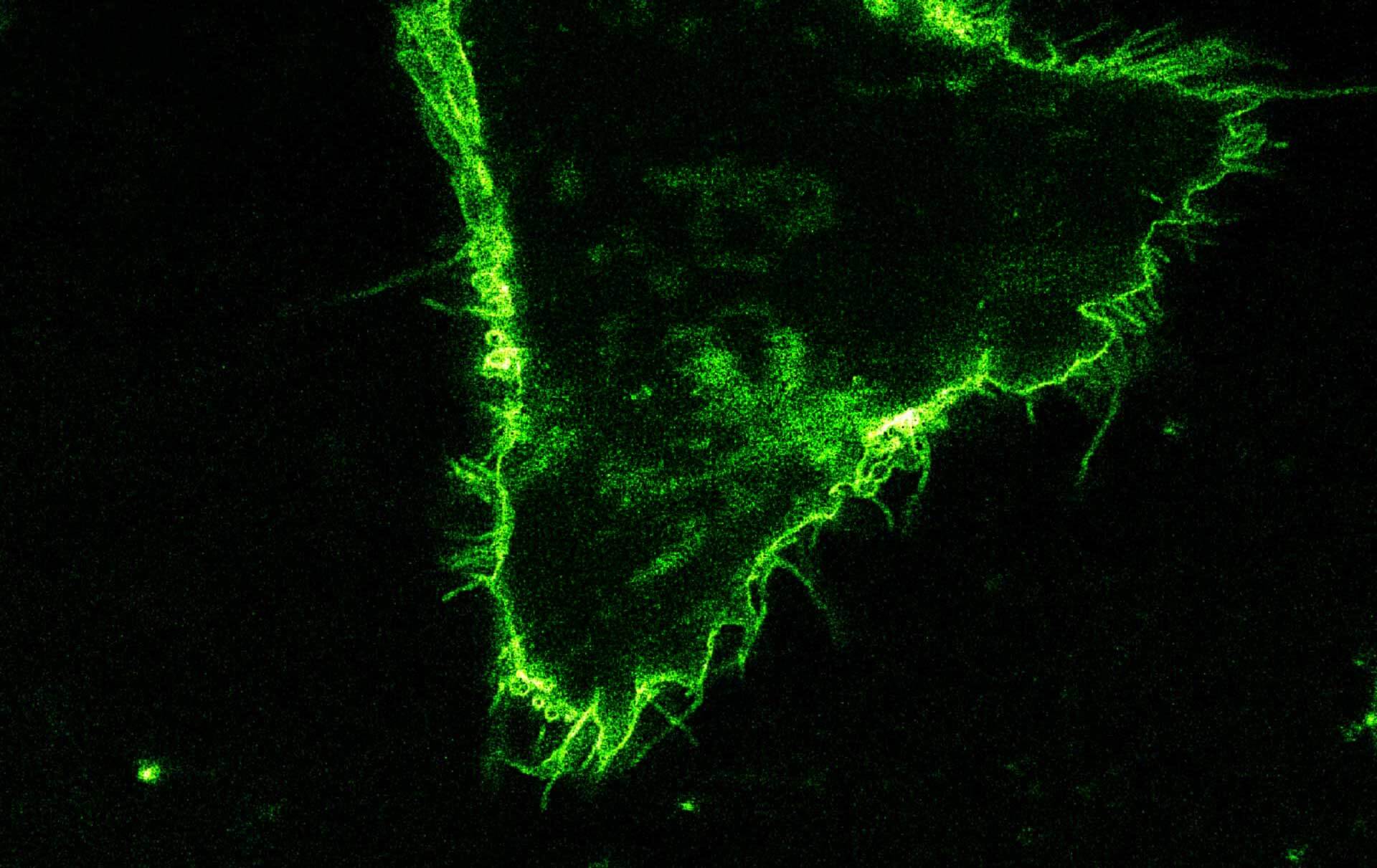
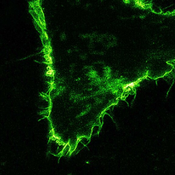
Description
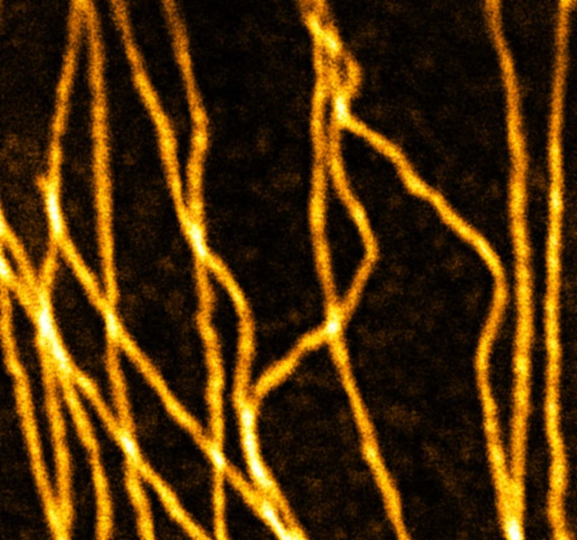
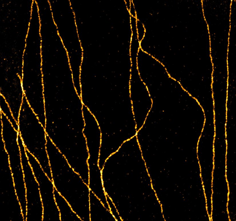
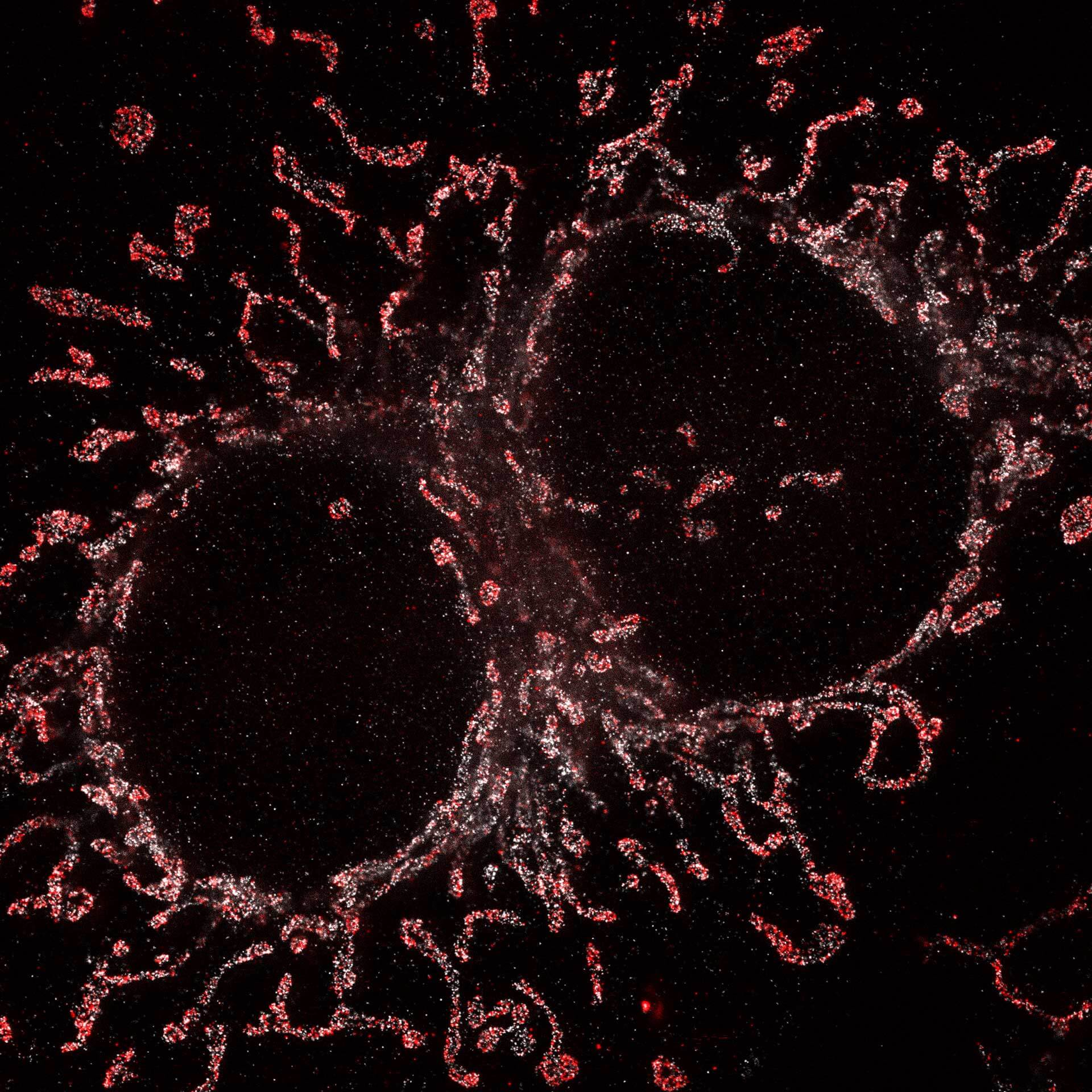
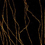
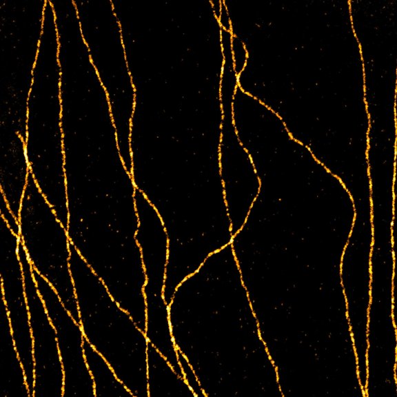
abberior STAR RED was coupled to polyclonal secondary nanobodies (alpaca VHH single domain antibodies) from Jackson ImmunoResearch and used to label tubulin in fixed mammalian cells via indirect immunofluorescence. The STED image was acquired with the STEDYCON system.
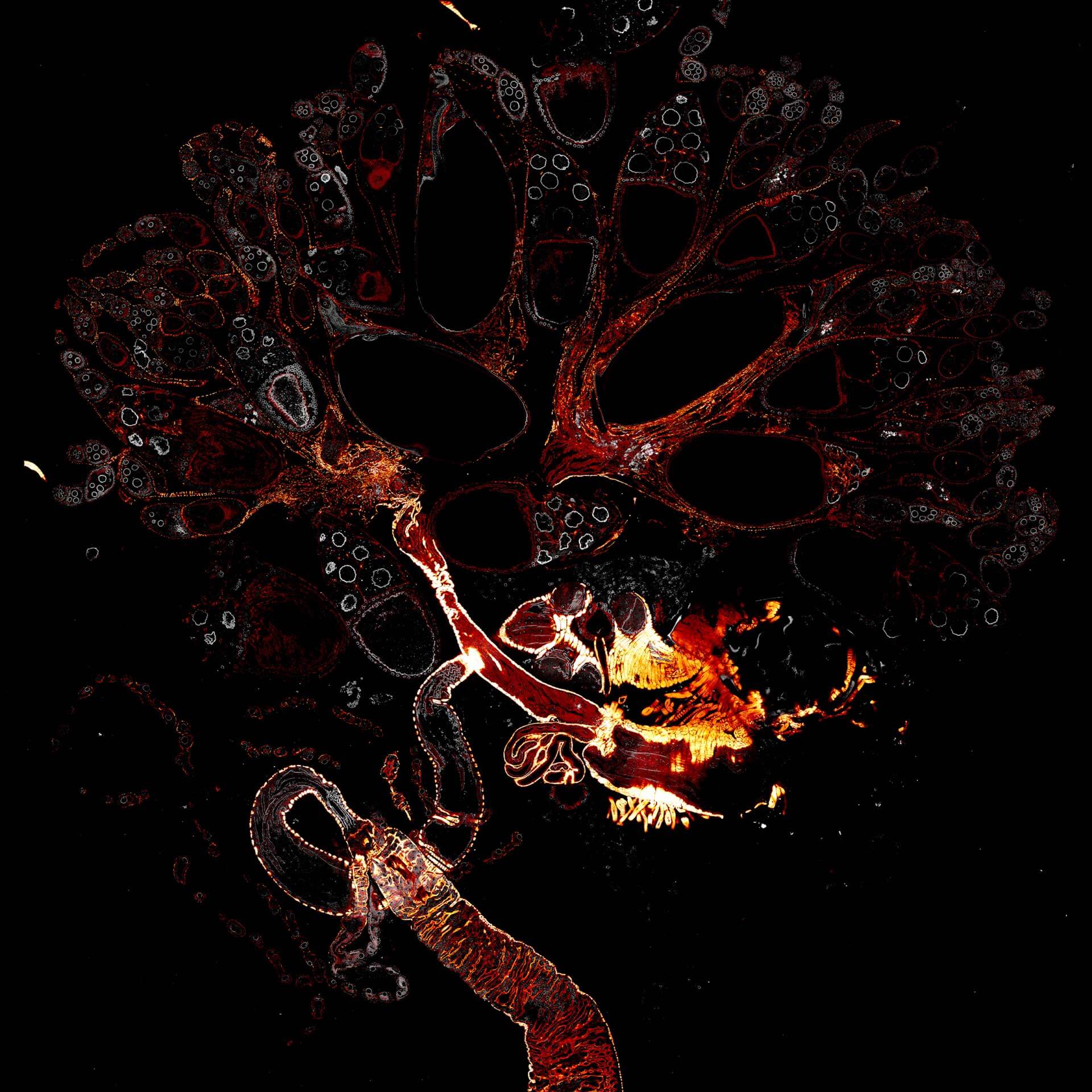
Description
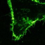
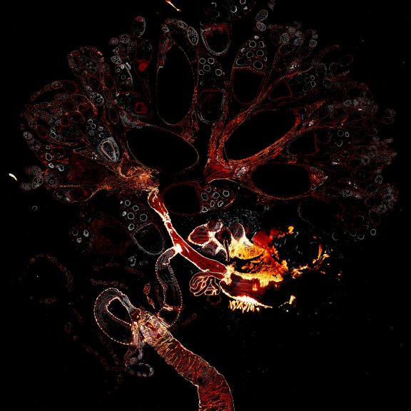
Drosophila female reproductive system stained for F-actin (red) with abberior STAR RED phalloidin. abberior STAR ORANGE is highlighting a nuclear pore protein (gray).
Image was acquired with the STEDYCON tiling feature and assembled with the SVI Huygens Stitcher.
Description
Two color live-cell STED and confocal image of a mammalian cultured cell stained with abberior STAR RED membrane (orange) and abberior LIVE 590 actin (cyan).
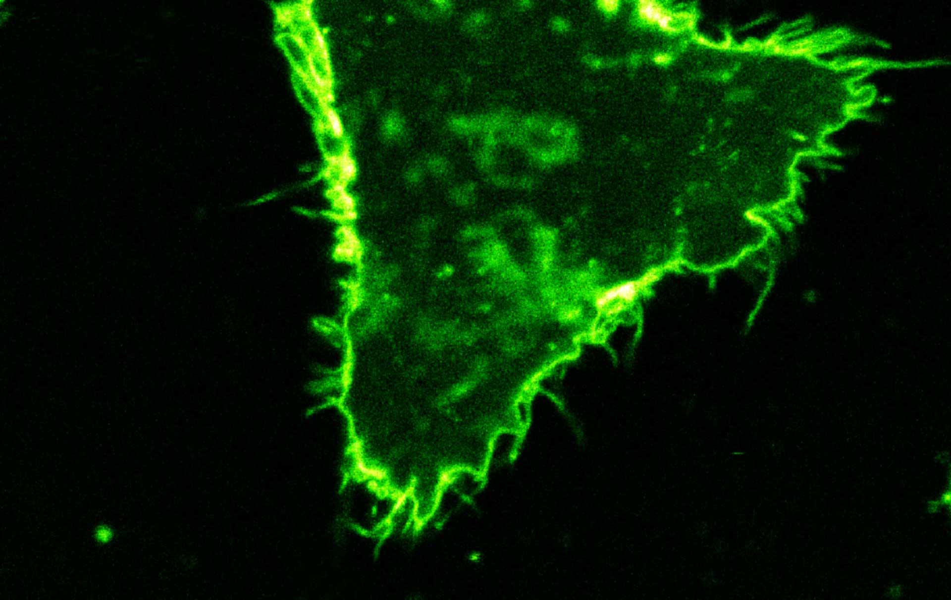
Description
Comparison of STED and confocal of a living mammalian cell stained with abberior STAR 488 membrane. Images were acquired with our FACILITY and STED @ 595 nm.

Description
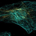
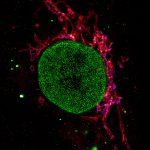
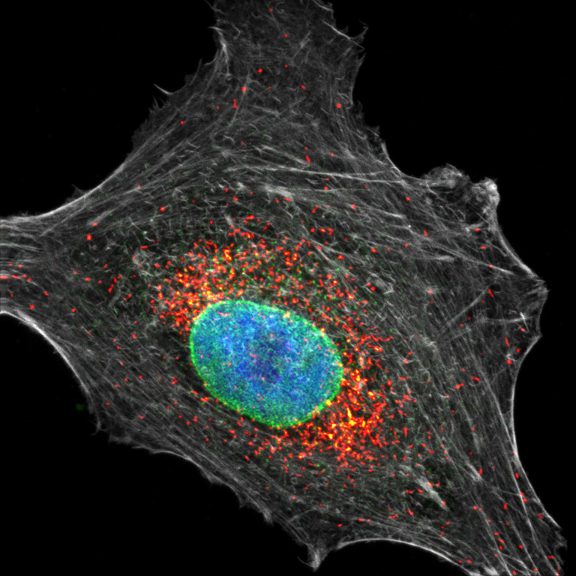
Four color confocal image of mammalian cultured cells. Peroxisomes were immunostained with abberior STAR RED (magenta) and nuclear pore proteins with abberior STAR 580 (green). F-actin fibers were highlighted with abberior STAR 488 phalloidin (gray).
The nucleus was visualized with DAPI (blue).
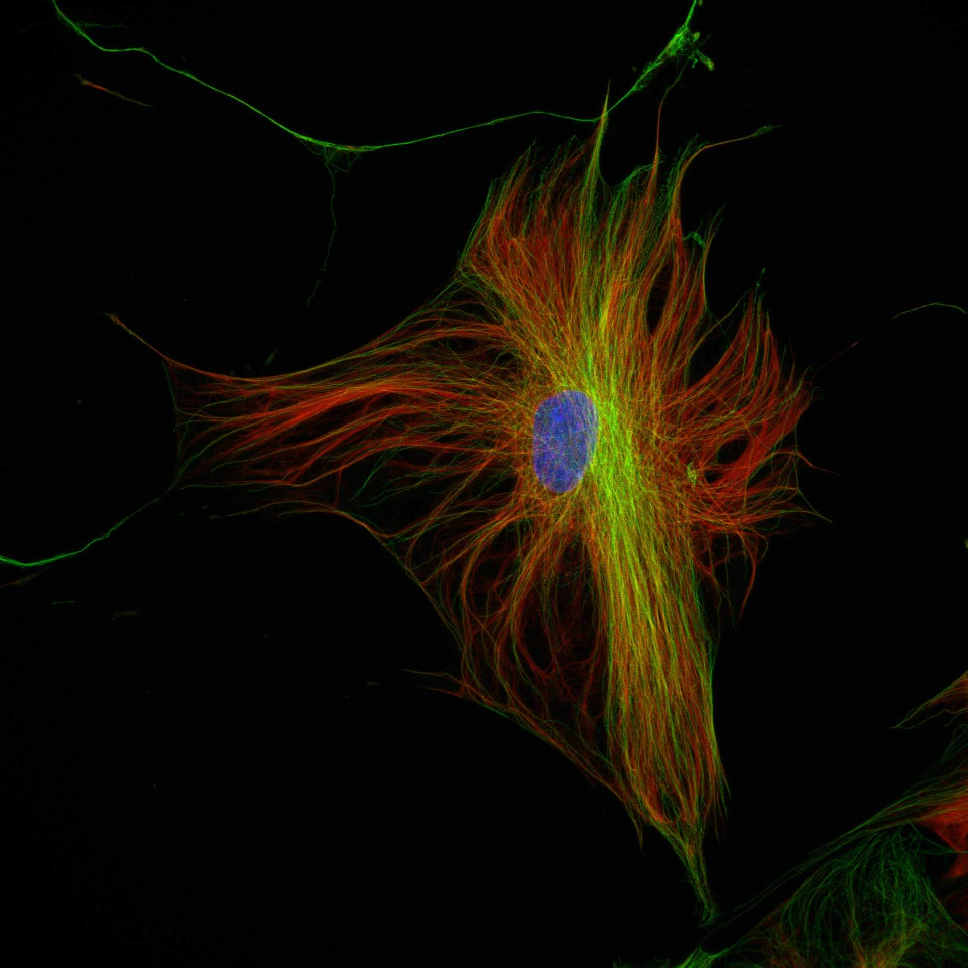
Description
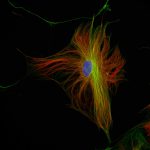
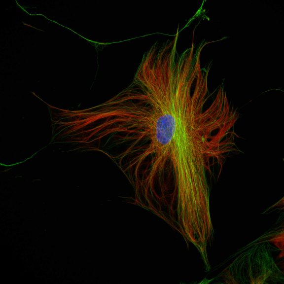
Three color confocal image of human fibroblast immunostained with abberior STAR 580 for vimentin (green) and with abberior STAR RED for tubulin (red).
DAPI was added to highlight the nucleus (blue).

Description
Three color STED and confocal image of a mammalian cultured cell immunostained for a nuclear pore protein in green and two golgi apparatus markers in red and blue.
abberior STAR RED highlights the golgi apparatus protein giantin (red) and abberior STAR ORANGE visualizes the cis-golgi protein GM130 (blue). NUP98 proteins were stained with abberior STAR GREEN (green).

Description
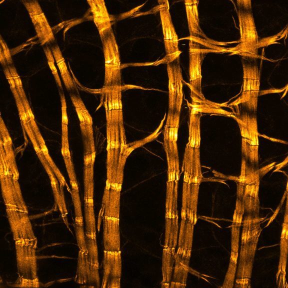
Drosophila male accessory gland stained for F-actin using abberior STAR 580 phalloidin.
Sample was prepared in cooperation with Dr. H. R. Shcherbata at MPl for Biophysical Chemistry, Göttingen, Germany.
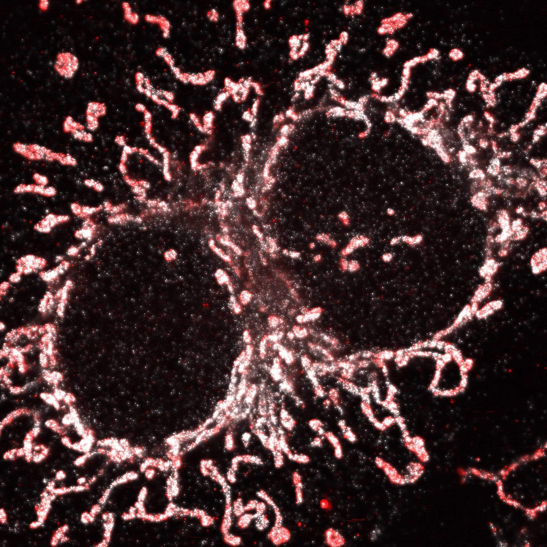
Description
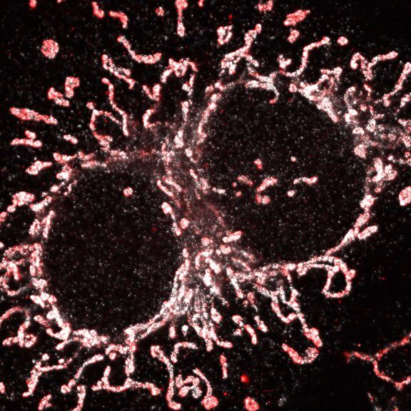
Cultured mammalian cell immunostained for an inner and outer mitochondrial membrane marker. Outer membrane is highlighted with abberior STAR RED (red) and the inner membrane with abberior STAR ORANGE (gray).
STED and confocal images were acquired with abberior’s FACILITY microscope.


abberior STAR
abberior STAR dyes enable background-free labeling in cells or tissue, to get the most out of your sample. They excel with high brightness and photostability, while conjugates of the dyes show an excellent solubility in aqueous buffers as PBS.
abberior STAR L
abberior STAR L dyes are abberior’s long Stokes-shift dye series. The common characteristic of the dyes is an exceptional long Stokes-shift between the excitation and emission maximum. Our abberior STAR L dyes are designed to provide bright fluorescence for multiplexed imaging. Conjugates of the dyes show a very good solubility in aqueous buffers as PBS to allow labeling of the structures of interest with a good signal-to-noise-ratio.
- Highest signal and photostability
- For unrivaled STED and confocal imaging