


abberior FLUX dyes
Our abberior FLUX dyes deliver the best performance in MINFLUX imaging. They bring together exceptional photostability, very high brightness, low background labeling with photoswitching kinetics optimized for single-molecule-localization microscopy.
created for nanometers

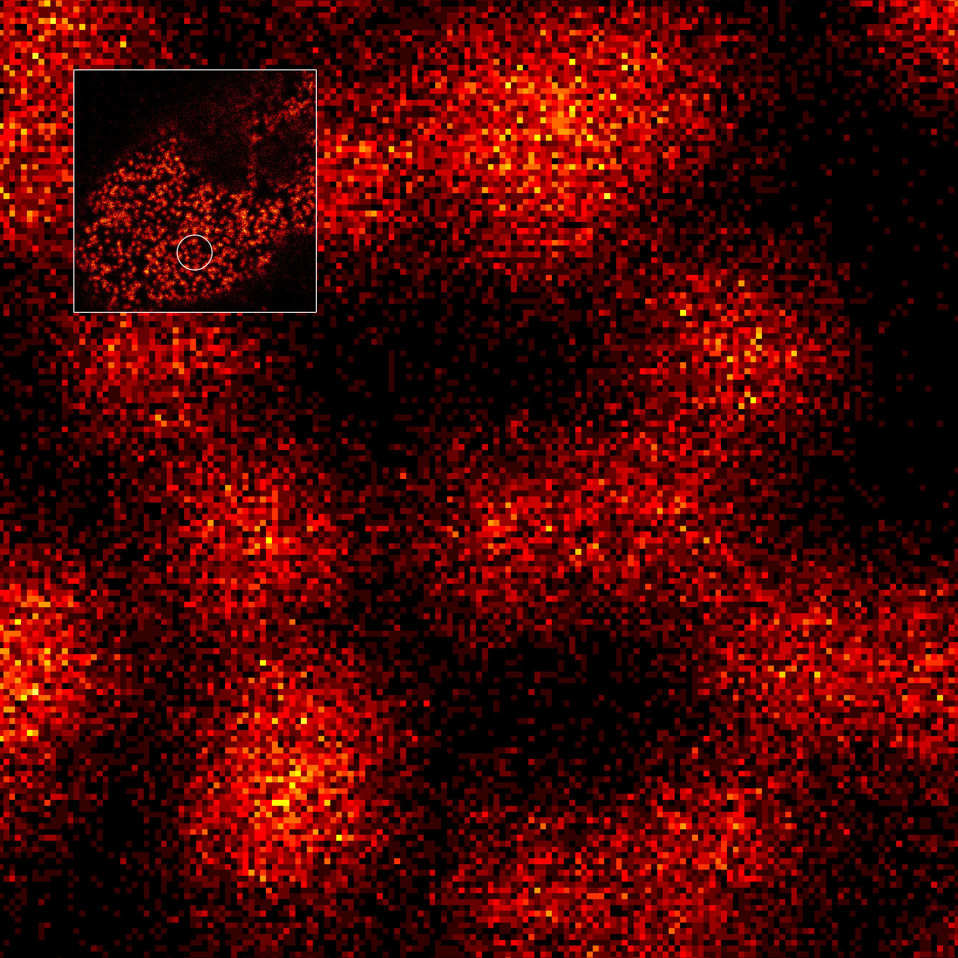
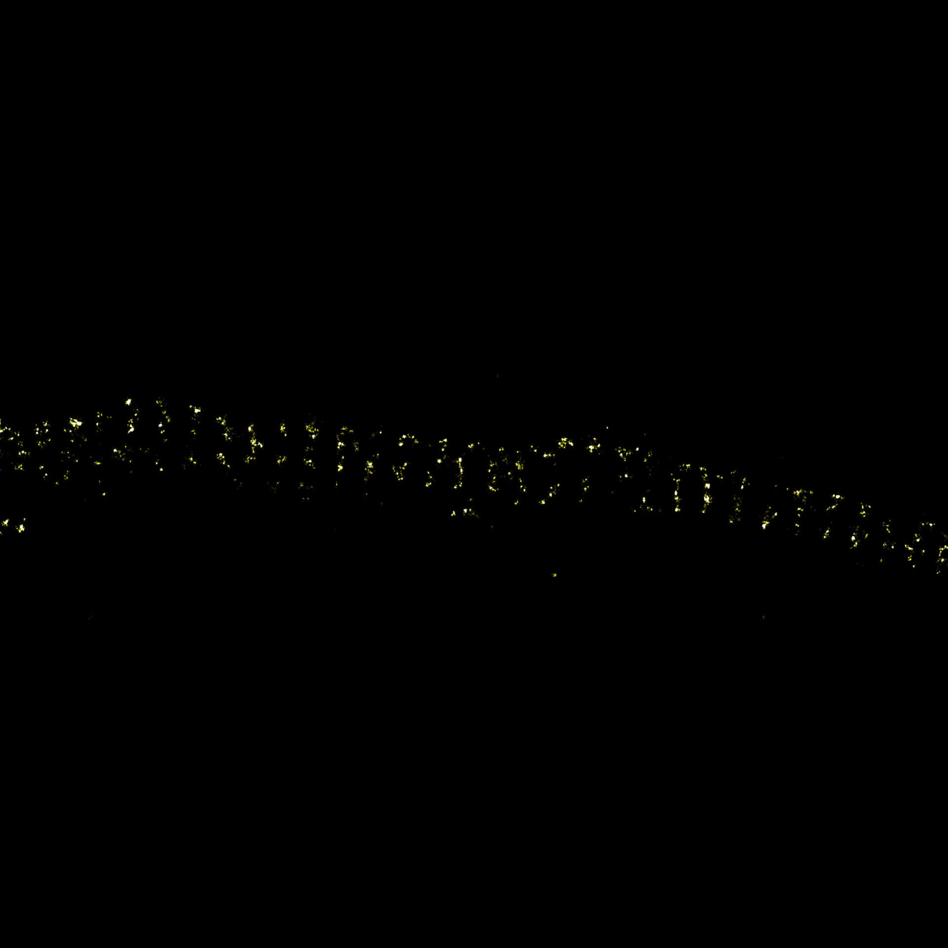
Description
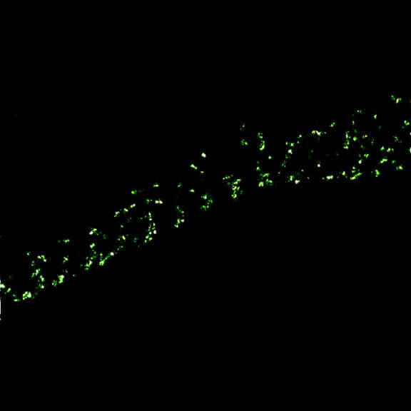
2D MINFLUX imaging of the peroxisomal membrane protein PMP70 labeled with abberior FLUX 640 in fixed mammalian cells using indirect immunofluorescence.
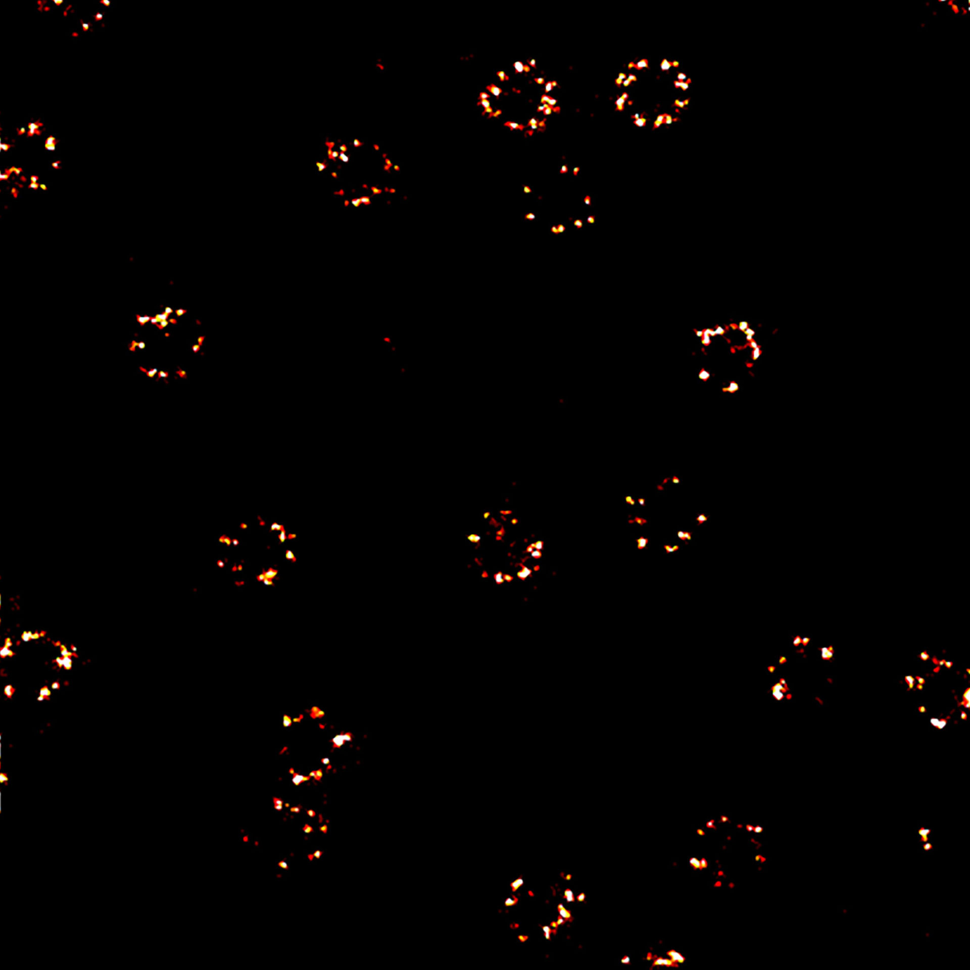
Description
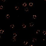
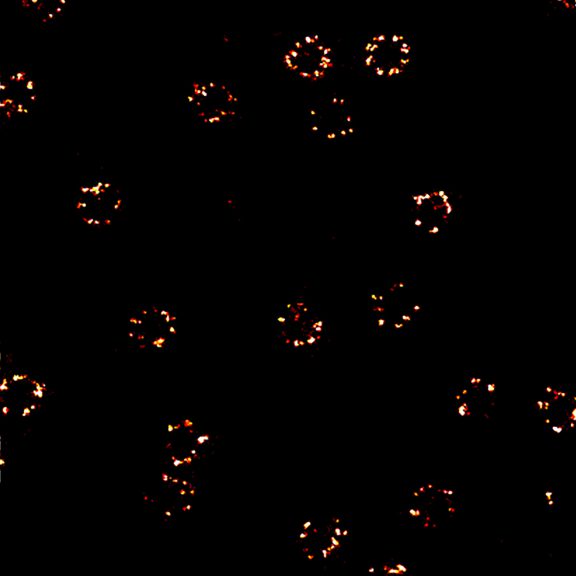
2D MINFLUX nanoscopy of the nuclear pore complex subunits. Fixed mammalian cells expressing SNAP-tag® NUP96 were labeled with abberior FLUX 647 SNAP. In contrast to confocal microscopy, 2D MINFLUX allows to visualize the shape and arrangement of individual subunits of the nuclear pore complex.
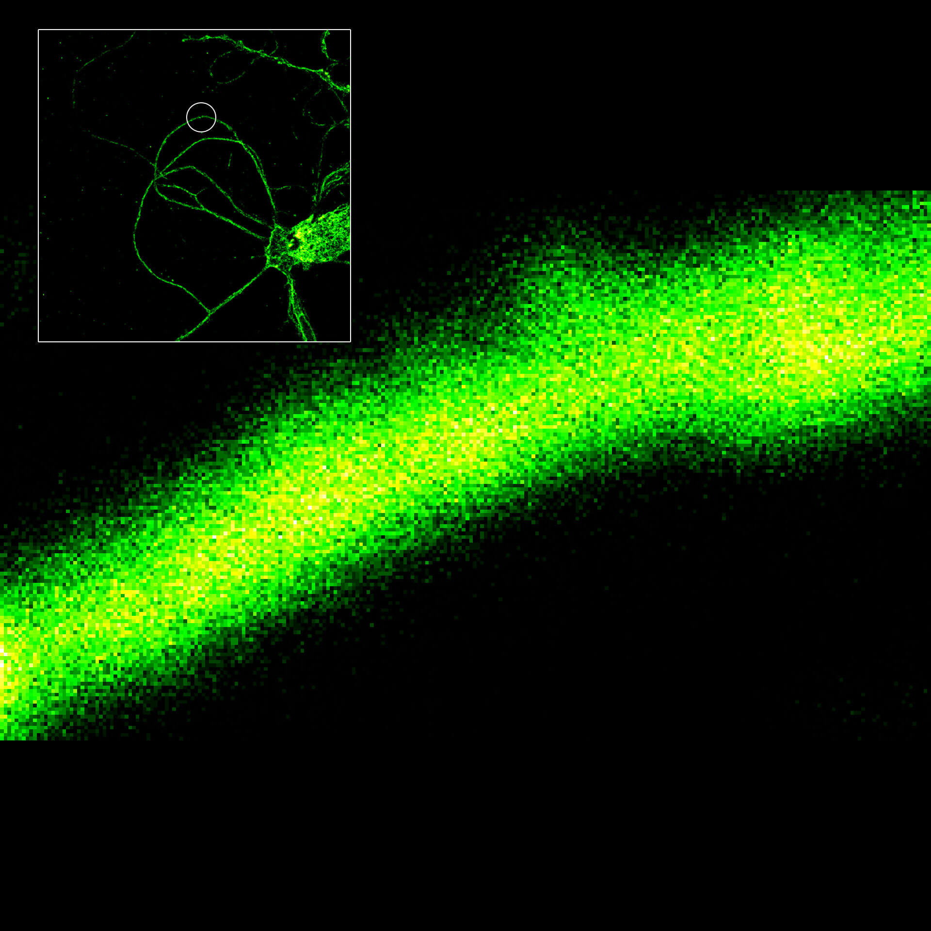
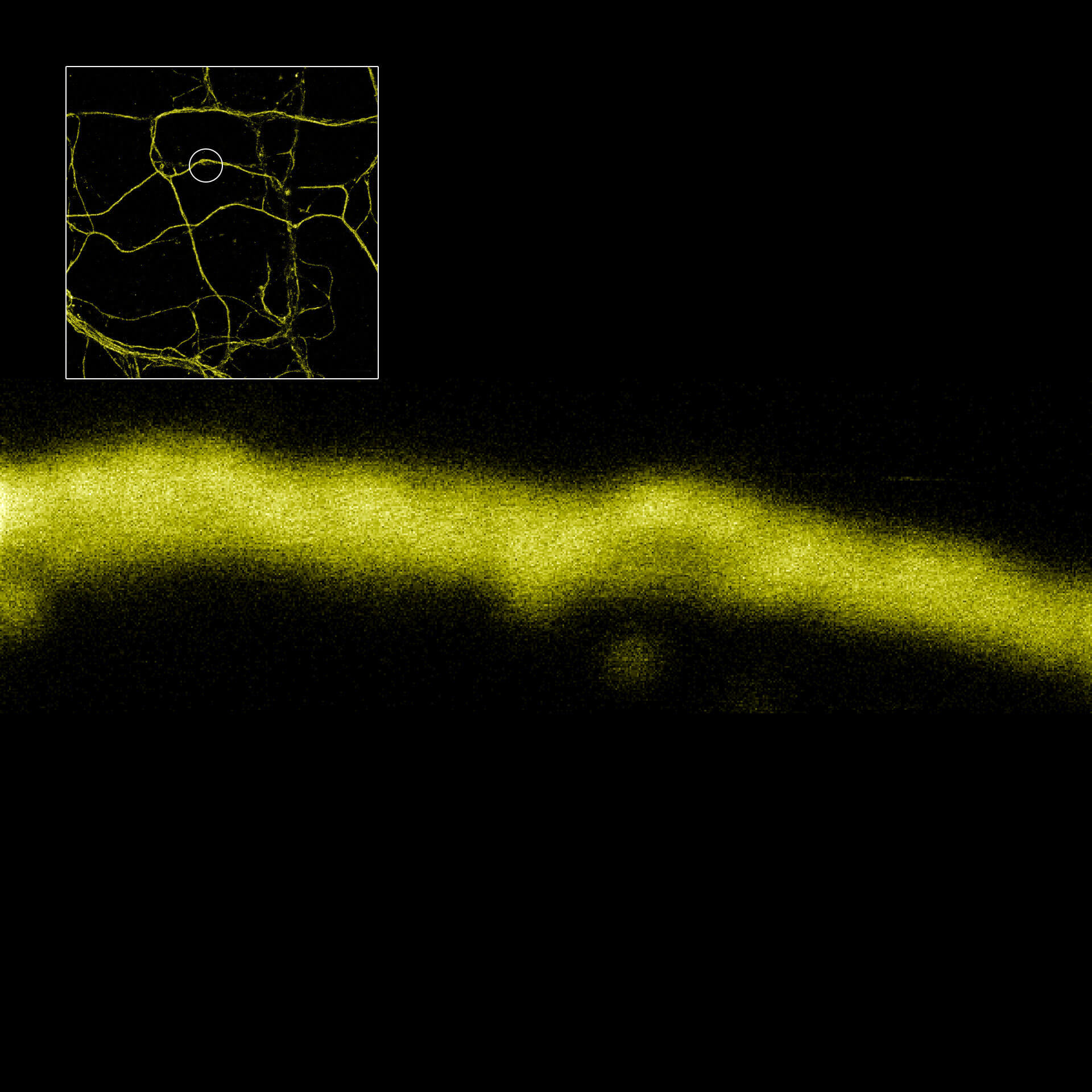
Description
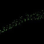
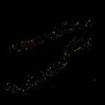
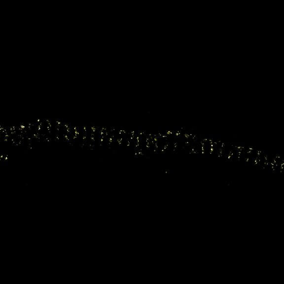
MINFLUX image of axonal βII spectrin labeled with abberior FLUX 660 in primary hippocampal neurons. Note the periodic arrangement of spectrin along the axon, and the absence of any details in the confocal counterpart image.
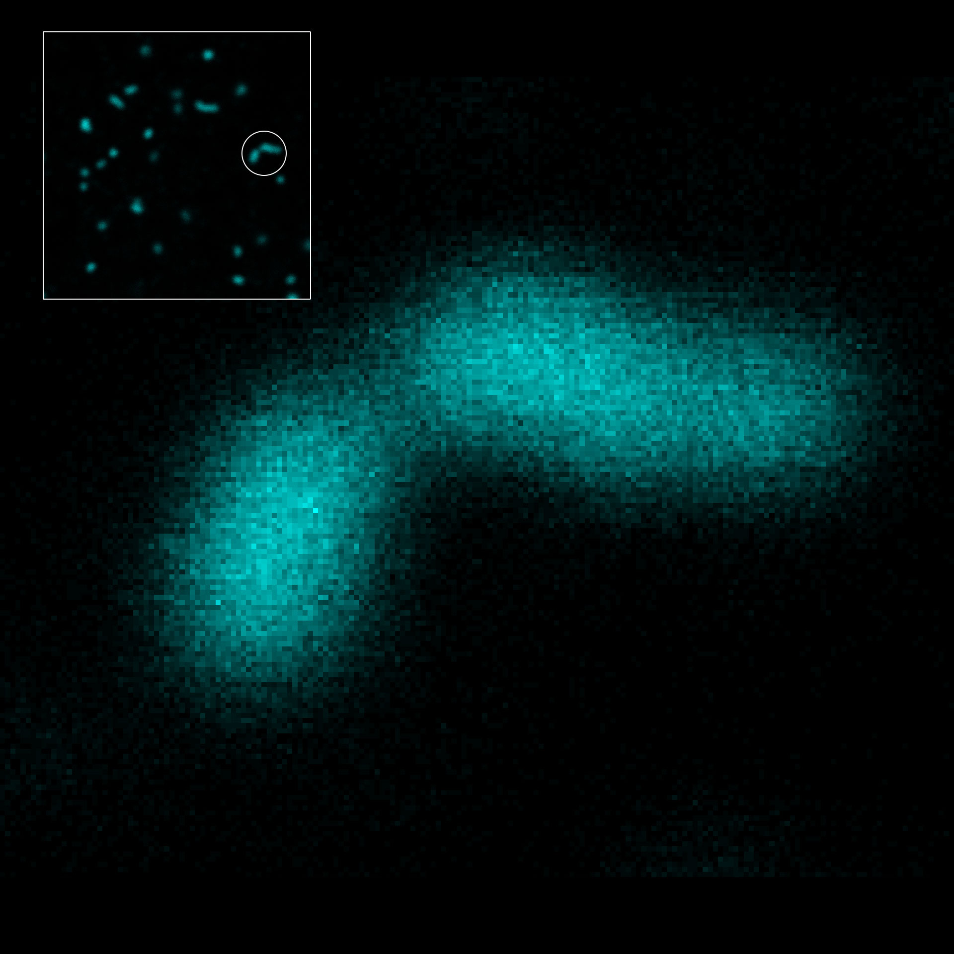
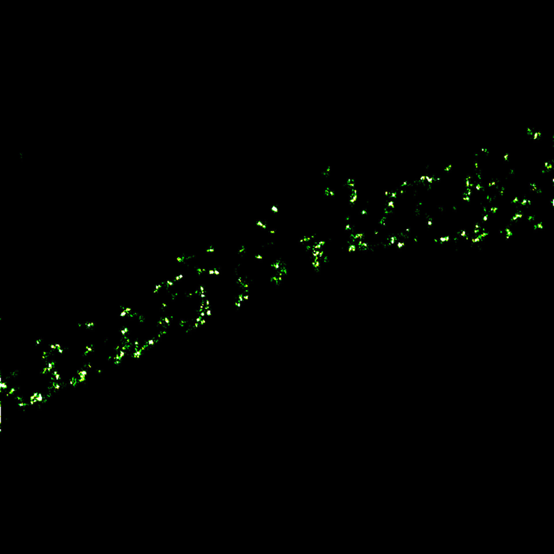
Description
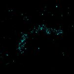
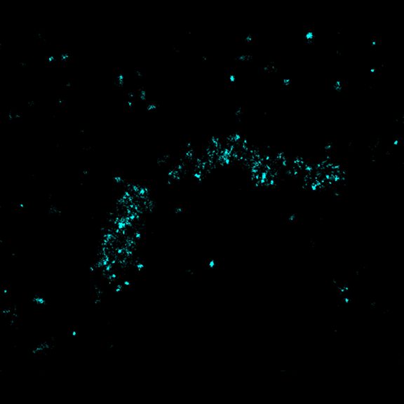
MINFLUX imaging of βII spectrin in a primary hippocampal neuron labeled with abberior FLUX 680 by indirect immunofluorescence. Please note the periodic arrangement of spectrin along the axon.
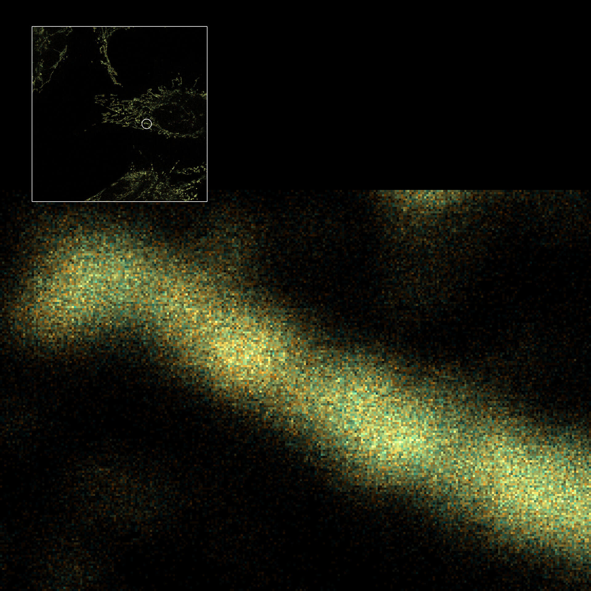
Description
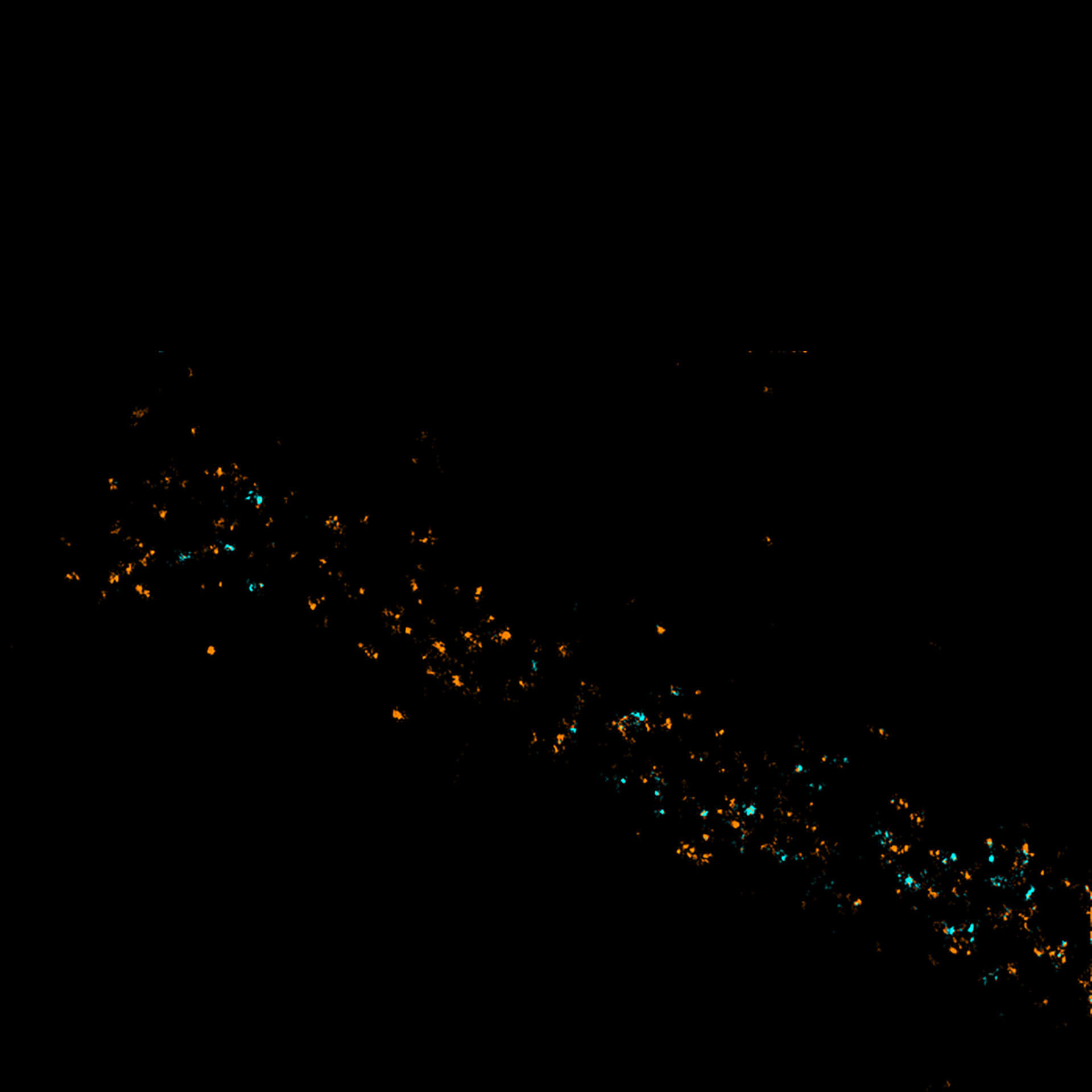
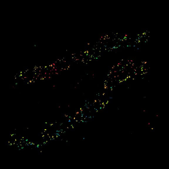
Two-color MINFLUX revealing an inner and outer mitochondrial membrane marker. Cultured mammalian cell labeled with indirect immunofluorescence using secondary antibodies coupled to abberior FLUX 640 (orange) and FLUX 680 (cyan). MINFLUX enables the visualization and separation of both structures.


abberior FLUX dyes
for unmatched resolution
abberior FLUX dyes are optimized for single-molecule localization and confocal microscopy. Conjugates of the dyes show excellent solubility in aqueous buffers and allow background-free labeling in cells or tissue, to get the most out of your sample.
- Highest signal and photostability
- For unrivaled MINFLUX imaging
- Best for single-molecule localization and confocal microscopy
- Optimized photoswitching kinetics
- For MINFLUX/STORM/GSD