STEDYCON:
A sleek, blue box transforms your widefield microscope into a confocal, superresolution STED, and lifetime instrument and your exploration of subcellular structures into a seamless, discovery-rich experience. Carefully designed with masterly engineering, STEDYCON breaks the stereotype of the finicky, hard-to-use scope. It opens new possibilities at the press of a button for any user and almost any location. How does it do it? The secret’s in the box.
ease-of-use in a shoebox
Human ingenuity
Human ingenuity commands admiration. We coax living entities – from elephants to viruses – into doing our bidding. We source out physical and mental work to machines. We sling people across the globe in thin-walled, stratospheric aircrafts. We receive pictures of the universe from a telescope 1.5 million kilometers from the Earth. Although it is the blink of an eye in the grand scheme of things, human history is a maelstrom of innovation.
All kinds of human motives drive that ingenuity. Need. Pleasure. Curiosity. Sharing. Greed. Fueling the adoption of its products, however, is one quality: ease-of-use. The press of a button that unleashes transactions, knowledge, and emotions is the name of the game.
Engineering ease-of-use into cutting-edge microscopy
The forces that drive technology adoption are no different in the science lab. Highly specialized instrumentation is limited to core facilities or niche research groups. Specific techniques may require an occasional unorthodox device in some labs, but a staple set of equipment and methods appears on nearly every benchtop, and its acquisition is driven by ease-of-use. Ease of installation. Ease of proficient use. Ease of results. By consequence, if you want to transform new technology into an everyday tool, you need to design it for one thing: ease-of-use.
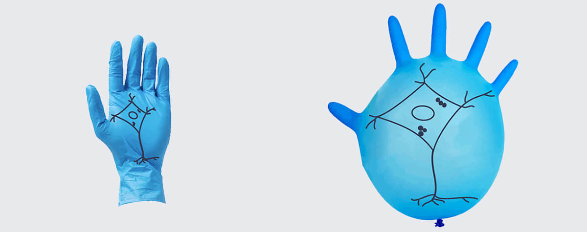
That thought sparked the engineering feat that brought superresolution microscopy into the average science lab. With stimulated emission depletion (STED) microscopy and other techniques already enabling sub-diffraction resolution, it seemed unreasonable that researchers curtail their ability to visualize sub-cellular structures by using diffraction-limited microscopes. Clearly, adding STED to the standard constellation of lab equipment would require more than outstanding resolution. The problem, developers reasoned, was that Nobel-prize technologies are like Formula 1 race cars that only high-performance drivers can maneuver. So, their strategy was to distill that Nobel-winning “race car” into a shoebox that everyone could “drive.” STED would become a powerful upgrade to any widefield microscope with plug-and-play operation, intuitive handling, and rapid outcomes. The product was STEDYCON.
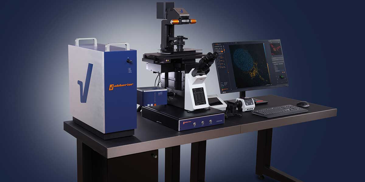
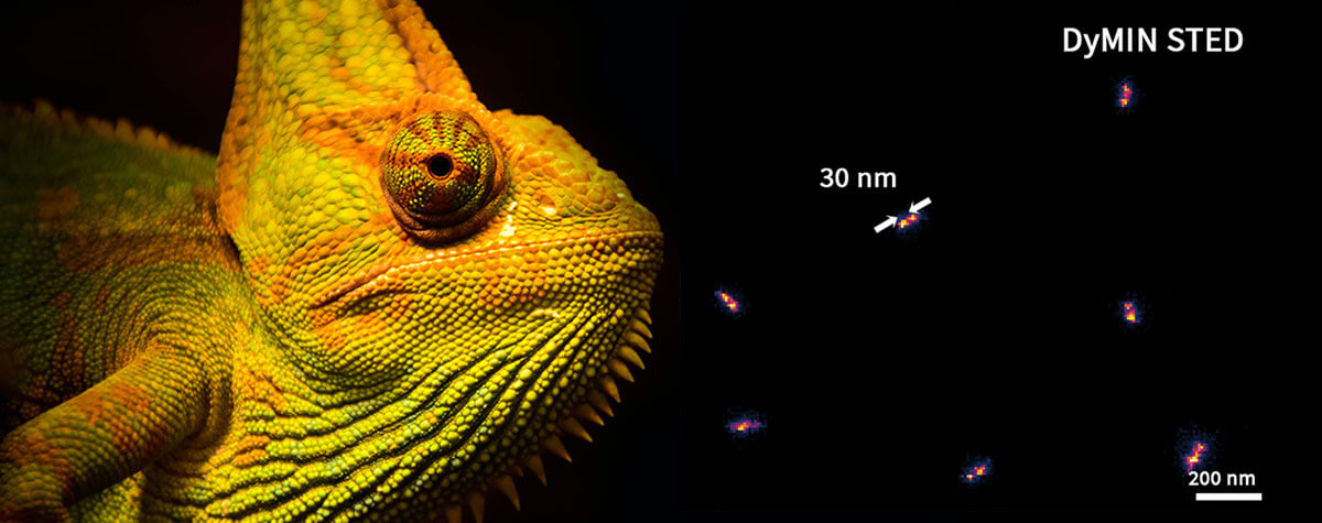
The STEDYCON upgrades your existing widefield system to a confocal and a superresolution STED microscope with a resolution down to 30 nm.
So, what does it look like?
At first glance, STEDYCON is a blue-and-metal casing about the size of a shoebox that sits on the camera port of a microscope. It is compact, unobtrusive, and sturdy. The plain exterior presages its ease-of-use but belies what lies within. Carefully planned engineering and design transform your microscope into a multicolor confocal and 2D STED system. It’s like plugging your scope into an amplifier and being rewarded immediately with power.
To begin with, the engineers tackled the big nuisances that make superresolution microscopy finicky. STEDYCON requires no alignment of the excitation and depletion lasers. They are aligned by design. With a novel optical arrangement that sends all laser beams through the same fiber, the system is more stable than other STED microscopes where beams travel separately. That inherent stability shortens the time to initiate imaging and simplifies installation, which takes only minutes. Furthermore, STEDYCON includes a scanner technology that allows it to operate with any widefield microscope, equalizing performance across platforms and precluding the need for dedicated equipment.
The engineers also revamped the autofocus. Unlike conventional optical instruments that use a dichroic mirror to couple the autofocus laser with the imaging beam path, STEDYCON runs the two beams parallel but segregated. Thus, autofocus in STEDYCON never interferes with imaging, which can produce fluorescence loss or distortions. With the simple press of a button, STEDYCON guarantees drift-free focus for extended imaging sessions up to several days. Also, as STEDYCON is equipped to control motorized stages, it automates the recording of multiple positions in a sample or a tile scan. The user is thus released from sitting at the microscope and can easily capture the whole sample or navigate across it to find objects of interest.
Secondly, STEDYCON makes superresolution microscopy a research technique for everyone. With minimal training, even novices to microscopy can produce high-quality STED images with a few intuitive manipulations of a browser-based control system with a user-friendly interface. Whichever the chosen procedure, users commonly visualize structures at a resolution of 30 nm. The same applies to lifetime imaging, which is fully integrated into the software with TIMEBOW.
Finally, STEDYCON makes no compromises on sensitivity or dynamic range of detection. The avalanche photo detectors (APDs) in STEDYCON have superior quantum efficiency. Thus, they reliably collect photons even under low-signal conditions, like when labeling densities are minimized to preserve physiological conditions in samples. This feature ensures clear images and exceptional signal-to-noise ratio, even under high-signal conditions.
The sway of ease-of-use
The greatest testament to the performance and value of STEDYCON is its growing user base. Being frame-agnostic, STEDYCON complements microscopes of various makes and models. Exceptionally stable, it breaks with convention by operating flawlessly in dedicated labs, trade fair floors, living rooms, and even campsites. And STEDYCON users are a new generation of innovators leveraging a level of microscopy previously reserved for “Formula 1 drivers”. Swayed by easy access to unprecedented detail, they add diversity to the where, when, and how superresolution is used and uncover a myriad of insights hidden behind the low resolution of standard microscopes. There, at the frontiers of science, those discoveries unlock new lines of research as the boundaries to how we study structure, movement, and interactions at the smallest scale are lifted. STEDYCON’s ease-of-use in a shoebox is a portal to breakthroughs in an infinite world.


