


Of unwanted guests and unwilling hosts – Virology


FAQ Video 38

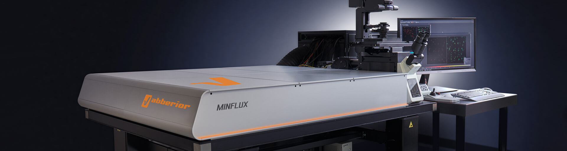
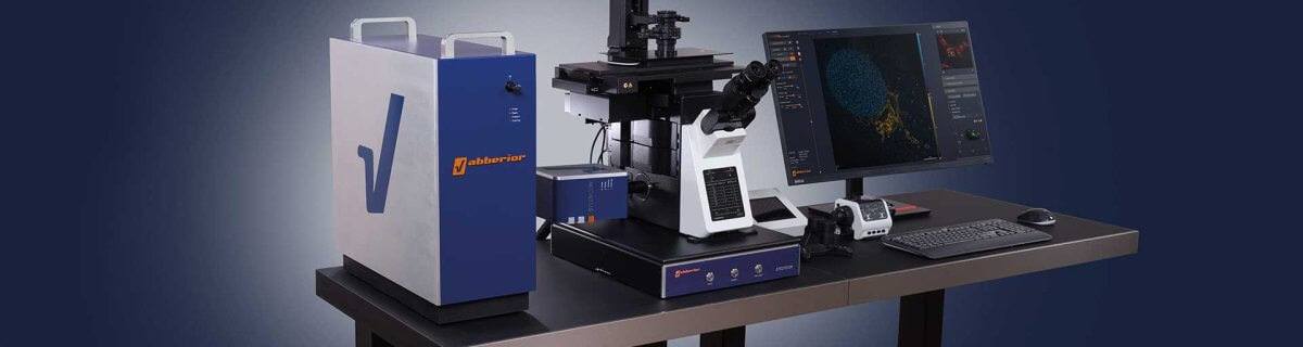
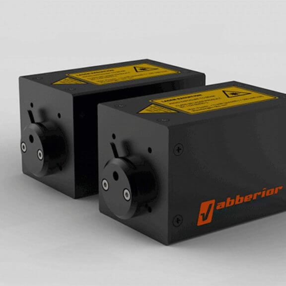
MINFLUX allows an everyday 3D resolution of 2 – 3 nm and offers about 100 times faster tracking than with a camera-based system.
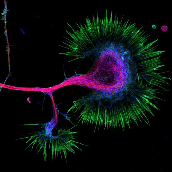
Viruses have long eluded light microscopy, as even the largest ones just reach the diffraction limit of visible light. This has fundamentally changed with the development of superresolution microscopy: STED extends the resolution down to about 20 nanometers and is particularly well-suited to identify sparse events like cell infection or to resolve structures within viruses like their genome or envelope. With MINFLUX, we are even talking single-digit nanometer localization precision, facilitating imaging of also the smallest viruses. Both techniques offer a significant advantage over electron microscopy, which has long dominated virological imaging: they can be used to image viruses and their components in living cells. This makes it possible to resolve dynamic processes relevant in virology, such as the penetration of viruses and phages into host cells and the maturation and assembly of virus particles.
Superresolution imaging techniques have already been used to gain new insights into the biology of HIV-1, Influenza A virus, HSV, Vaccinia Virus, AAV, Nipah Virus, Adenovirus, and many more.
Talk to a scientist >Bringing viruses into view
with superresolution
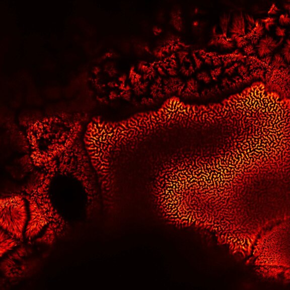
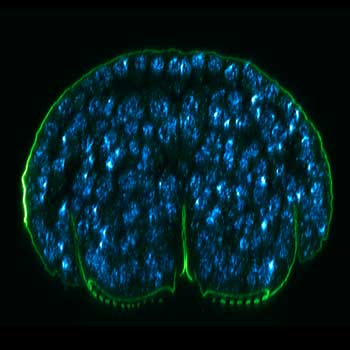
These SARS-CoV-2 viruses (magenta, abberior STAR RED) and cellular actin (green, STAR ORANGE) were imaged in infected Vero E6 cells with STED. One might think that techniques like STED and MINFLUX were invented for virology, given that the size of viruses and their components lies precisely within the size span of superresolution microscopy.
Test your sample >Every Jack has his Jill and every application an abberior solution
We at abberior are experts in superresolution. Our microscopes feature the best technology and optics for cutting-edge STED and MINFLUX imaging, combined with exceptional user-friendliness. They can be flexibly adapted to your particular demands. Add abberior’s fluorescent dyes and labels optimized for applications from confocal to MINFLUX, and you are ideally equipped to take virus imaging to the next level.
MINFLUX – unrivaled resolution and speed
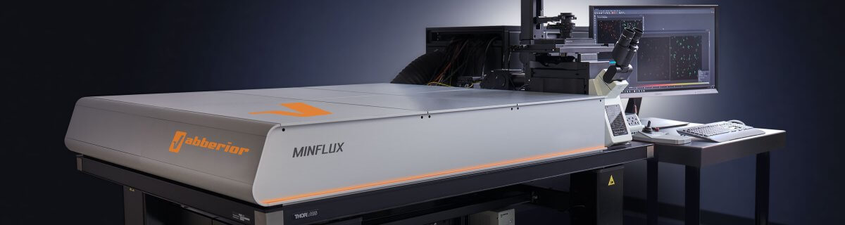
The MINFLUX platform offers an unprecedented array of imaging possibilities and allows you to resolve viruses and even single molecules like capsid proteins along all three dimensions. This unmatched resolution capability combined with unprecedented speed reveals sample details never seen before and helps to dissect fast and dynamic cellular processes in space and time. With MINFLUX, it is now possible to follow single virus particles over time with unprecedented precision and speed. Details >
INFINITY – forever cutting edge
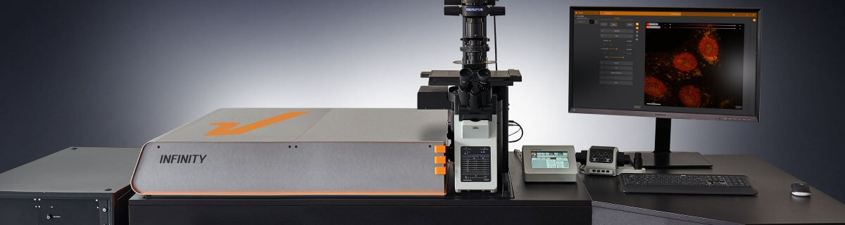
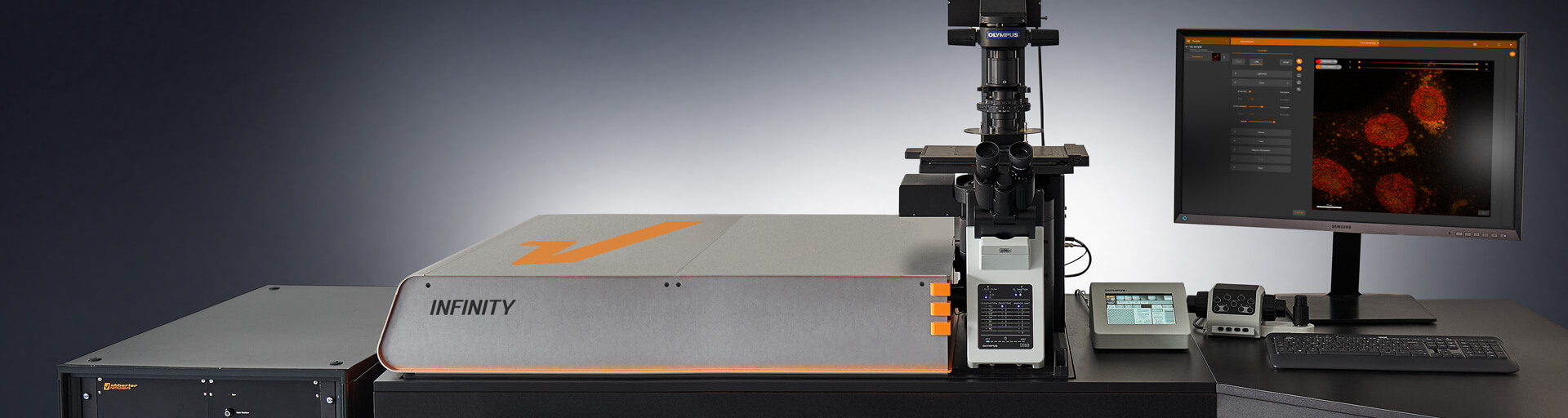
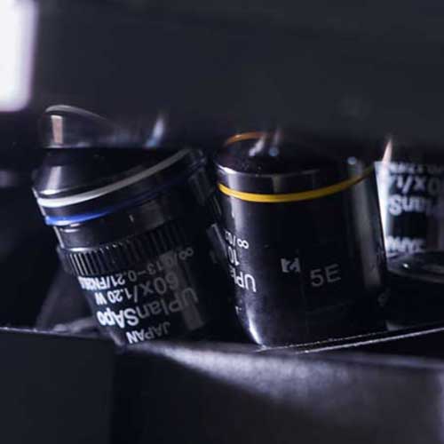
The INFINITY platform is the most customizable platform for all things microscopy and may be adapted to your particular demands in virological imaging. Optimize your microscope for the imaging of anything from single virus particles on a coverslip to infected cells or tissue. INFINITY is always up to the challenge. Just tell us what you need and we will build you a customized, continuously upgradable system specialized for your research. Details >
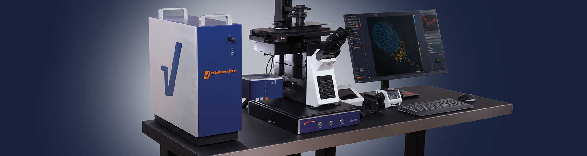
MIRAVA POLYSCOPE – one for all and all for one

The MIRAVA® POLYSCOPE® combines every resolution – from millimeters down to 3 nm – in a singularly unique system. MIRAVA unites four microscopy technologies to cover an unprecedented resolution spectrum, extending over several orders of magnitude from diffraction-limited imaging all the way to true molecular resolution. Our LiGHTBOX software allows beginners to intuitively arrive at a top-notch image within three clicks, while also giving experts full control over the instrument. Details >
STEDYCON 2 – confocal, STED, lifetime… WOW!
The STEDYCON upgrades your existing widefield microscope to a confocal. STED, and lifetime machine with a resolution down to 30 nm. All that’s required is a free camera port and a good objective lens. With its super-intuitive user interface, the STEDYCON provides an intelligent microscope platform that enables everyone to acquire superb superresolution images after only minutes of training. Details >
Cutting-edge tools
to solve the riddles
Choose a superpower for your experiment – our modules
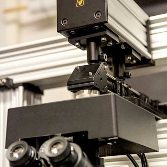
Every MINFLUX, INFINITY, and FACILITY microscope from abberior can be equipped with different modules, powerful functional units that expand the system’s core functionality to reach new imaging heights. You may, for instance, use TIMEBOW lifetime imaging to separate and remove the autofluorescence of lipofuscin from images. Your deep tissue images will be crisp and clear from top to bottom thanks to RAYSHAPE aberration correction with a deformable mirror. FLEXPOSURE adaptive illumination puts light only where it has an effect and may reduce the light burden by orders of magnitude, facilitating extended live cell imaging and volume scans without bleaching or phototoxic effects. Our modules can do all of this and even more! And if you can’t find what you need on our website, get in touch with us, and we’ll develop it for you.
MATRIX Detector
Many eyes see more than one. The MATRIX detector drastically improves signal-to-background ratio, resolution, and dynamic range.
TIMEBOW Imaging
TIMEBOW lifetime imaging for stunning results at confocal and STED super-resolution.

FLEXPOSURE Illumination
Brings down the light dose on your sample and lables dramatically. Key ingredient for volume and live-cell superresolution.
RAYSHAPE Mirror
Dynamic aberration correction with a deformable mirror over about 200 µm z-range. 140 digital actuators adjust the mirror surface within milliseconds.


Custom Solutions
We offer solutions for even the most challenging applications. Everything that can be done, we will do.
Broaden your knowledge
of microscopes, dyes, and superresolution
Light microscopy and superresolution are a broad field. Even for advanced users, there is always something new to learn and knowledge gaps to fill. The articles in our knowledge base are each devoted to a single topic, which they illuminate and explain in detail. Read and learn!
Tell me more >
PALM and STORM are often used as synonyms, and in fact they have a lot in common. But there are slight differences that can be important for your application. And then there are other superresolution techniques, too – like STED and MINFLUX. Details >

For centuries, conventional light microscopy was and continues to be the workhorse of labs to visualize cells and cellular details. But the advent of electron microscopy brought about a new level of detail. Let’s take a closer look at the two techniques. Details >
How to correct for aberrations in light microscopy

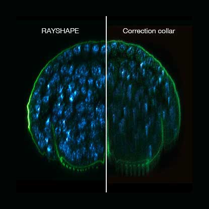
Aberrations can give microscopists a hard time. They belong to microscopy like pathogens belong to life. There are ways to bring diverted rays back on track, but some are better than others. The question is: deformable mirror or correction collar? Details >

Every technique that allows to observe cells is more or less invasive and fluorescence microscopy is no exception. Many imaging situations profit from a reduction in light dose as provided by FLEXPOSURE adaptive illumination. Details >

MINFLUX reaches unprecedented spatio-temporal resolution in light microscopy and provides 2D and 3D localization precisions in the single-digit nanometer range. Details >
Why do superresolution microscopists love alpacas?

It is a very simple yet very important fact: the localization precision of any superresolution microscope can only be as good as the size of the fluorescent staining allows. In other words, when your fluorescent dye is too big or too far away from the protein you want to label, you will never be able to reach a resolution that is higher than this offset. The good news is: there are ways to reduce the offset between target protein and fluorescent label. And one of these are nanobodies. Details >

A little insight into the advances in virus research made possible by STED microscopy and a hint to were the journey might go. Details >
Superresolution for biology: when size, time, and context matter

The spatial resolution achievable with today’s light microscopes has unveiled life at the scale of individual molecules. Size is no longer a barrier to seeing biology at the most fundamental level. But life is not static. It emerges from movement and change. How do superresolution technologies hold up to the challenges of documenting dynamic biological mechanisms? Details >
Additional information and articles

RAYSHAPE webinar – dynamic aberration correction for deep tissue imaging
Hello RAYSHAPE! Bye-bye, aberrations!
RAYSHAPE special
RAYSHAPE dynamic aberration correction with a deformable mirror preserves resolution and increases brightness. 140 actuators adjust mirror surface within milliseconds for over 200 µm z-range.

Summer Symposia: Adaptive Optics
All about aberration correction in STED and how the outstanding capabilities of abberior’s Adaptive Optics helps overcome the challenges of complex samples.

TIMEBOW Lifetime Imaging – gives time color
Explore abberior’s TIMEBOW lifetime toolbox with our application experts Julia Menzel and Jan-Gero Schloetel.

FLEXPOSURE special
Fluorescence requires light. Light inevitably bleaches fluorophores. What a dilemma. abberior has the solution: FLEXPOSURE! It brings down the light dose by up to two orders of magnitude, without compromising…
MINFLUX webinar – Tracking with unprecedented spatio-temporal resolution!
Thanks MINFLUX it’s possible to track the movement of individual molecules in the cell, e.g. kinesin. See what EMBL scientists have achieved.

Adaptive STED in Neuroscience Research
Prof. Hiroshi Kawabe presents his research with STED for neuroscience applications and Martin Meschkat explains the benefits MATRIX array detection.

Summer Symposia: MINFLUX superresolution post Nobel
Stefan Hell talks about his post Nobel development MINFLUX, reaching single digit molecular resolution and allows tracking with unprecedented spatio-temporal resolution.