


Live and let live – Live Cell Imaging


NEWS
Bye, bye, bleaching! Start “endless“ live-cell imaging with our exchangeable HaloX® labels!
Tailor your experiments to the max – abberior Halo labels
NEW abberior LIVE ORANGE mito – a probe for cristae structure in living mitochondria!
NEW abberior LIVE click dyes
New Publication: Two-color live-cell STED nanoscopy by click labeling with cell-permeable fluorophores
New abberior STAR membrane probes for live-cell STED
Live cell imaging is used in virtually all areas of molecular biology. It places unique demands on imaging conditions and microscope. Controlling the environment of the sample is pivotal to ensure physiological conditions during imaging: temperature, humidity, and CO2 concentration need to be kept constant. Furthermore, a good autofocus unit is a must, and scanning and detection have to work fast to resolve dynamic cellular processes in time. The microscope should be equipped with a water immersion objective and possibly also a dipping objective. Fluorescence lifetime imaging (FLIM) is a plus. And to facilitate longer measurements and time-lapse microscopy, it’s key to keep the light dose as low as possible to avoid phototoxic effects that might damage the sample. Last but not least, you need to use membrane-permeable, non-toxic, and at the same time bright fluorescent dyes.
No matter whether you want to image in confocal or STED mode – abberior has the solution for your live cell imaging experiment. With us, you will find the ideal dye, a suitable microscope system, and the matching technical modules for your purpose; all with the aim of recording the best possible data while treating the sample with care.
Talk to a scientist >It’s alive!
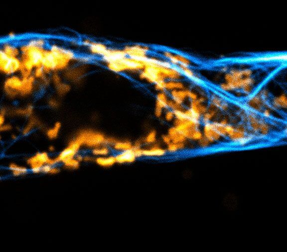
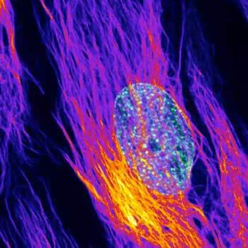
STED image of a mammalian cell
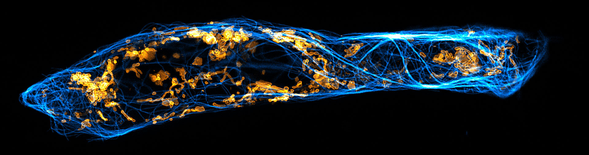
STED live cell image of a mammalian cell expressing a SNAP-tag OMP25 fusion protein in the outer mitochondrial membrane. OMP25 is visualized by abberior LIVE 610 SNAP ligand (orange), tubulin is labeled with abberior LIVE 550 tubulin (cyan).
Test your sample >Capture the moment
Imaging living cells may be trickier than working with fixed specimens. But when you have a suitable microscope and apply the right dyes, you are already halfway there and may generate stunning images and videos like those below. Flip through and enjoy!
Description
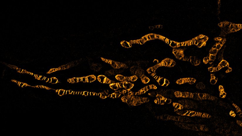
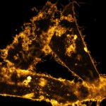
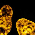
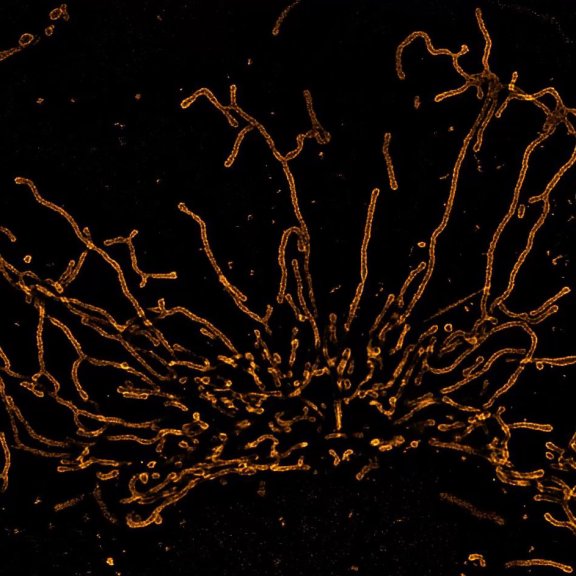
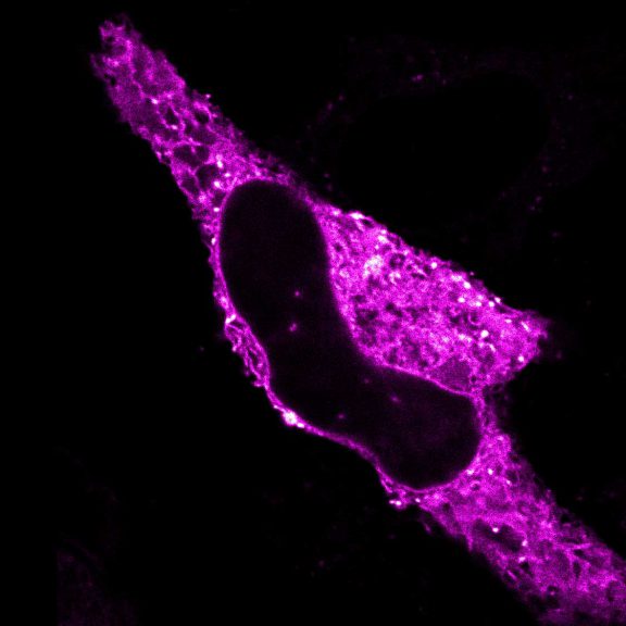
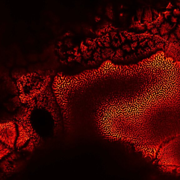
Living HeLa cells stained with the mitochondrial membrane marker abberior LIVE ORANGE mito, visualizing both outer and inner membranes. Confocal and STED images where deconvolved with TRUESHARP image boosting.
Modules:
Description
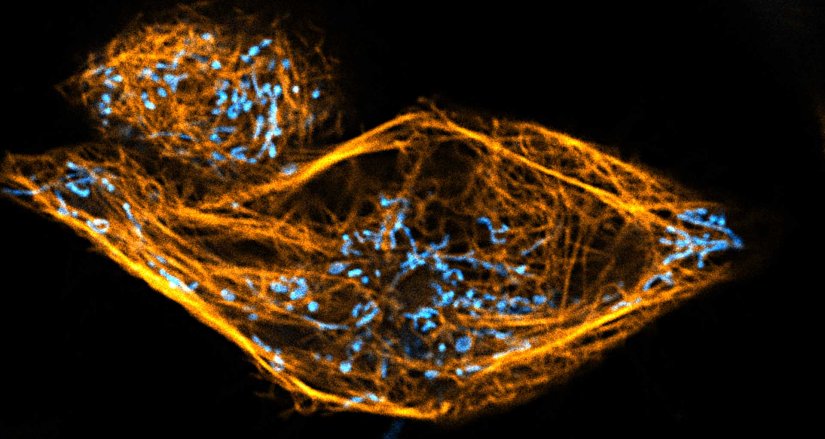
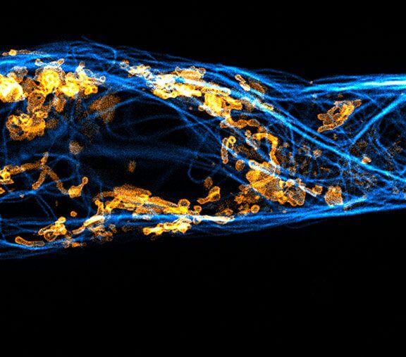
Two color live-cell confocal and STED image of a mammalian cell directly labeled with abberior LIVE 510 mito (cyan), and LIVE RED tubulin. This image was acquired with a STEDYCON microscope.
Description
Description
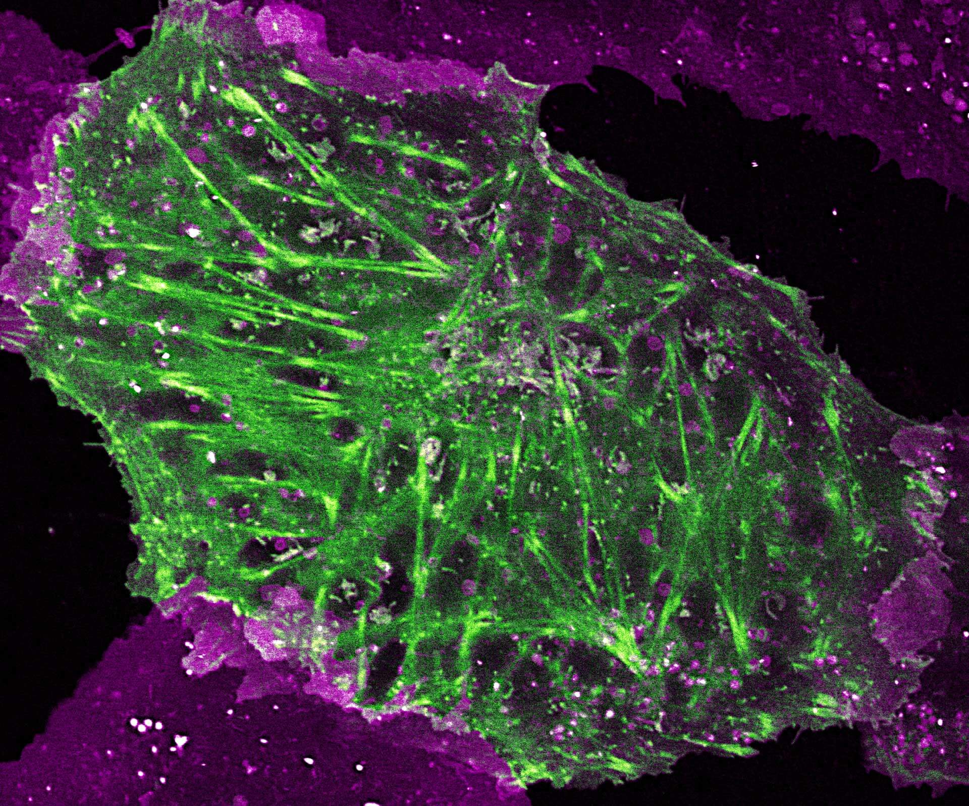
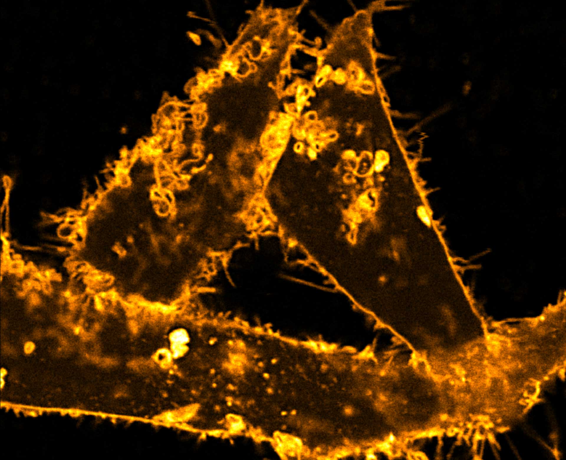
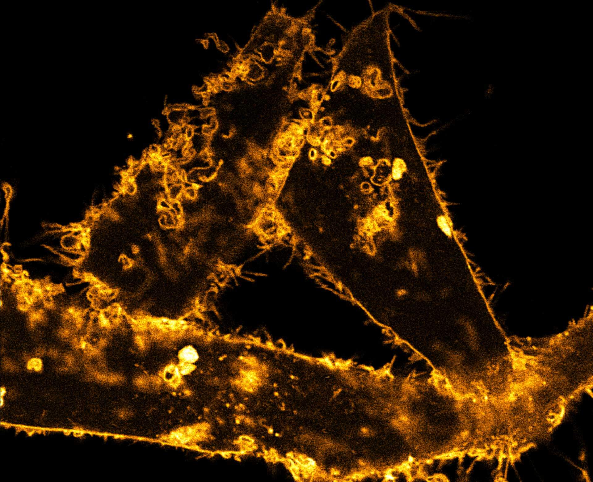
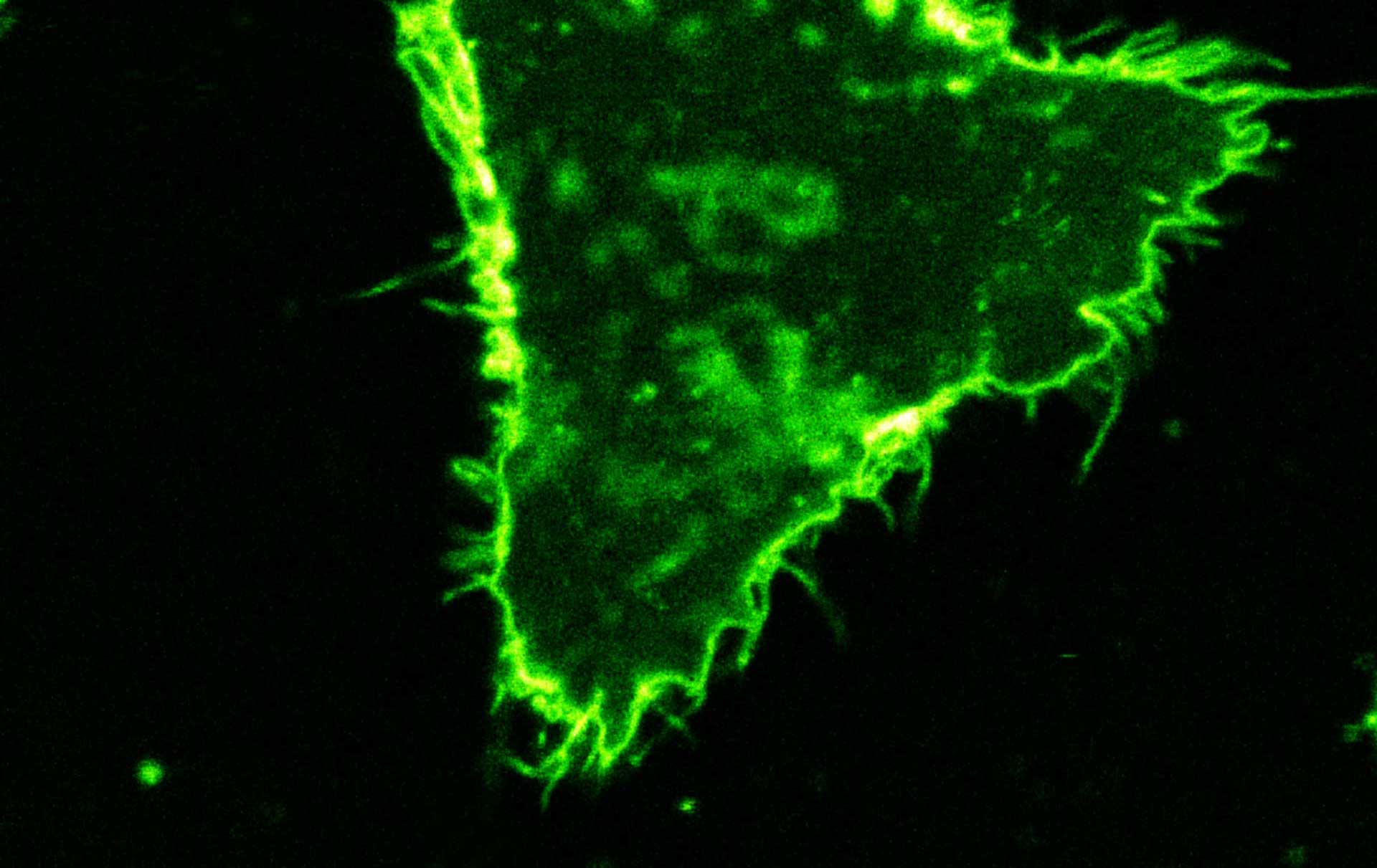
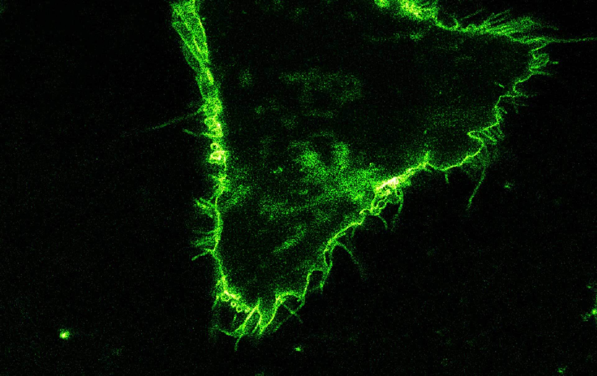
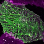
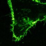
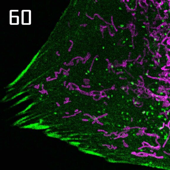
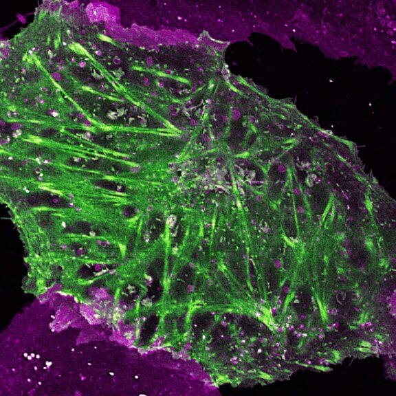
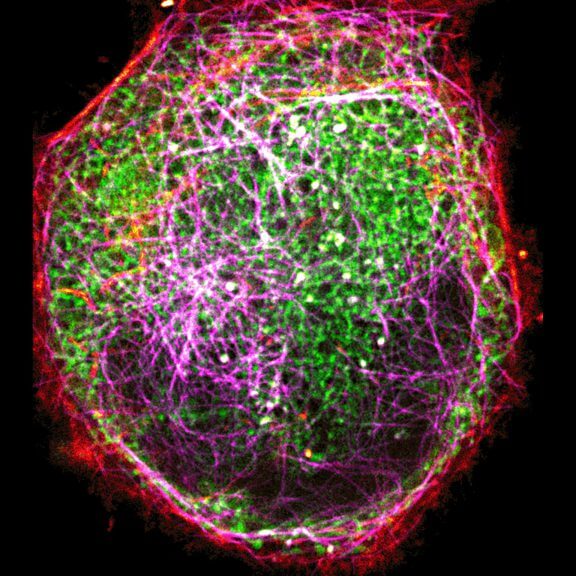
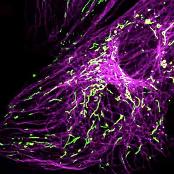
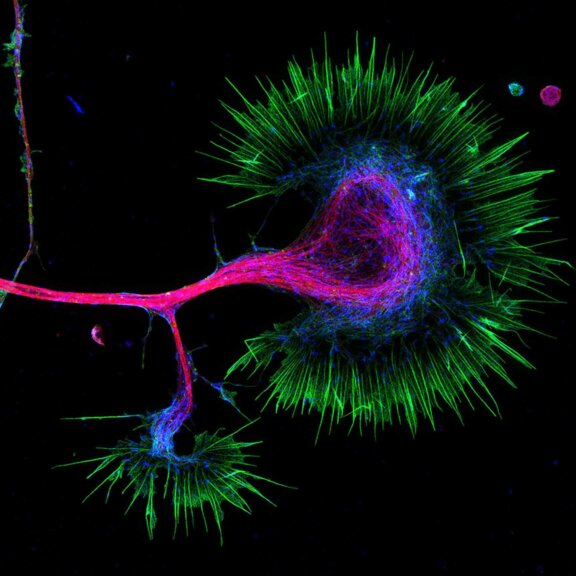
2-color STED image of a living cell expressing actin K118TAG incorporating TCO*A labeled with abberior LIVE 590 click (green). Plasma membrane is highlighted with abberior STAR RED membrane (magenta).
Description
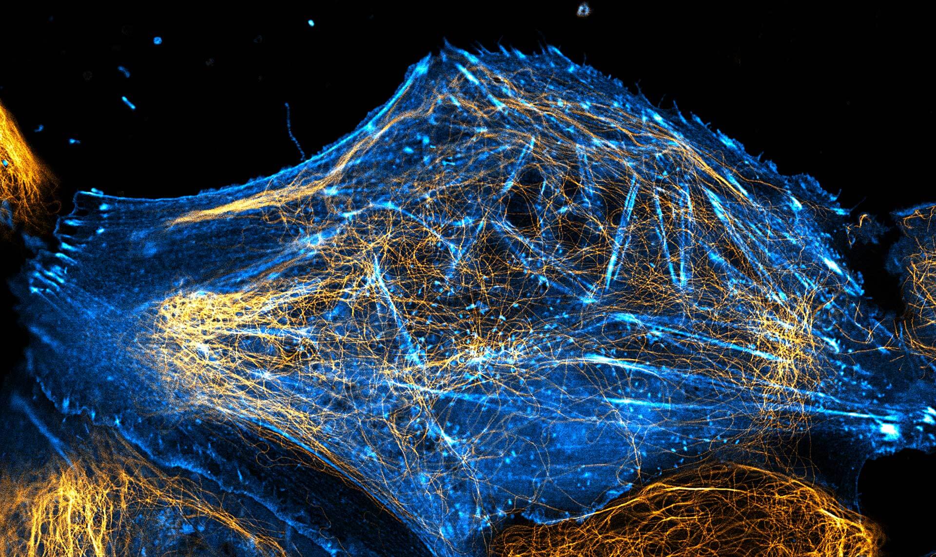
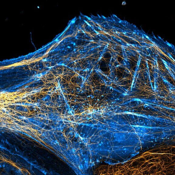
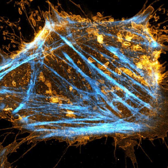
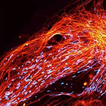
2-color STED image of a living cell expressing actinK118TAG incorporating TCO*A labeled with abberior LIVE 550 click (cyan). Tubulin filaments were stained with our direct probe abberior LIVE 610 tubulin (yellow).
Description
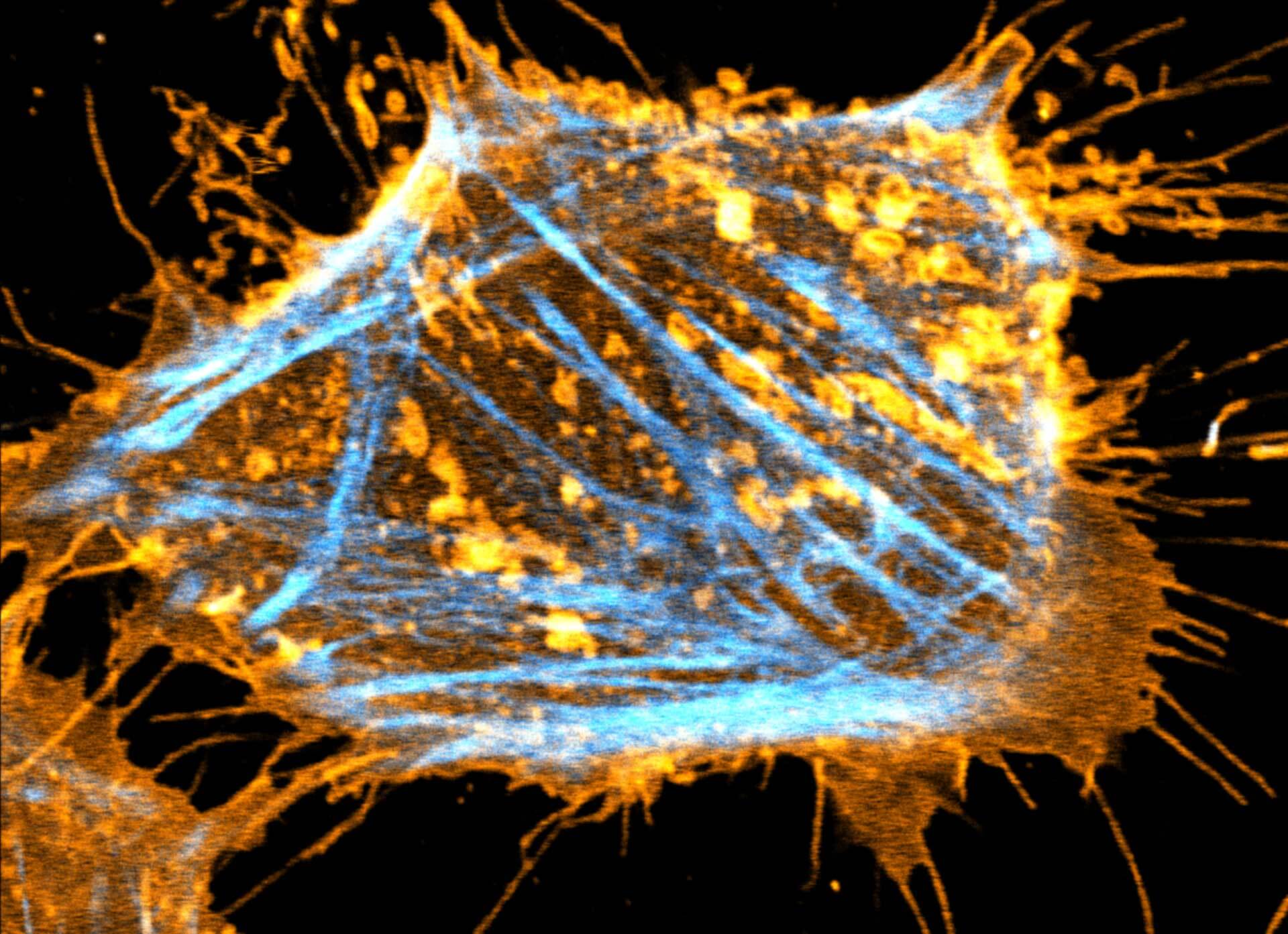
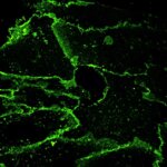
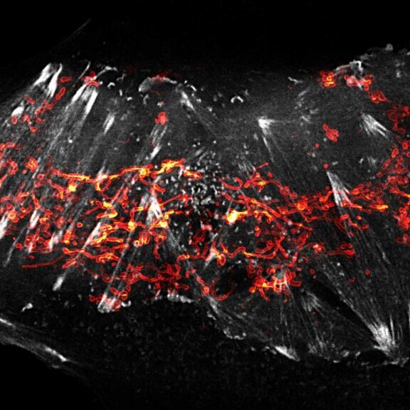
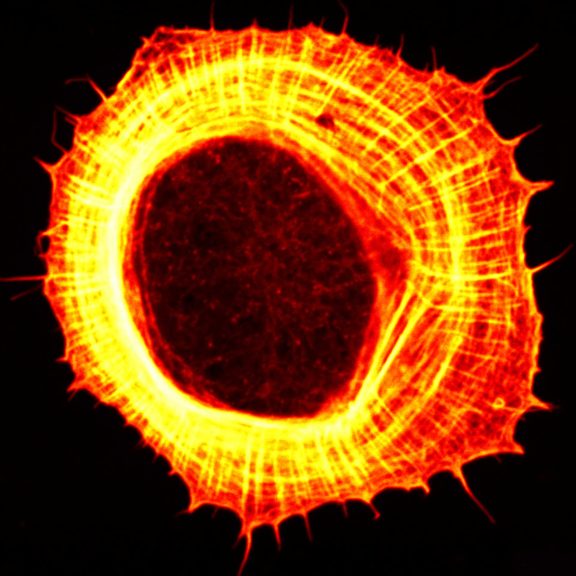
Two color live-cell STED and confocal image of a mammalian cultured cell stained with abberior STAR RED membrane (orange) and abberior LIVE 590 actin (cyan).
Description
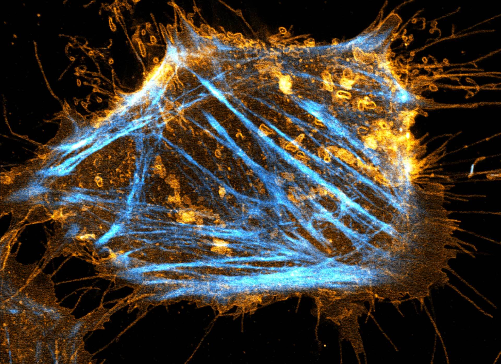
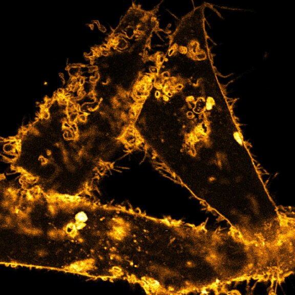
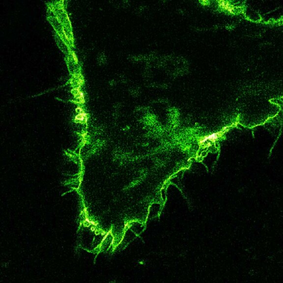
Comparison of STED and confocal of a living mammalian cell stained with abberior STAR ORANGE membrane.
Description
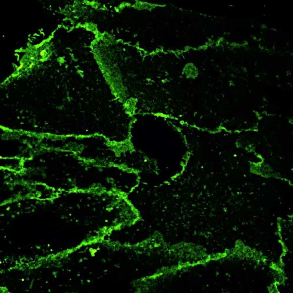
Comparison of STED and confocal of a living mammalian cell stained with abberior STAR 488 membrane. Images were acquired with our FACILITY and STED @ 595 nm.
Description
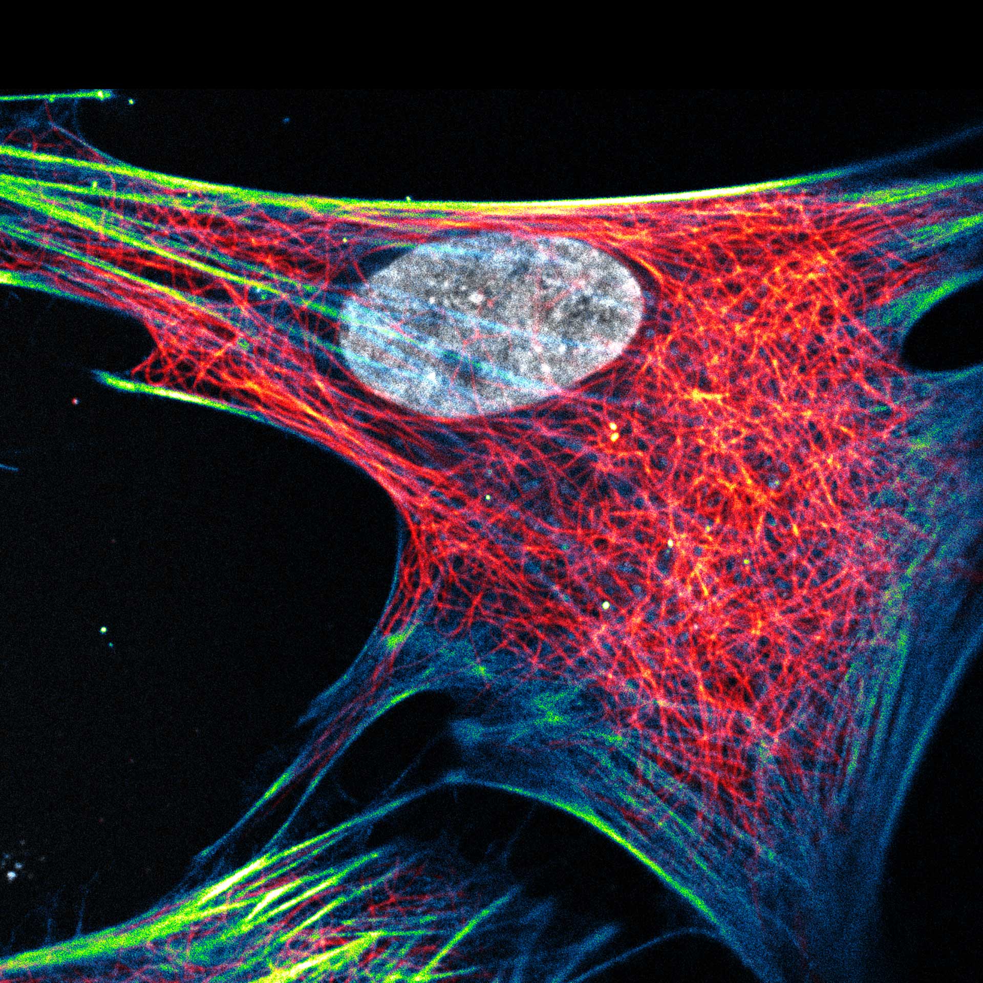
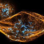
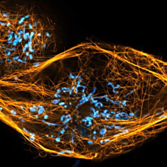
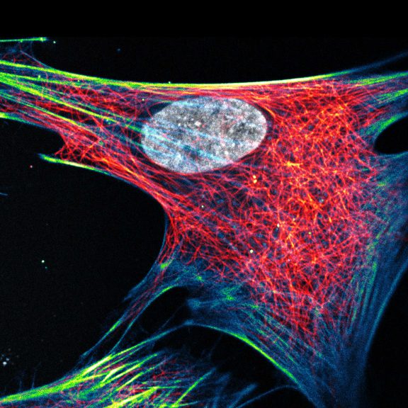
Composite of three color live-cell image of an adherent mammalian cell. This living cell was directly labelled with abberior LIVE 510 actin (blue/green), LIVE 560 DNA (gray) and LIVE 610 tubulin. This image was acquired with a STEDYCON microscope.

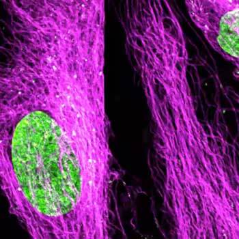
Description
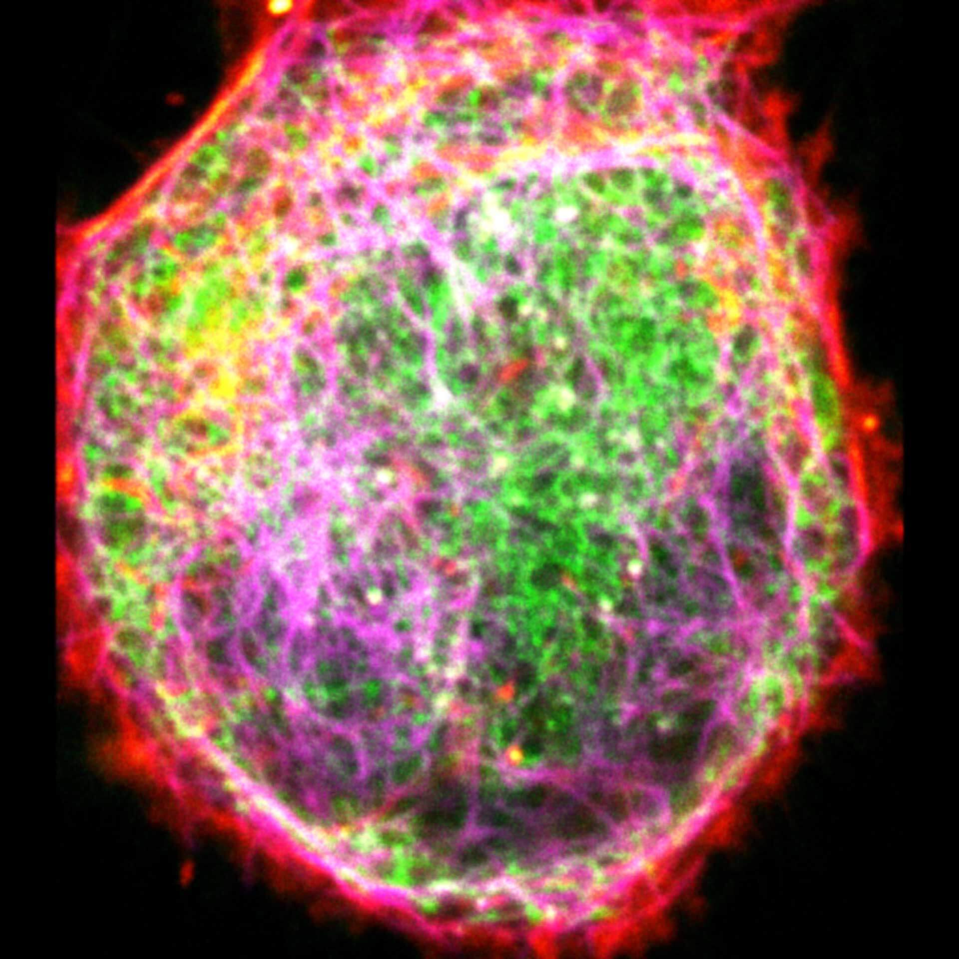
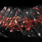
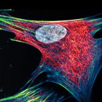
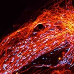
Three color live-cell STED at 775 nm: living cell labelled with abberior LIVE 460L (ER, green), LIVE 560 tubulin (magenta) and LIVE 610 actin (red).
Description
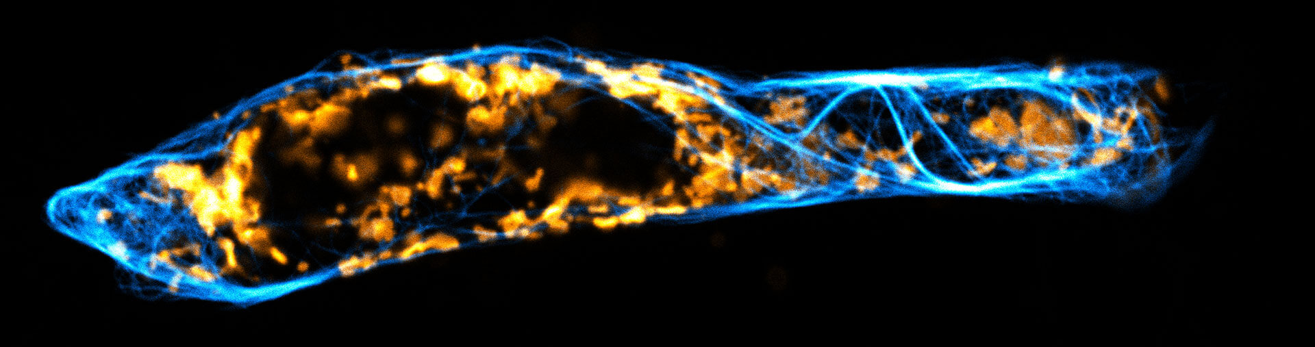
Two color live-cell confocal and STED image of a mammalian cell expressing a SNAP-tag® OMP25 fusion protein decoration the outer membrane of mitochondria. OMP25 is visualized by our new abberior LIVE 610 SNAP ligand (orange). Tubulin filaments are highlighted with abberior LIVE 550 tubulin (cyan).
The SNAP-OMP25 plasmid was a gift from Francesca Bottanelli, FU Berlin.
Description
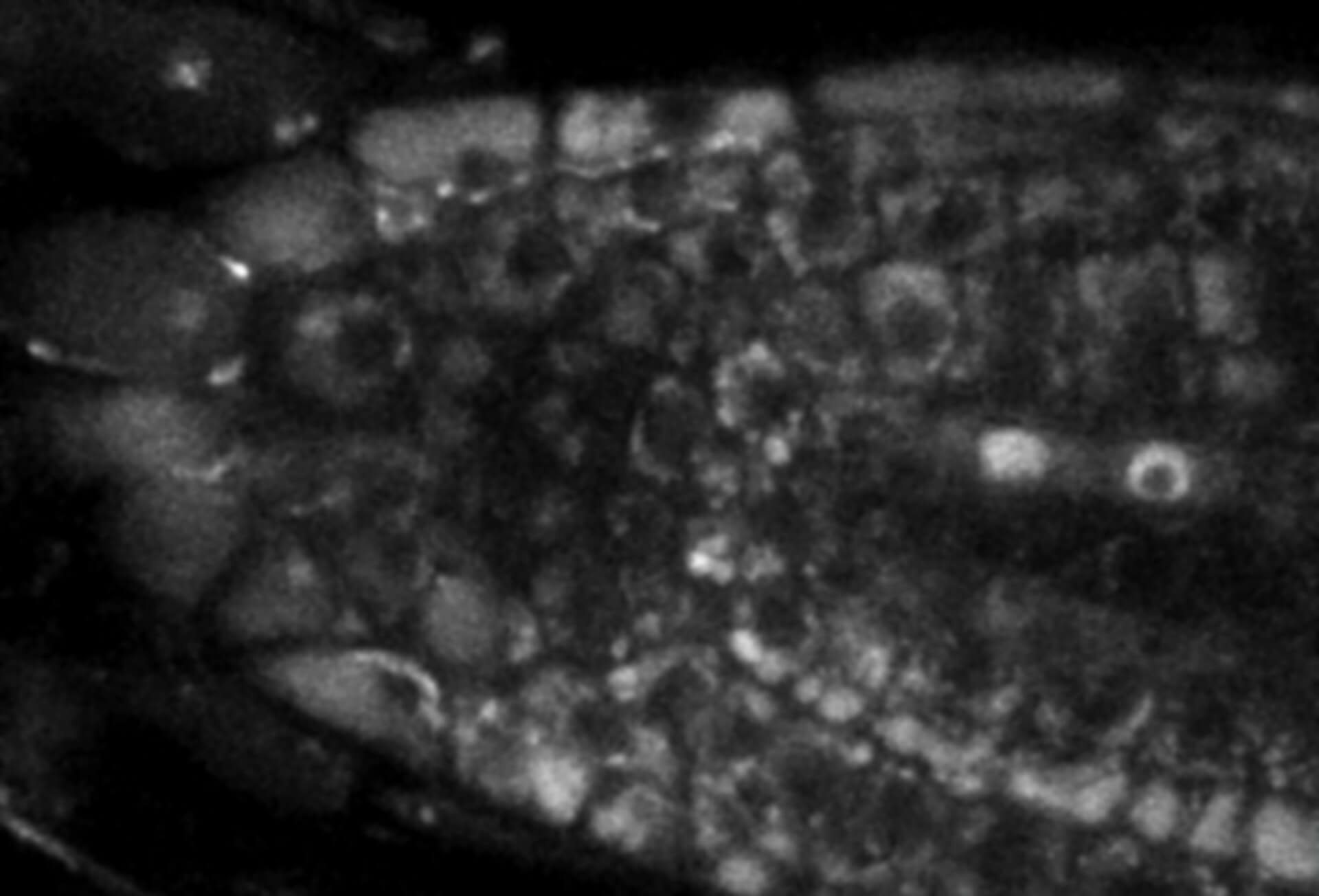
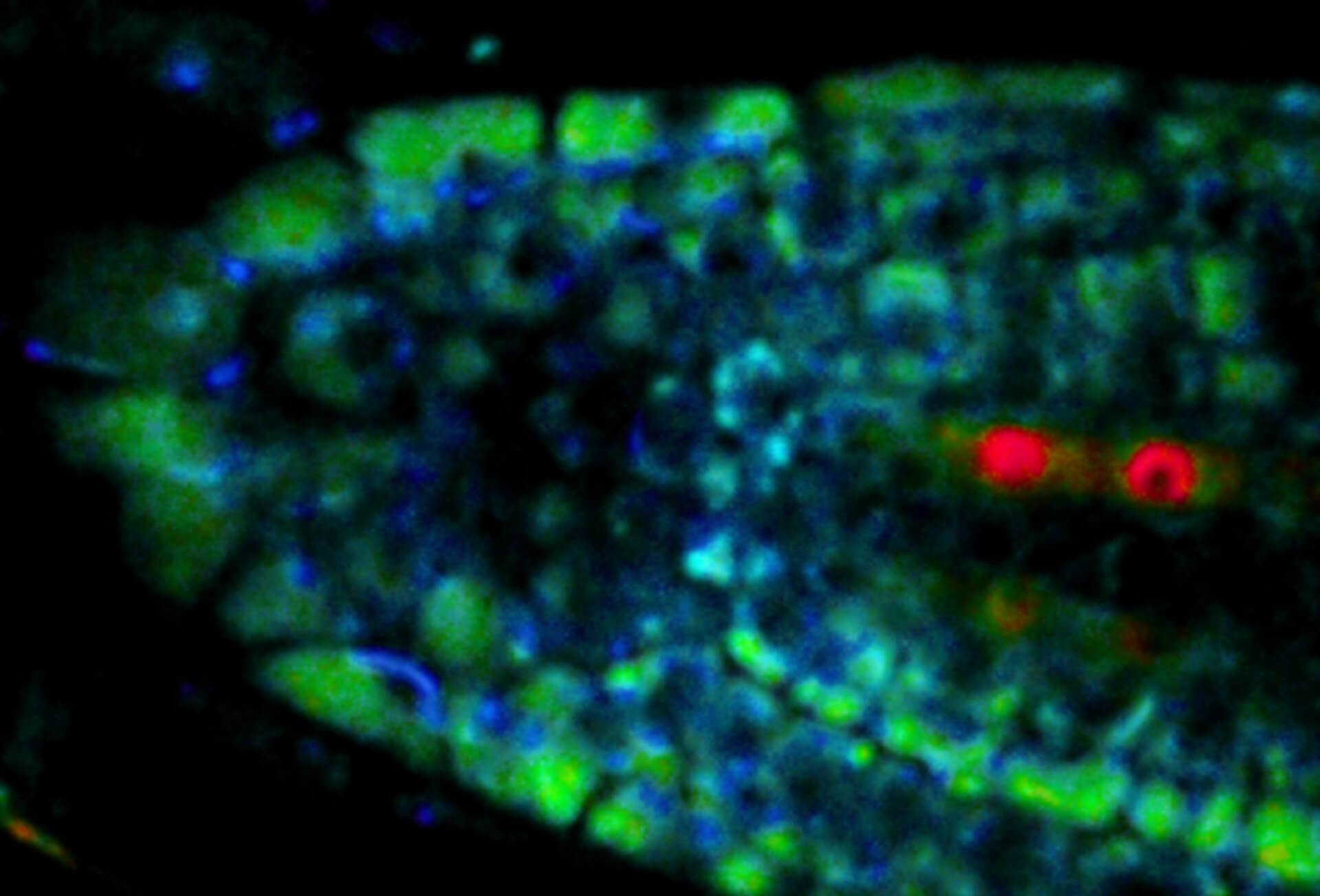
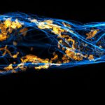
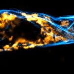
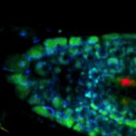
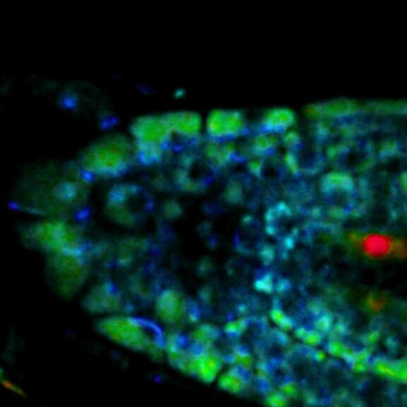
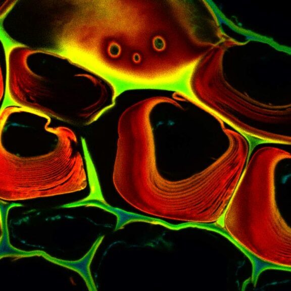
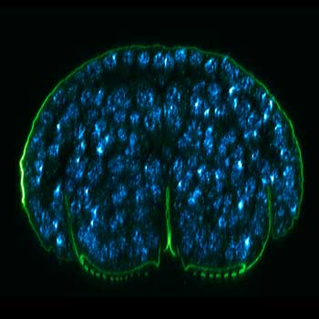
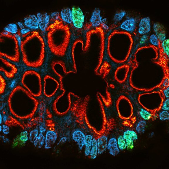
Live-cell sample of an Arabidopsis root tip suspended in water recorded with FACILITY. A subset of cells expresses a YFP construct. Lifetime imaging with TIMEBOW shows shifts in YFP fluorescence lifetime caused by the proteins nano-environment.
We thank Dr. Fábián Attila and Soós Vilmos (ATK, Brunszvik, HU) for providing this sample. Contact: soos.vilmos@atk.hu.
Modules:


Live from the cell stage –
the best microscopes for live cell imaging
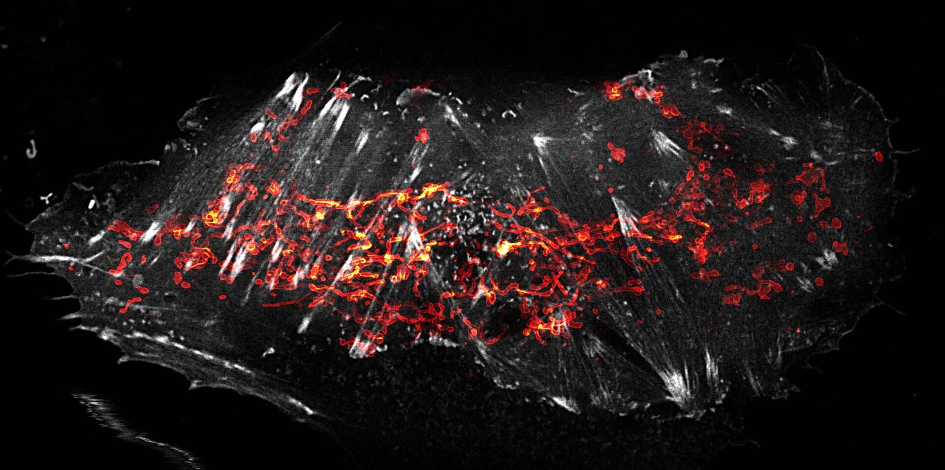
Two-color STED image of a living cell expressing actinK118TAG incorporating TCO*A labeled with abberior LIVE 550 click (gray) and SNAP-tag in the outer mitochondrial membrane labeled with abberior LIVE 610 SNAP (red). abberior offers microscopes ideally suited for extended imaging sessions, live intracellular molecule tracking with unprecedented spatial and temporal resolution, and other in vivo applications.
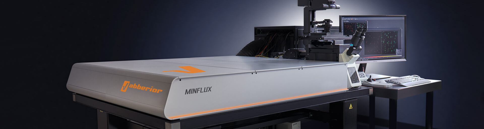
Get a demo >MINFLUX – unrivaled resolution and speed
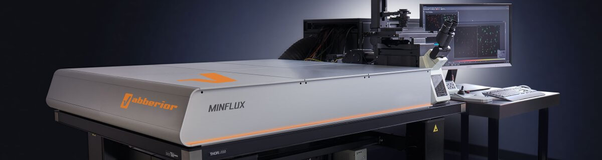
The MINFLUX platform offers an unprecedented array of imaging possibilities and allows you to resolve structures as small as a molecule, along all three dimensions. This unmatched resolution capability combined with unprecedented speed reveals sample details never seen before helps to dissect fast and dynamic cellular processes in space and time. MINFLUX is the world’s most powerful fluorescence microscope. Details >
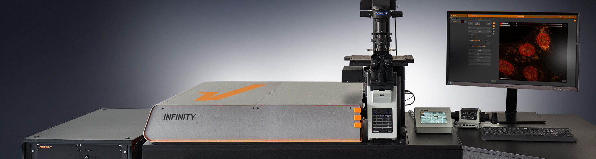
INFINITY – forever cutting edge
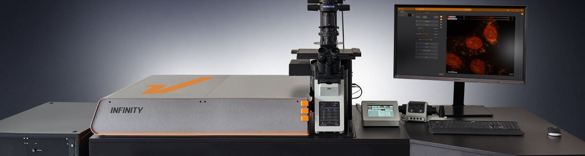
The INFINITY platform is the most customizable platform for all things microscopy and may be adapted to your particular demands in live cell imaging. INFINITY is always up to the challenge. Just tell us what you need and we will build you a customized, continuously upgradable system specialized for your research. Details >
MIRAVA POLYSCOPE – one for all and all for one

The MIRAVA® POLYSCOPE® combines every resolution – from millimeters down to 3 nm – in a singularly unique system. MIRAVA unites four microscopy technologies to cover an unprecedented resolution spectrum, extending over several orders of magnitude from diffraction-limited imaging all the way to true molecular resolution. Our LiGHTBOX software allows beginners to intuitively arrive at a top-notch image within three clicks, while also giving experts full control over the instrument. Routine live cell imaging is a snap with MIRAVA. Details >
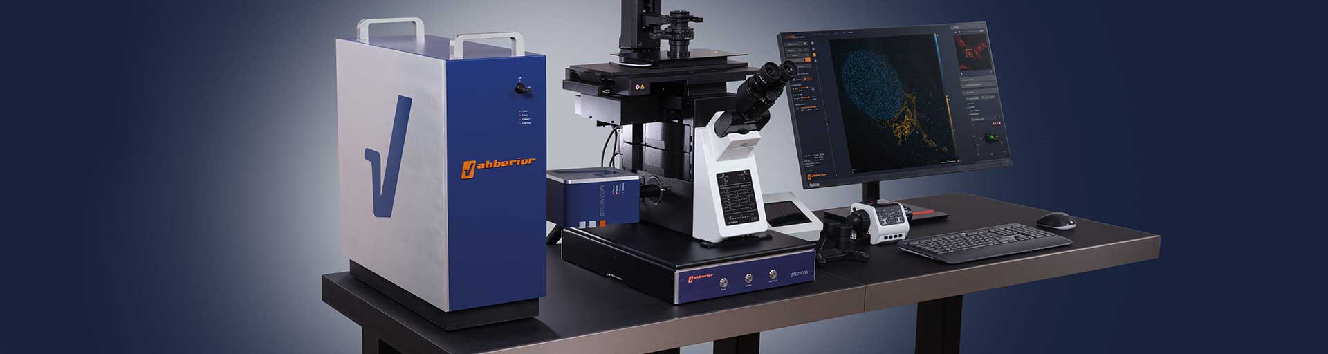
STEDYCON 2 – confocal, STED, lifetime… WOW!

The STEDYCON upgrades your existing widefield microscope to a confocal, STED, and lifetime machine with a resolution down to 30 nm. All that’s required is a free camera port and a good objective lens. With its super-intuitive user interface, the STEDYCON provides an intelligent microscope platform that enables everyone to acquire superb superresolution images after only minutes of training. Define your live cell experiment and enjoy fine imaging at the push of a button! Details >
Enjoy
winning combinations
Every one of our modules adds another superpower to your microscope. When combined, they perform even better, like in this live-cell sample of an Arabidopsis root tip suspended in water and imaged with FACILITY in confocal mode. A subset of cells expresses a YFP construct. TIMEBOW imaging reveals shifts in YFP fluorescence lifetime caused by the protein’s nano-environment. MATRIX array detection improves signal-to-background ratio and resolution.
Sample courtesy: Dr. Fábián Attila and Soós Vilmos, ATK, Brunszvik, Hungary
Ask for detailed information >Choose a superpower for your experiment – our modules
Every MINFLUX, INFINITY, and FACILITY microscope can be upgraded with specific modules to reach new imaging heights. Many of those modules specifically address challenges of live cell imaging and can make all the difference for your experiment. They help to reduce the light burden on your sample, enhance resolution, reduce background signal, provide fluorescence lifetime information, and much more. And if you can’t find what you need on our website, get in touch with us, and we’ll develop it for you.
MATRIX Detector
Many eyes see more than one. The MATRIX detector drastically improves signal-to-background ratio, resolution, and dynamic range.
TIMEBOW Imaging
TIMEBOW lifetime imaging for stunning results at confocal and STED super-resolution.
FLEXPOSURE Illumination
Brings down the light dose on your sample and lables dramatically. Key ingredient for volume and live-cell superresolution.
RAYSHAPE Mirror
Dynamic aberration correction with a deformable mirror over about 200 µm z-range. 140 digital actuators adjust the mirror surface within milliseconds.


Custom Solutions
We offer solutions for even the most challenging applications. Everything that can be done, we will do.
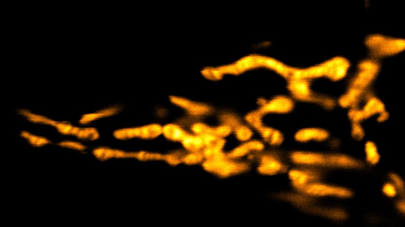
Broaden your knowledge
of microscopes, dyes, and superresolution
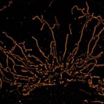
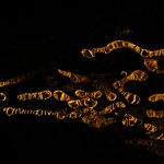
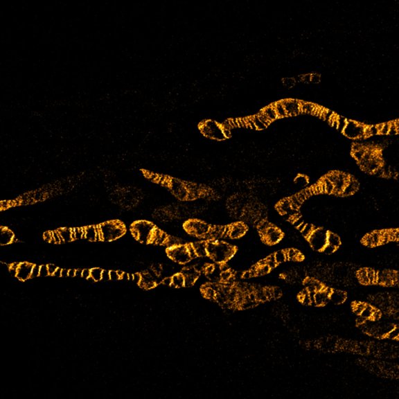
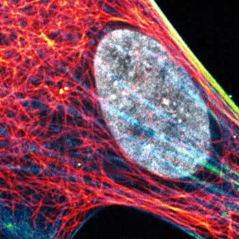
Have you ever wondered what’s behind images like this one of a living cell revealing mitochondrial cristae (stained with abberior LIVE ORANGE mito, yellow)? How do the properties of a dye help to reduce bleaching? What ways are there to lower the light burden on a sample? How does optical sectioning work? Our knowledge base is your chance to close any gaps in fluorescence microscopy expertise you might have!

Fluorescent labeling strategies have become more and more sophisticated and offer ever-new options to improve microscopic imaging. Among the latest are exchangeable HaloTag ligands that put an end to photobleaching for good. Details >

Today’s high-end fluorescence microscopy is unthinkable without lasers. Reason enough to take a closer look at these sophisticated light sources. Details >

Every technique that allows to observe cells is more or less invasive and fluorescence microscopy is no exception. Many imaging situations profit from a reduction in light dose as provided by FLEXPOSURE adaptive illumination. Details >

Ideal imaging conditions are often compromised by imperfections in the optical path. These can severely compromise a microscope’s performance, unless they are eliminated by RAYSHAPE’s deformable mirror. Details >
Superresolution for biology: when size, time, and context matter

The spatial resolution achievable with today’s light microscopes has unveiled life at the scale of individual molecules. Size is no longer a barrier to seeing biology at the most fundamental level. But life is not static. It emerges from movement and change. How do superresolution technologies hold up to the challenges of documenting dynamic biological mechanisms? Details >
Additional information and articles
FLEXPOSURE special
Fluorescence requires light. Light inevitably bleaches fluorophores. What a dilemma. abberior has the solution: FLEXPOSURE! It brings down the light dose by up to two orders of magnitude, without compromising…

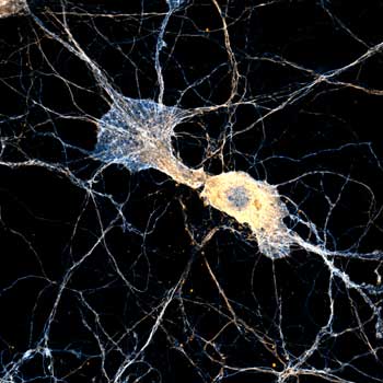
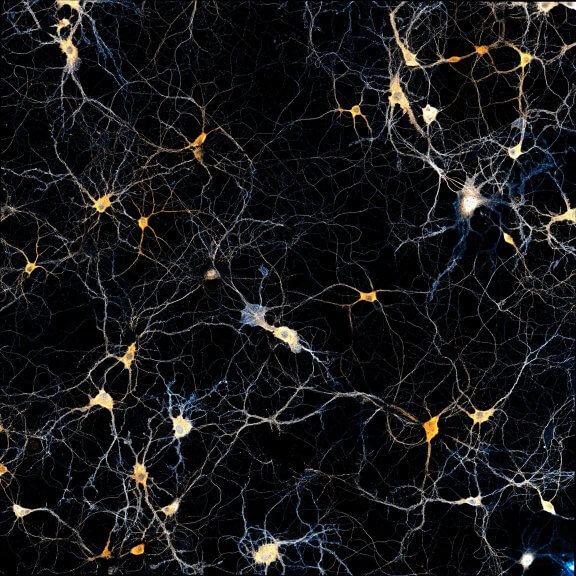
Adaptive STED in Neuroscience Research
Prof. Hiroshi Kawabe presents his research with STED for neuroscience applications and Martin Meschkat explains the benefits MATRIX array detection.
MATRIX special
Get rid of the haze with MATRIX array detection, because it’s all about separating the focal plane’s signal from the rest.

Bye, Bye bleaching! Hello exchangable HaloTag®
The exchangeable nature of abberior LIVE HaloX makes this probe very attractive for multiframe live-cell applications. Watch the webinar!
abberior LIVE HaloX
Exchangeable fluorescent labels for the HaloTag® system and “endless” live-cell imaging without bleaching.
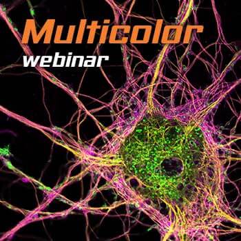
Multicolor STED Imaging
Do you often wonder what strategies are available for multicolor STED experiments? Our experts will share their secrets!
abberior LIVE special
Excellent biocompatibility is the big plus of abberior LIVE dyes, they are gentle and bright at the same time for best STED images – see for yourself!
Summer-Symposia: Live-Cell STED
Francesca Bottanelli and Florian Grimm explain why high-res live cell microscopy is an excellent method to study molecular processes on the nanometer scale.
Live-Cell Imaging – Next Level Labeling Techniques
Francesca Bottanelli uses novel labeling strategies for multicolor live-cell STED imaging and Florian Grimm is focusing on optimized live dyes for STED and confocal microscopy.
Live-cell imaging – nanometer-sized fluorescent probes & tags
Ivana Nikić-Spiegel & Gražvydas Lukinavičius are giving a deeper inside of the design & use of nanometer-sized molecular probes for labeling living probes.