


Get rid of the haze with
MATRIX array detection
It’s all about separating the focal plane’s signal from the rest. With confocal and other STED microscopes, unwanted signal coming from outside the focal plane is blocked by a pinhole before it can reach the detector. However, this only works up to a certain degree and the pinhole generally diminishes the signal. As a result, background light often fogs the image.
So the question is: How can we get rid of the haze? There are numerous post-processing algorithms available, but these merely guess an approximation of the background signal and are prone to producing computational artefacts.
abberior’s MATRIX array detection physically measures the background separately and thus can truly extract the in-focus signal.
Description
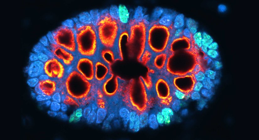
Paraffin section of gut biopsy stained for Ki67 (abberior STAR ORANGE), Muc2 (abberior STAR RED), and DAPI.
Modules:

Description
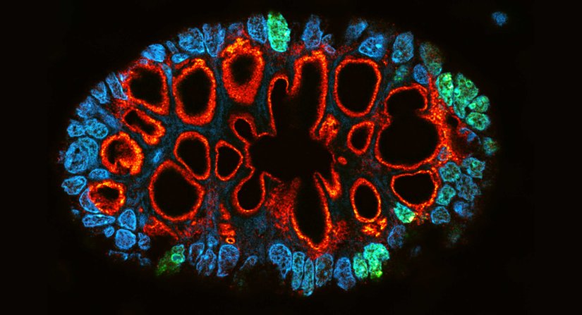
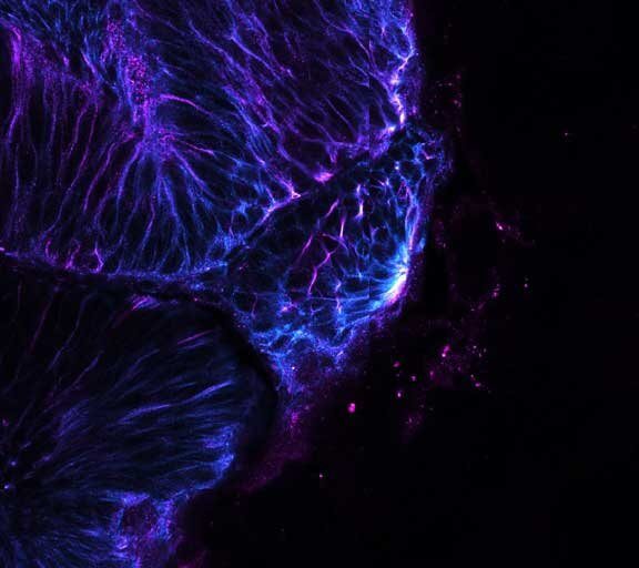
Gain in both signal-to-background ratio and resolution: MATRIX detection dramatically improves a conventional STED image of the zebrafish olfactory epithelium, resulting in a perfect and crystal-clear image revealing even more detail than STED alone can.
Modules:
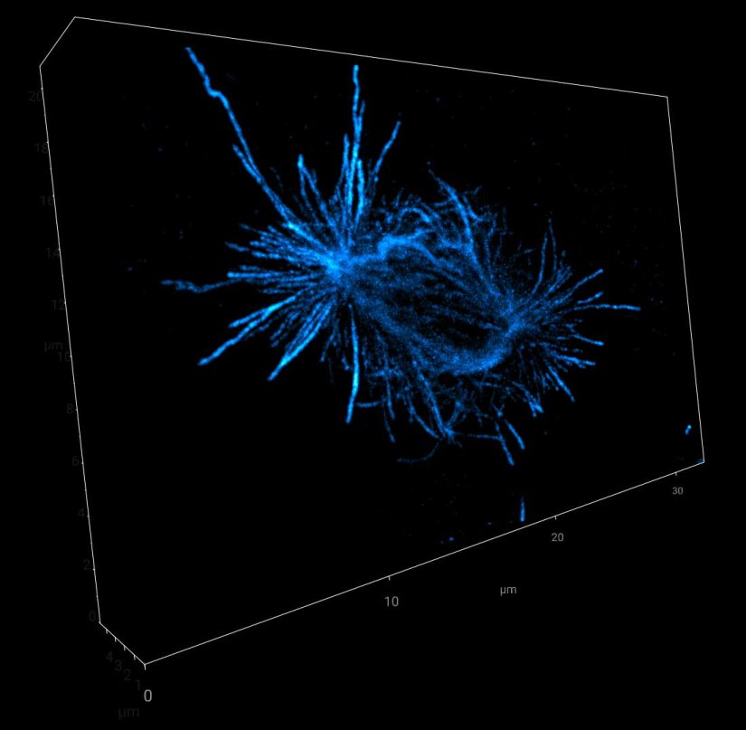
Description
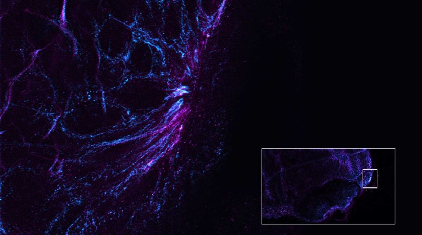
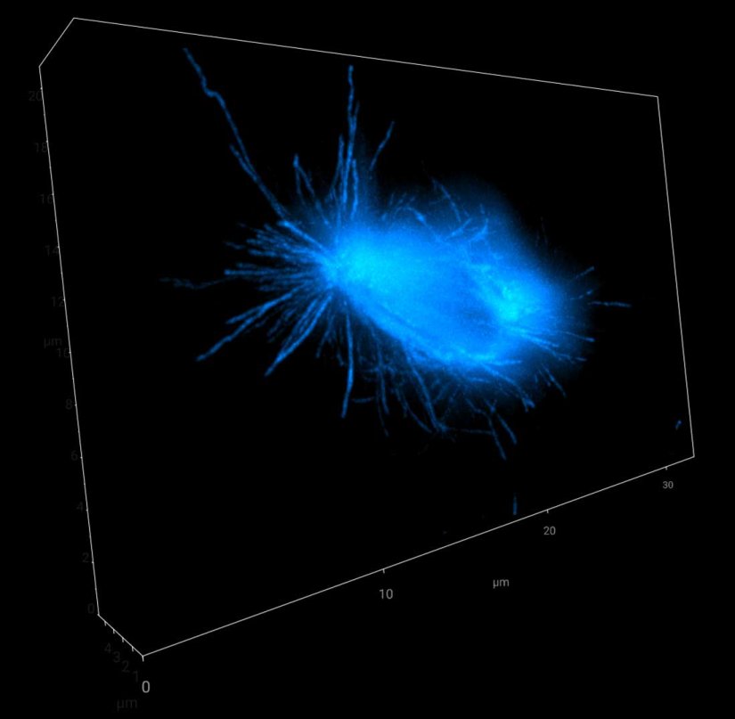
Comparison of a 3D volume of cellular tubulin filaments:
Signal from outside the focal plane recorded by a single detector often fogs the image.
MATRIX records the out-of-focus signal separately so that it can be removed from the overall signal, resulting in an image with superior signal-to-background ratio.
Modules:


If you are interested in MATRIX array detection and how to get rid of the haze, please contact us.
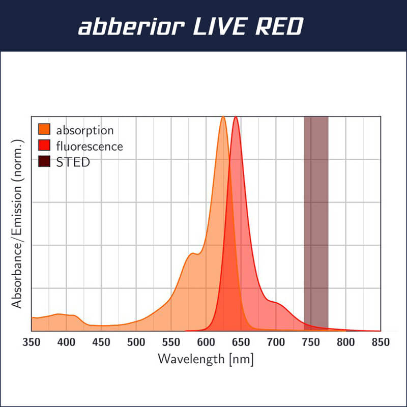
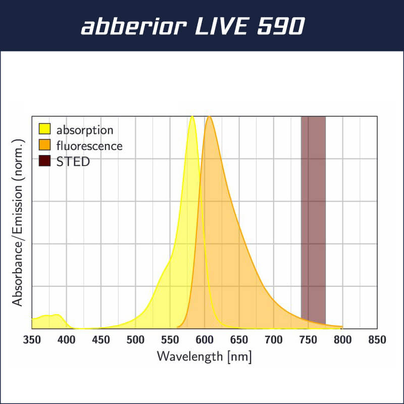
If you would like to test our perfectly fitting dyes for tissue imaging, you can order a free test kit consisting of our dyes abberior LIVE RED DNA (2 nmol) and abberior LIVE 590 Tubulin (2 nmol).
A self-experiment with the MATRIX principle

MATRIX detector
Focus on your thumb and close one eye.

MATRIX detector
Now close your eye and open the other one. The scence behind your thumb will jump, allowing you to tell foreground (your thumb) from background. As you see, many eyes see more than one.

Point detector
To a single point detector, foreground and background look exactly the same. They cannot be physically discriminated.
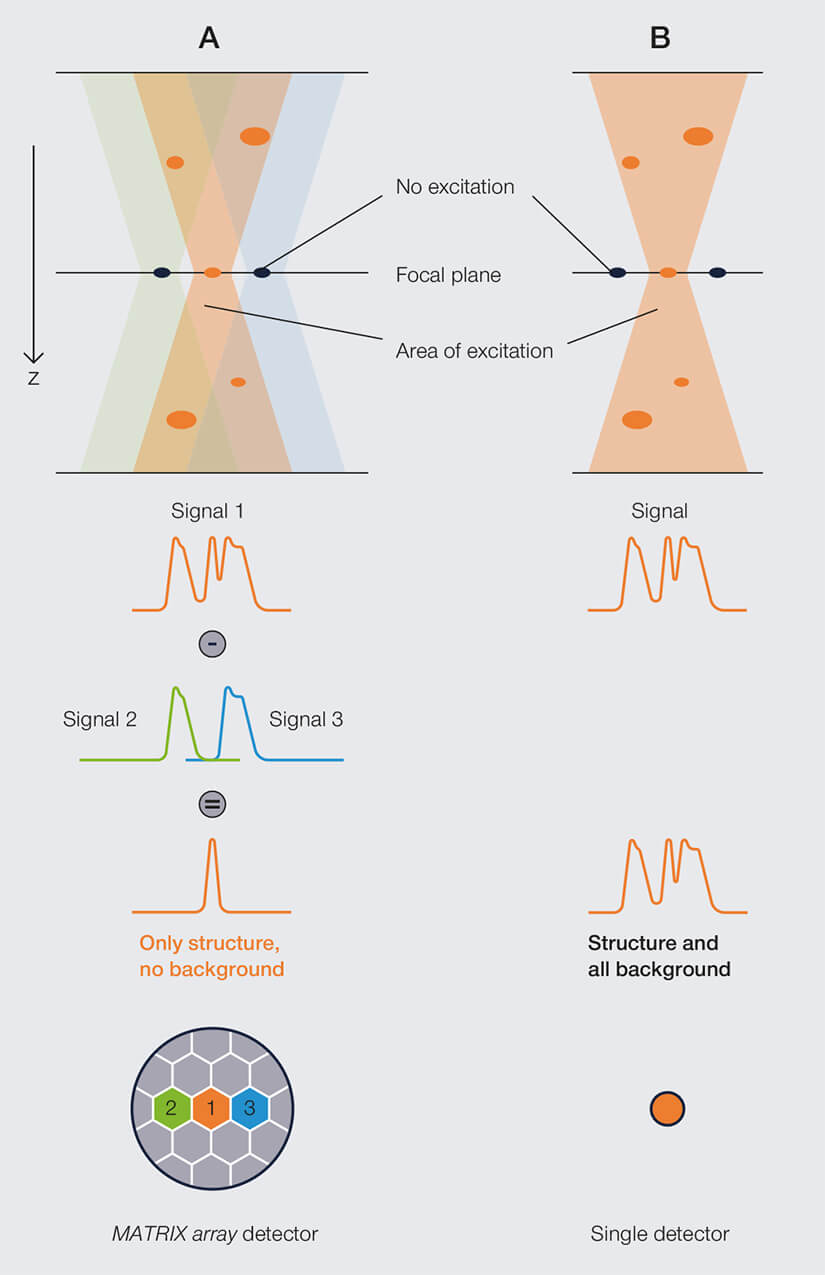
When a single detector receives a light signal, there is no way to tell whether it originates from the focal plane or from out-of-focus structures, because it looks exactly the same (B).
In contrast, each of the over 20 elements of the MATRIX detector “look” at a slightly different spot in the sample (A). Some of the elements record both in-focus and background signal (signal 1 in the figure), while others only receive out-of-focus signal (signal 2 and 3 – note that the excitation double-cone only overlaps with the central detector element).
This separate recording makes it possible to remove out-of-focus signal from signal 1, leaving the final image without background, sharp and clear.