


Exploring life’s inner workings – Physiology


FAQ Video 37

“How does TIMEBOW lifetime imaging work and why is MATRIX array detection a perfect match?”

NEWS
TIMEBOW, MATRIX, STED and confocal imaging can be freely combined
TIMEBOW gives time a color
Watch Recording: TIMEBOW lifetime imaging for stunning results with confocal and STED superresolution!
Article in Physics Today about MINFLUX and tracking kinesin.
abberior rocked @ EMBL – Imaging down to single-molecule resolution: STED and MINFLUX nanoscopy
How do individual proteins, macromolecular machines, cells, or tissues work together? How do they interact? And how is this interaction controlled? These are the questions at the core of physiological research.
Given these questions, it is no wonder that light microscopy is a key technique in physiology. It is used to image specimen of vastly different length scales: from the whole organism over tissues or organs to individual cell and intracellular features such as protein assemblies or organelles. The latter’s size is often below the resolution limit of conventional light microscopy, but superresolution techniques like STED and MINFLUX nowadays overcome this limit and reveal ever-new details about cellular and organismal physiology.
abberior’s STED provides resolution down to 20 nm in 2D and 75 nm in 3D, and MINFLUX takes it another step further with single-digit nanometer resolution and molecular tracking at frequencies up to 10 kHz. Both techniques are well-suited for live cell imaging, which is essential for physiological research. Add abberior’s fluorescent dyes and labels optimized for applications from confocal to MINFLUX, and you are ideally equipped to make your phyiological imaging experiments a success.
Talk to a scientist >Adaption is key
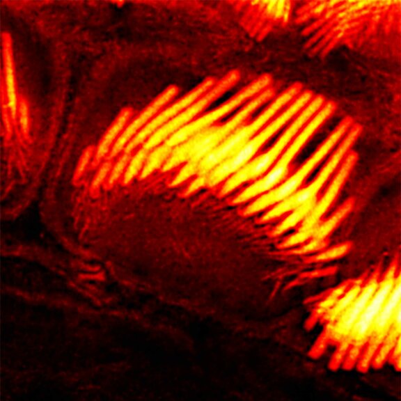
Physiology is often about fine differences. And in light microscopy, these only become visible with superresolution, like in this STED image of an actin stain of mouse inner ear hair cells using abberior STAR RED phalloidin.
The sample was provided by Christian Vogl, InnerEarLab, University Medical Center Göttingen, Germany.
Test your sample >Capture the moment
There is more to life than meets the naked eye. Fortunately there are microscopes! And even more fortunately, they are nowadays able to resolve structures of only a few nanometers, and in living cells and tissues, bringing to light details decisive for various physiological processes. We collected some of them in our gallery.
Description
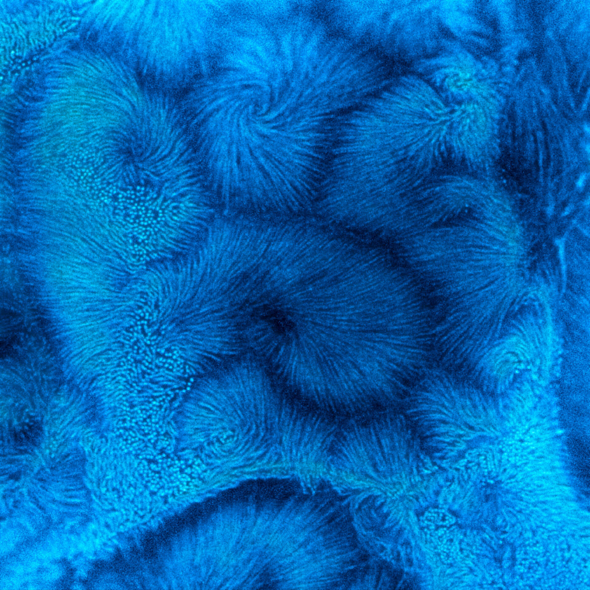
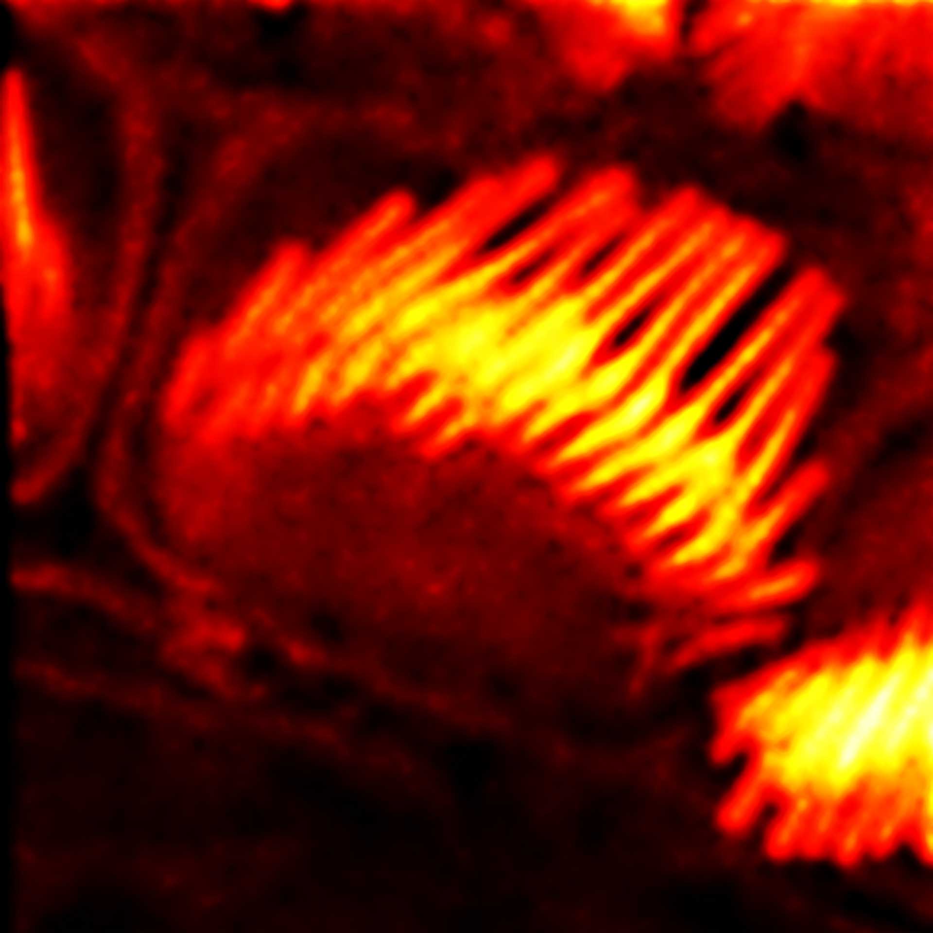
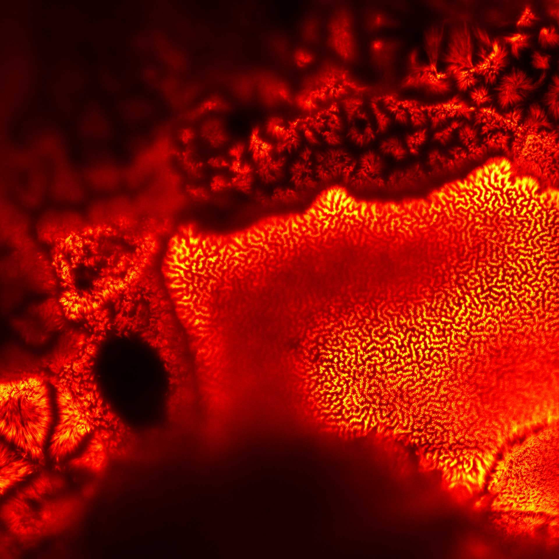
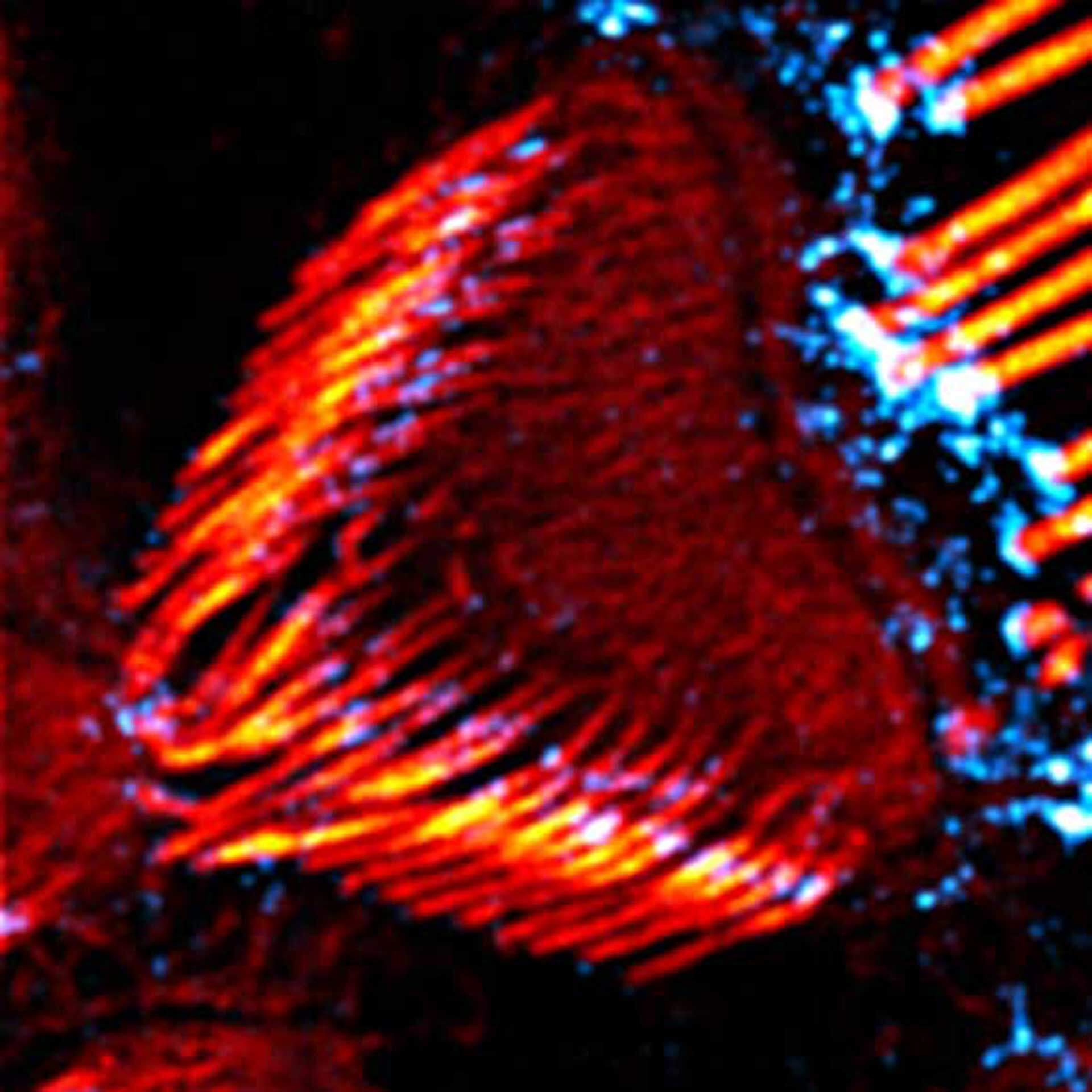
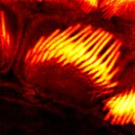
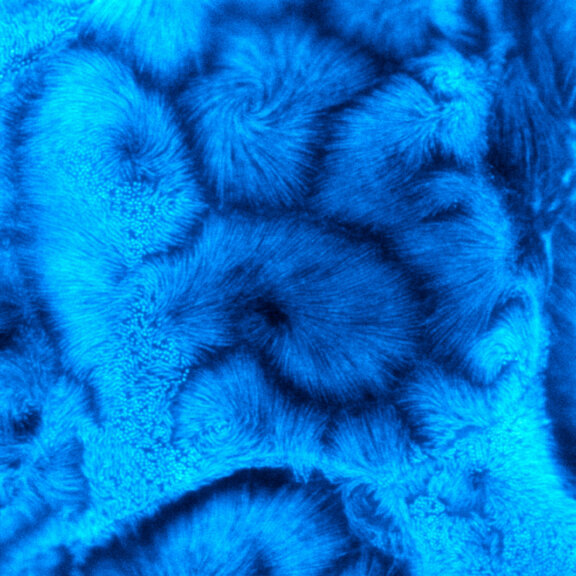
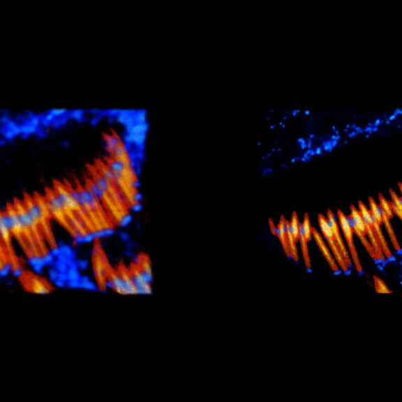
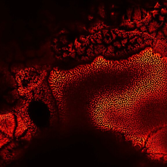
TIMEBOW STED vs MATRIX + TIMEBOW STED
Actin in microvilli of CaCo2 cells recorded with FACILITY. Fixed cells were stained with abberior STAR RED phalloidin. Imaging of the intense-background sample is optimized by MATRIX detection by directly subtracting unfocussed data. To boost resolution TIMEBOW is used, keeping STED laser power low. Lifetime information enables the selective exclusion of STED-affected emission at the rim of each measurement.
We thank Prof. Dorothee Günzel and Jörg Piontek (Charité, Berlin, DE) for providing this sample.
Modules:
Description
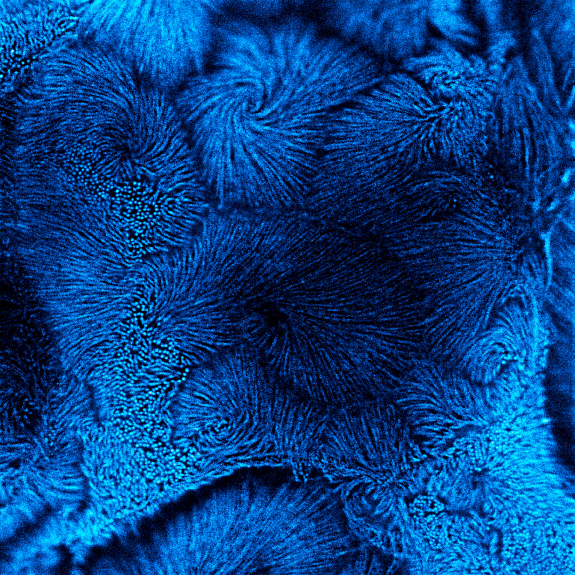
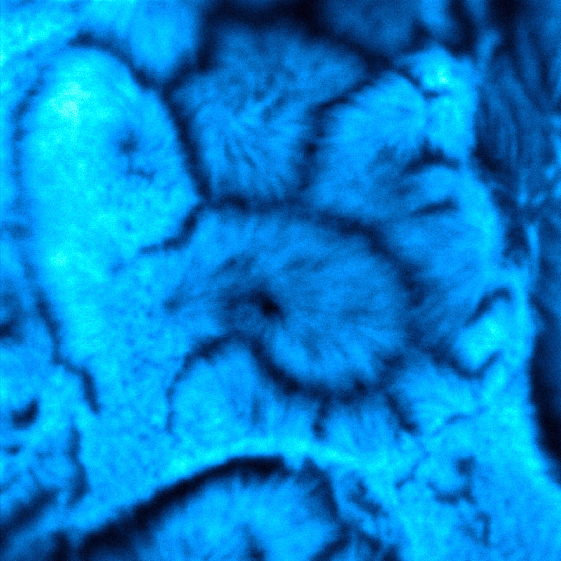
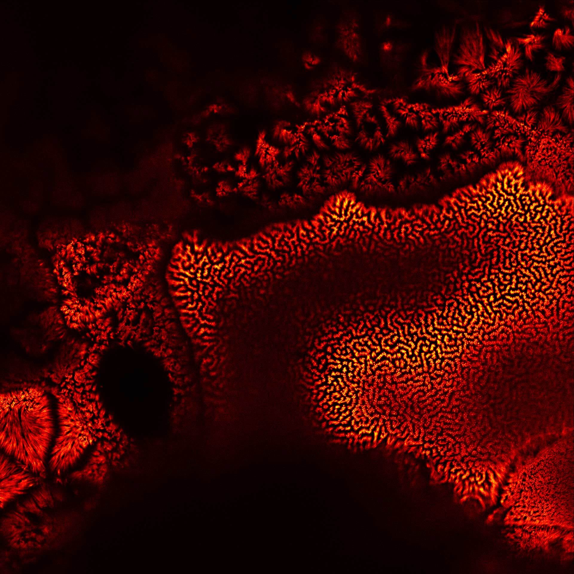
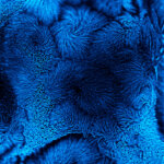
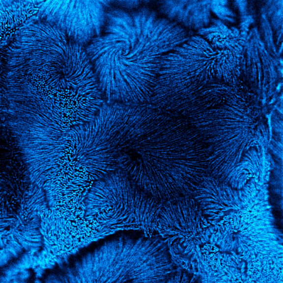
Confocal vs MATRIX + TIMEBOW STED
Actin in microvilli of CaCo2 cells recorded with FACILITY. Fixed cells were stained with abberior STAR RED phalloidin. Imaging of the intense-background sample is optimized by MATRIX detection by directly subtracting unfocussed data. To boost resolution TIMEBOW is used, keeping STED laser power low. Lifetime information enables the selective exclusion of STED-affected emission at the rim of each measurement.
We thank Prof. Dorothee Günzel and Jörg Piontek (Charité, Berlin, DE) for providing this sample.
Modules:

Description
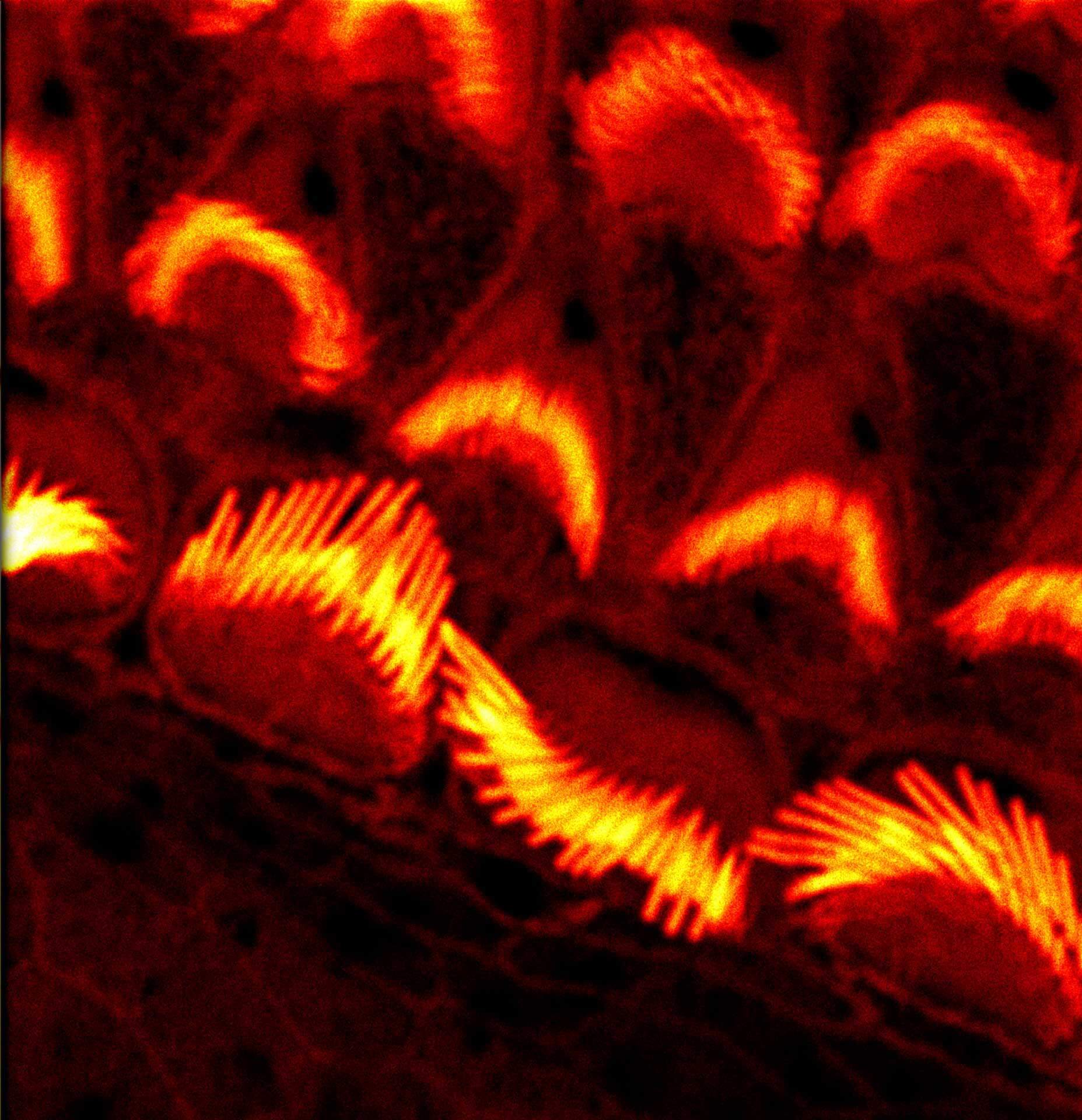
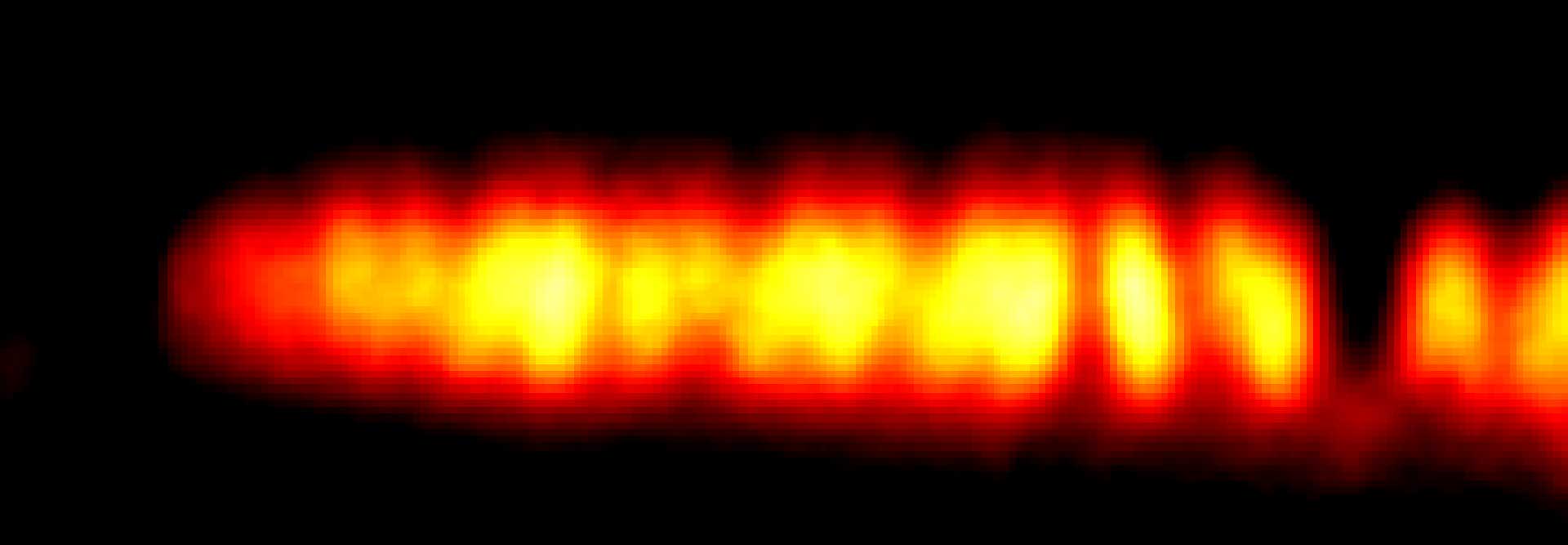
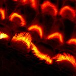
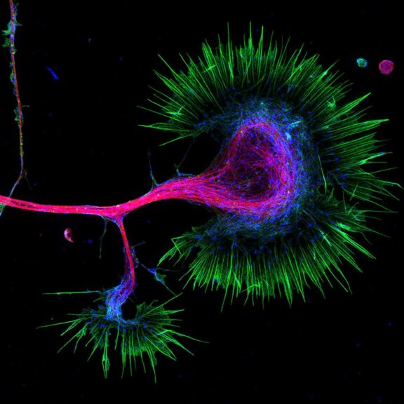
Actin stain of mouse inner ear hair cells using abberior STAR RED phalloidin.
Samples were prepared by Dr. Christian Vogl, InnerEarLab, UMG Göttingen, Germany.
Description
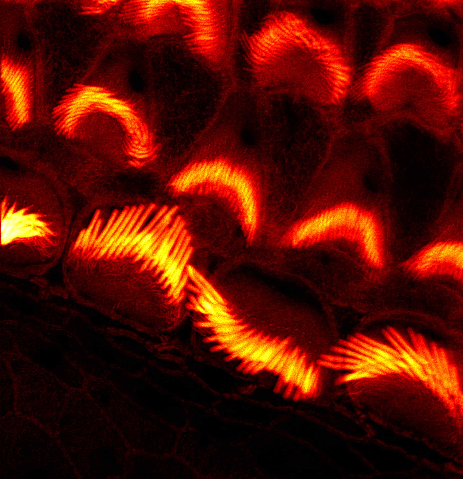
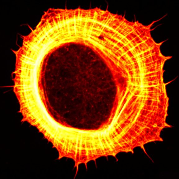
Actin stain of mouse inner ear hair cells using abberior STAR RED phalloidin. Zoom in for full effect.
Samples were prepared by Dr. Christian Vogl, InnerEarLab, UMG Göttingen, Germany.
Modules:
Description
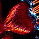
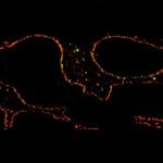
Actin in microvilli of Caco2 cells. Fixed cells were stained with abberior STAR RED phalloidin. MATRIX detection removes background signal and dramatically improves optical sectioning for 2D-STED.
Samples by Prof. Dorothee Günzel & Roman Mannweiler, Institut für Klinische Physiologie, Charité – Universitätsmedizin Berlin.
Modules:

Description
Actin stain of mouse inner ear hair cells using abberior STAR RED phalloidin.
Samples were prepared by Dr. Christian Vogl, InnerEarLab, UMG Göttingen, Germany.
Description
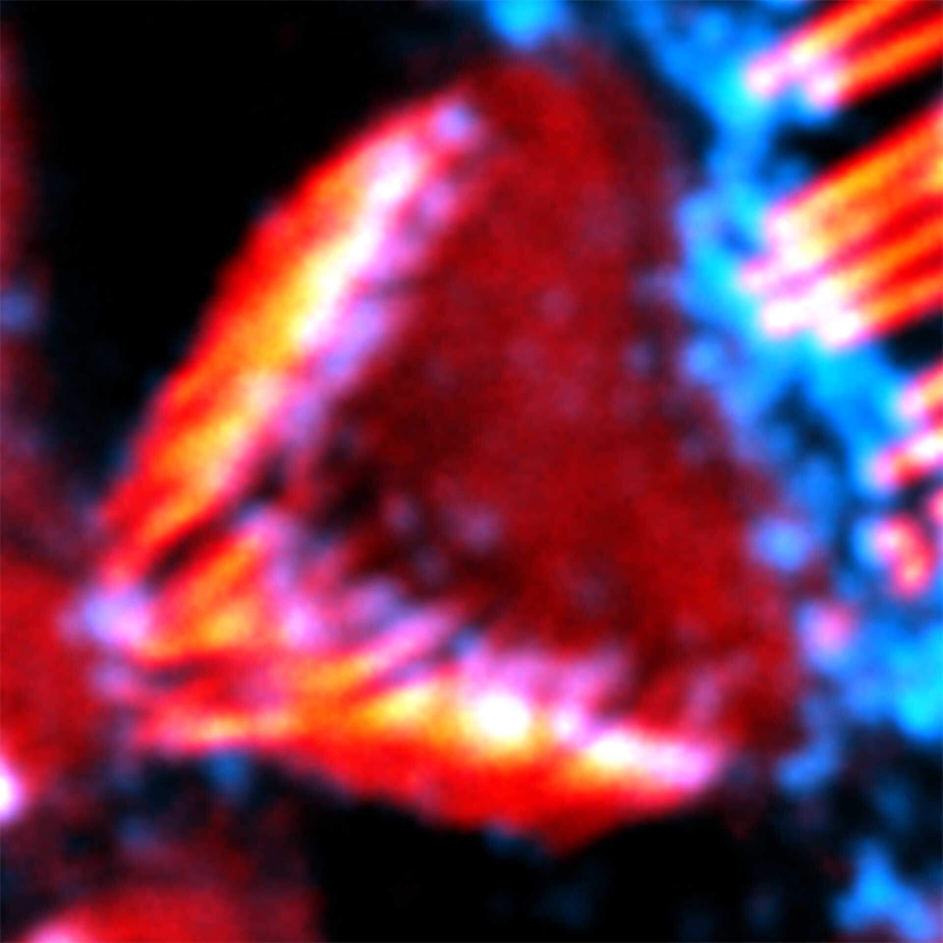
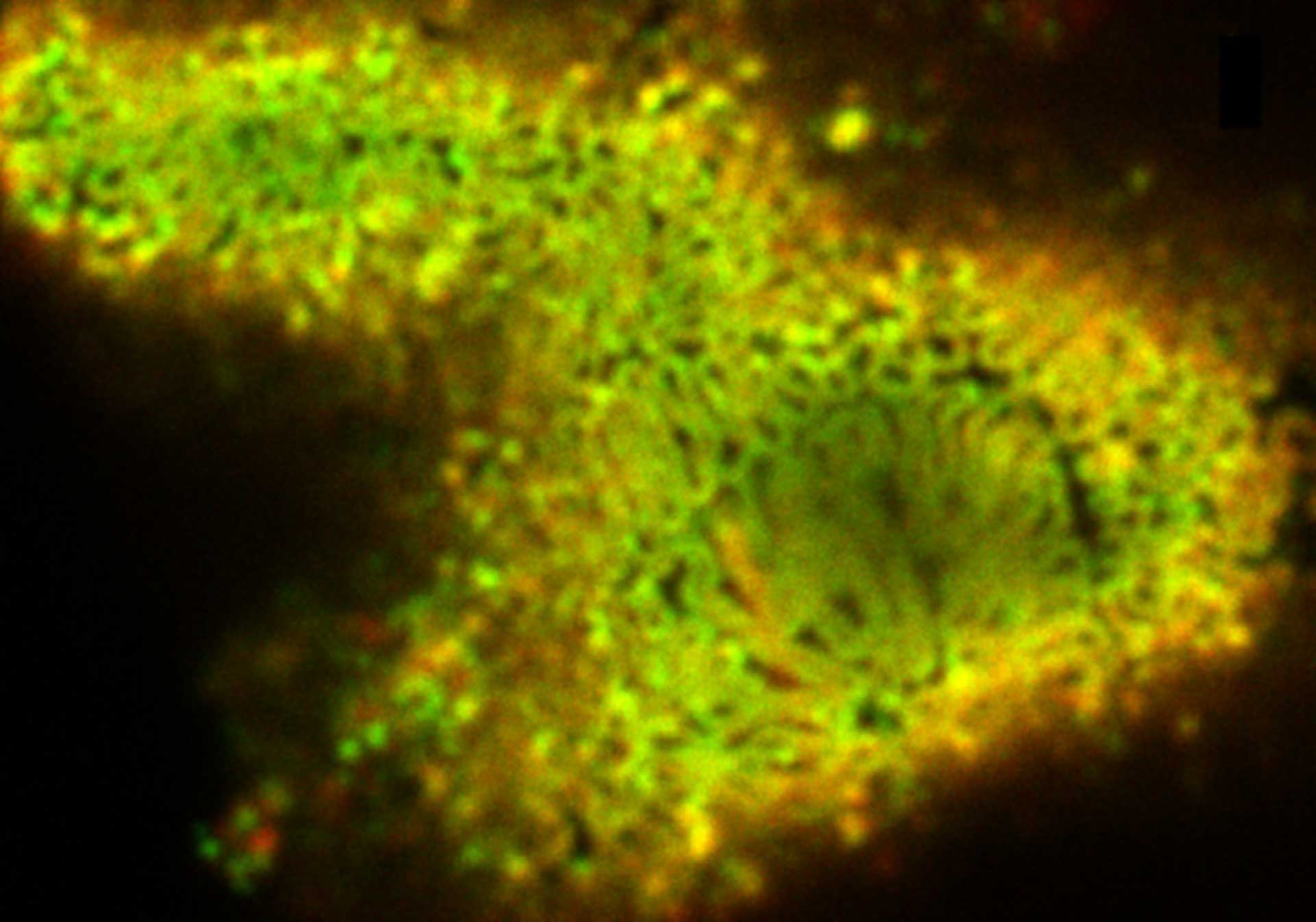
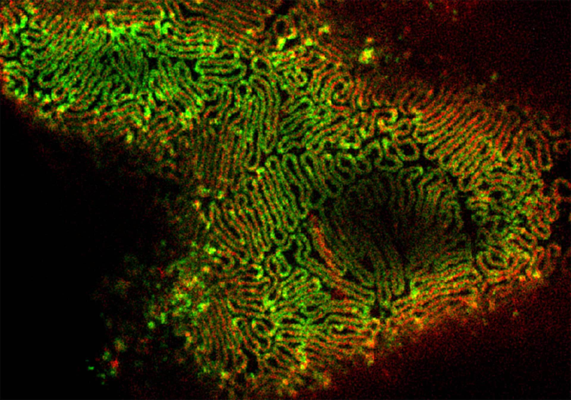
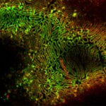
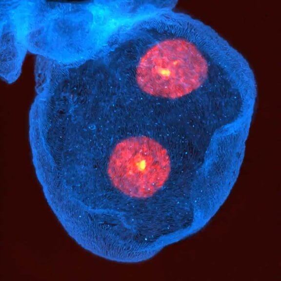
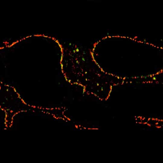
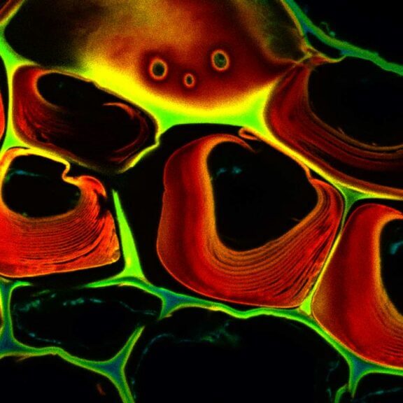
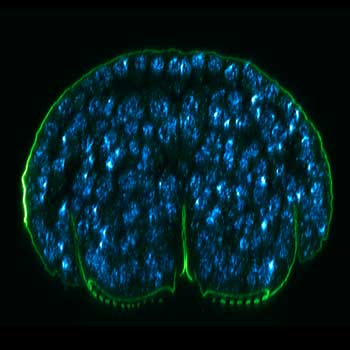
2-color 2D STED image of a cleared adult kidney sample of a rat. Shown is an image of a renal corpuscle showing Nephrin (red, abberior STAR 635P) structures inbetween the Podocin slits (green, AlexaFluor594).
Sample was prepared by D. Unnersjö Jess and H.G. Blom @ KTH Stockholm, Sweden.
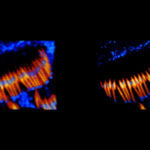
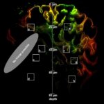
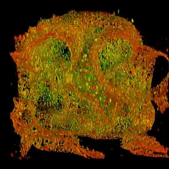
Description
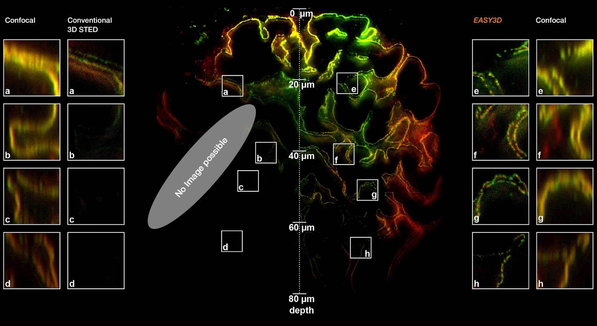
Adaptive optics EASY3D enables deep imaging of cleared adult kidney samples. Comparison of a XZ-slice deep into a renal corpuscle using conventional 3D STED imaging and an EASY3D imaging. EASY3D allowed imaging up to 80 µm into cleared rat kidney tissue using an oil objective. Without adaptive optics, the mismatch between immersion oil and sample leads to complete signal loss. Labels: Nephrin (red, Abberior STAR635P) and Podocin (green, AlexaFluor594).
Sample was prepared by D. Unnersjö Jess and H.G. Blom @ KTH Stockholm, Sweden.
Modules:


From blurs to brilliance –
superresolution for physiology
abberior microscopes facilitate superb imaging. Their optics, developed for the high demands of superresolution STED, also bring confocal imaging to a new level. Shown is a 2D STED image of a renal corpuscle in a cleared adult rat kidney sample stained for Nephrin (red, abberior STAR 635P) and structures inbetween the Podocin slits (green, AlexaFluor594).
The sample was prepared by D. Unnersjö Jess and H.G. Blom, KTH, Stockholm, Sweden.
On top of confocal and STED microscopes, abberior is the only commercial provider of MINFLUX, which achieves single-digit nanometer localization precision.
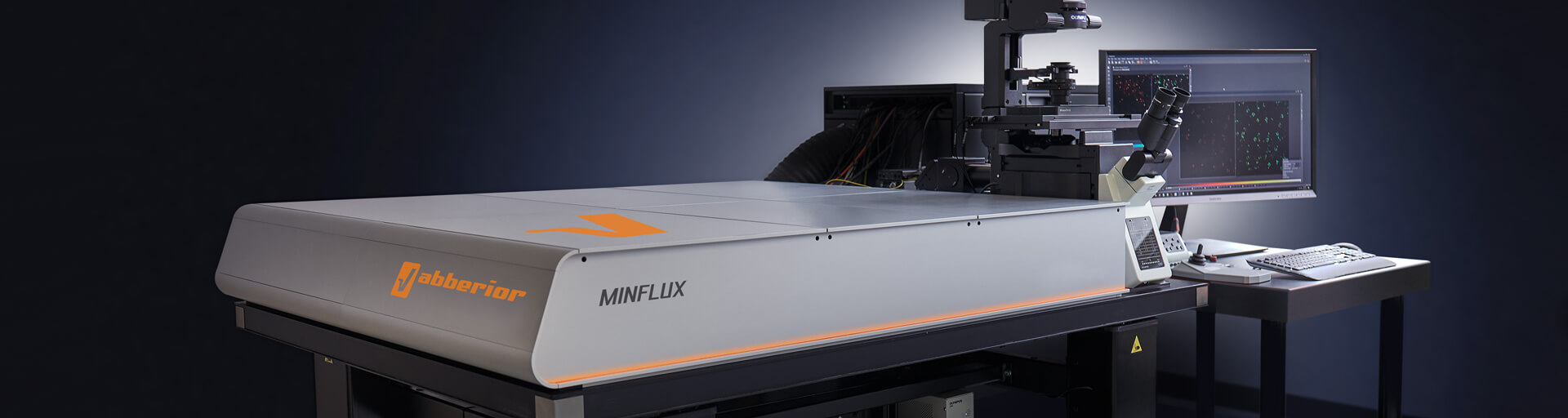
Get a demo >MINFLUX – unrivaled resolution and speed
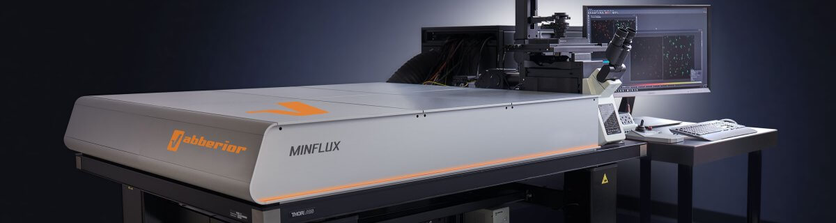
The MINFLUX platform offers an unprecedented array of imaging possibilities and allows you to resolve structures as small as a molecule, along all three dimensions. This unmatched resolution capability combined with unprecedented speed reveals sample details never seen before and helps to dissect fast and dynamic cellular processes in space and time. MINFLUX is the world’s most powerful fluorescence microscope. Details >
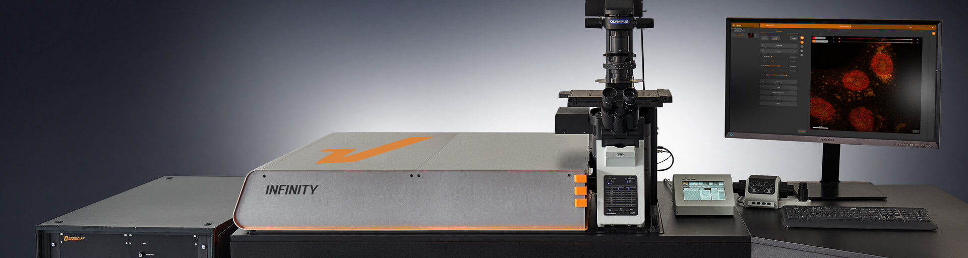
INFINITY – forever cutting edge
The INFINITY platform is the most customizable platform for all things microscopy and may be adapted to your particular demands in physiological imaging. INFINITY is always up to the challenge. Just tell us what you need and we will build you a customized, continuously upgradable system specialized for your research. Details >
MIRAVA POLYSCOPE – one for all and all for one

The MIRAVA® POLYSCOPE® combines every resolution – from millimeters down to 3 nm – in a singularly unique system. MIRAVA unites four microscopy technologies to cover an unprecedented resolution spectrum, extending over several orders of magnitude from diffraction-limited imaging all the way to true molecular resolution. Our LiGHTBOX software allows beginners to intuitively arrive at a top-notch image within three clicks, while also giving experts full control over the instrument. Details >
STEDYCON 2 – confocal, STED, lifetime… WOW!

The STEDYCON upgrades your existing widefield microscope to a confocal, STED, and lifetime machine with a resolution down to 30 nm. All that’s required is a free camera port and a good objective lens. With its super-intuitive user interface, the STEDYCON provides an intelligent microscope platform that enables everyone to acquire superb superresolution images after only minutes of training. Details >
A winning combination
This image of actin in microvilli of CaCo2 cells would not be as crisp and clear as it is without the superpower of two particular modules installed in FACILITY. With MATRIX array detection, background was physically separated from in-focus light and removed to drastically increase contrast. By providing lifetime information about the fluorescence signal. TIMEBOW-STED boosted resolution while keeping STED power low.
The sample was provided by Dorothee Günzel and Jörg Piontek, Charité – Universitätsmedizin Berin, Germany.
Ask for detailed information >Choose a superpower for your experiment – our modules
We offer many modules to overcome the specific imaging challenges in your research. Use TIMEBOW to boost STED resolution and to monitor changes in the cellular nano-environment of your fluorophores. FLEXPOSURE adaptive illumination puts light only where it has an effect and may reduce the light burden by orders of magnitude, keeping your cells happy during extended live cell imaging and volume scans. MATRIX Detection allows you to physically measure and remove background signal. Our modules can do all of this and even more! And if you can’t find what you need on our website, get in touch with us, and we’ll develop it for you.
MATRIX Detector
Many eyes see more than one. The MATRIX detector drastically improves signal-to-background ratio, resolution, and dynamic range.
TIMEBOW Imaging
TIMEBOW lifetime imaging for stunning results at confocal and STED super-resolution.
FLEXPOSURE Illumination
Brings down the light dose on your sample and lables dramatically. Key ingredient for volume and live-cell superresolution.
RAYSHAPE Mirror
Dynamic aberration correction with a deformable mirror over about 200 µm z-range. 140 digital actuators adjust the mirror surface within milliseconds.


Custom Solutions
We offer solutions for even the most challenging applications. Everything that can be done, we will do.
Broaden your knowledge
of microscopes, dyes, and superresolution
Actin staining of mouse inner ear hair cells with abberior STAR RED phalloidin.
The sample was provided by Christian Vogl, InnerEarLab, University Medical Center Göttingen, Germany.
Tell me more >
PALM and STORM are often used as synonyms, and in fact they have a lot in common. But there are slight differences that can be important for your application. And then there are other superresolution techniques, too – like STED and MINFLUX. Details >

Confocal microscopy offers superior optical sectioning. But what is that exactly? And what about other ways to get rid of the background, such as array-based detectors like the MATRIX? Details >

Fluorescent labeling strategies have become more and more sophisticated and offer ever-new options to improve microscopic imaging. Among the latest are exchangeable HaloTag ligands that put an end to photobleaching for good. Details >

Every technique that allows to observe cells is more or less invasive and fluorescence microscopy is no exception. Many imaging situations profit from a reduction in light dose as provided by FLEXPOSURE adaptive illumination. Details >
Why do superresolution microscopists love alpacas?

It is a very simple yet very important fact: the localization precision of any superresolution microscope can only be as good as the size of the fluorescent staining allows. In other words, when your fluorescent dye is too big or too far away from the protein you want to label, you will never be able to reach a resolution that is higher than this offset. The good news is: there are ways to reduce the offset between target protein and fluorescent label. And one of these are nanobodies. Details >
Superresolution for biology: when size, time, and context matter

The spatial resolution achievable with today’s light microscopes has unveiled life at the scale of individual molecules. Size is no longer a barrier to seeing biology at the most fundamental level. But life is not static. It emerges from movement and change. How do superresolution technologies hold up to the challenges of documenting dynamic biological mechanisms? Details >
STEDYCON: ease-of-use in a shoebox

A sleek, black-and-orange box transforms your widefield microscope into a confocal and a superresolution STED instrument and your exploration of subcellular structures into a seamless, discovery-rich experience. Carefully designed with masterly engineering, STEDYCON breaks the stereotype of the finicky, hard-to-use scope. It opens new possibilities at the press of a button for any user and almost any location. How does it do it? The secret’s in the box. Details >
Additional information and articles
RAYSHAPE webinar – dynamic aberration correction for deep tissue imaging
Hello RAYSHAPE! Bye-bye, aberrations!
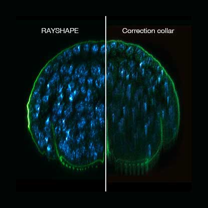
RAYSHAPE special
RAYSHAPE dynamic aberration correction with a deformable mirror preserves resolution and increases brightness. 140 actuators adjust mirror surface within milliseconds for over 200 µm z-range.

TIMEBOW Lifetime Imaging – gives time color
Explore abberior’s TIMEBOW lifetime toolbox with our application experts Julia Menzel and Jan-Gero Schloetel.

FLEXPOSURE special
Fluorescence requires light. Light inevitably bleaches fluorophores. What a dilemma. abberior has the solution: FLEXPOSURE! It brings down the light dose by up to two orders of magnitude, without compromising…

Summer Symposia: Adaptive Optics
All about aberration correction in STED and how the outstanding capabilities of abberior’s Adaptive Optics helps overcome the challenges of complex samples.
MINFLUX webinar – Tracking with unprecedented spatio-temporal resolution!
Thanks MINFLUX it’s possible to track the movement of individual molecules in the cell, e.g. kinesin. See what EMBL scientists have achieved.