


Crisp and clear from head to tail – Zoology


FAQ Video 37

“How does TIMEBOW lifetime imaging work and why is MATRIX array detection a perfect match?”
Light microscopy is a cornerstone tool in zoology, particularly in developmental biology and embryology, but also in physiology and genetics. In zoology, imaging spans several length scales: it starts at the whole organism, continues with tissues and organs, followed by individual cells, and eventually is used to resolve intracellular features such as protein assemblies or organelles. Organoids play an increasingly important role. Light microscopy faces several challenges here, including intransparent structures, autofluorescence, and loss of signal in deep tissue.
The good news is: whatever the model organism and type of specimen, abberior has the solution! We offer confocal and STED microscopes with superior performance and equipped with objective lenses ranging from low to high magnification as well as add-on modules tailored to your particular imaging demands. And we are the only manufacturer of MINFLUX, the fluorescent microscope with unsurpassed spatiotemporal resolution. Our product portfolio is completed by a comprehensive selection of fluorescent dyes and labels suitable for various applications.
Talk to a scientist >Catching the beauty of complexity
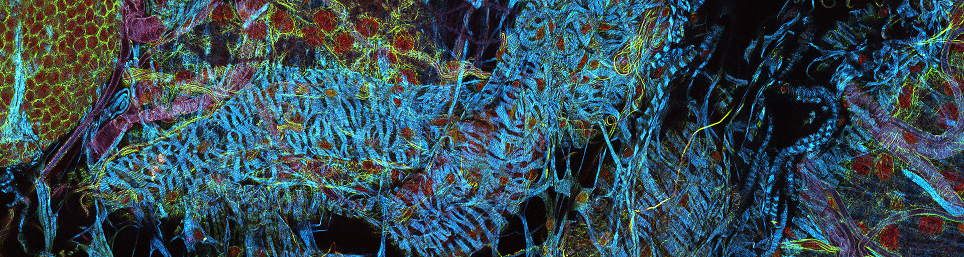
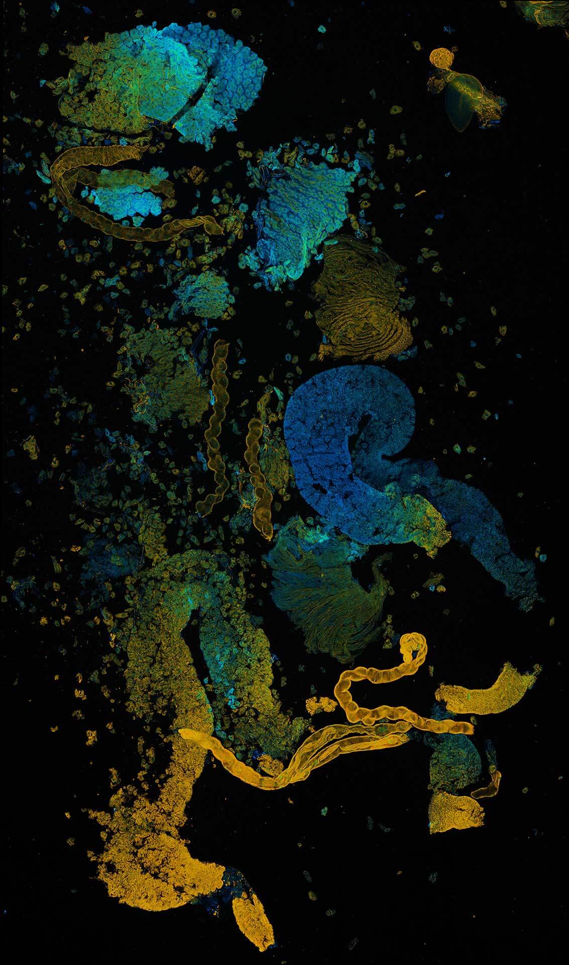
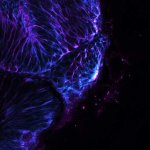
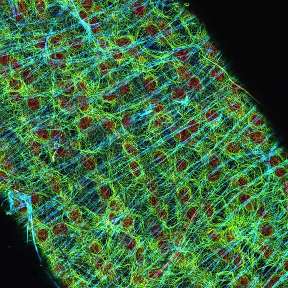
Zoology faces the challenge (or has the privilege) to dissect the complexity of organismal life. This quest occasionally produces images of stunning artfulness, like this section of Drosophila ovaries stained for tubulin (yellow, abberior STAR RED), DNA (red, LIVE 590 DNA), and F-actin (cyan, STAR GREEN phalloidin).
Test your sample >Zooming in on zoology
The diversity of sample and target structure is a major challenge in zoological microscopy. Our gallery gives you an impression of what this means. You will find images of entire embryos, organs like the Drosophila ovary, tissue sections of inner ear hair cells or trachea, and subcellular components such as the neuromuscular junction. Browse through and enjoy!
Description
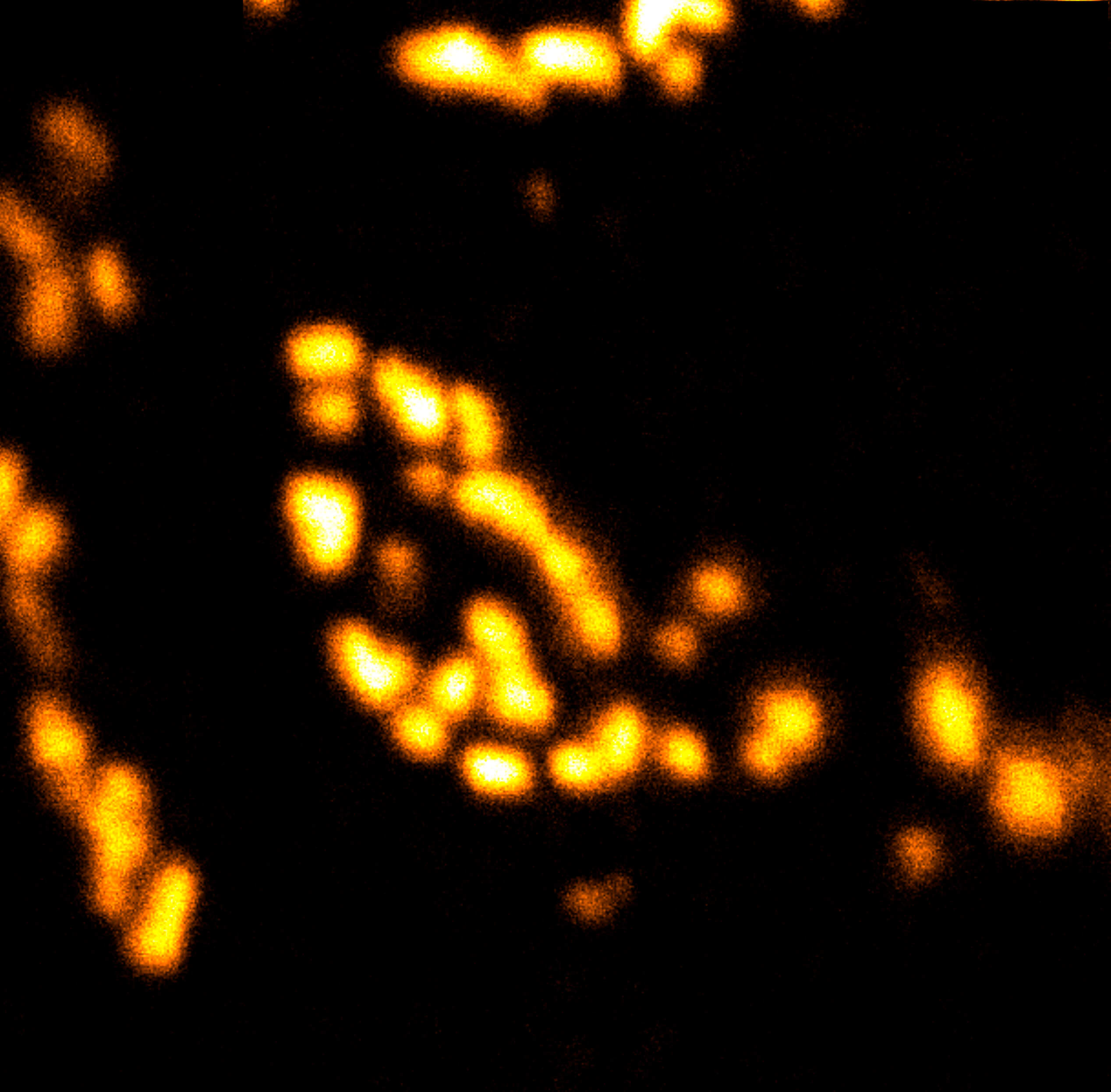
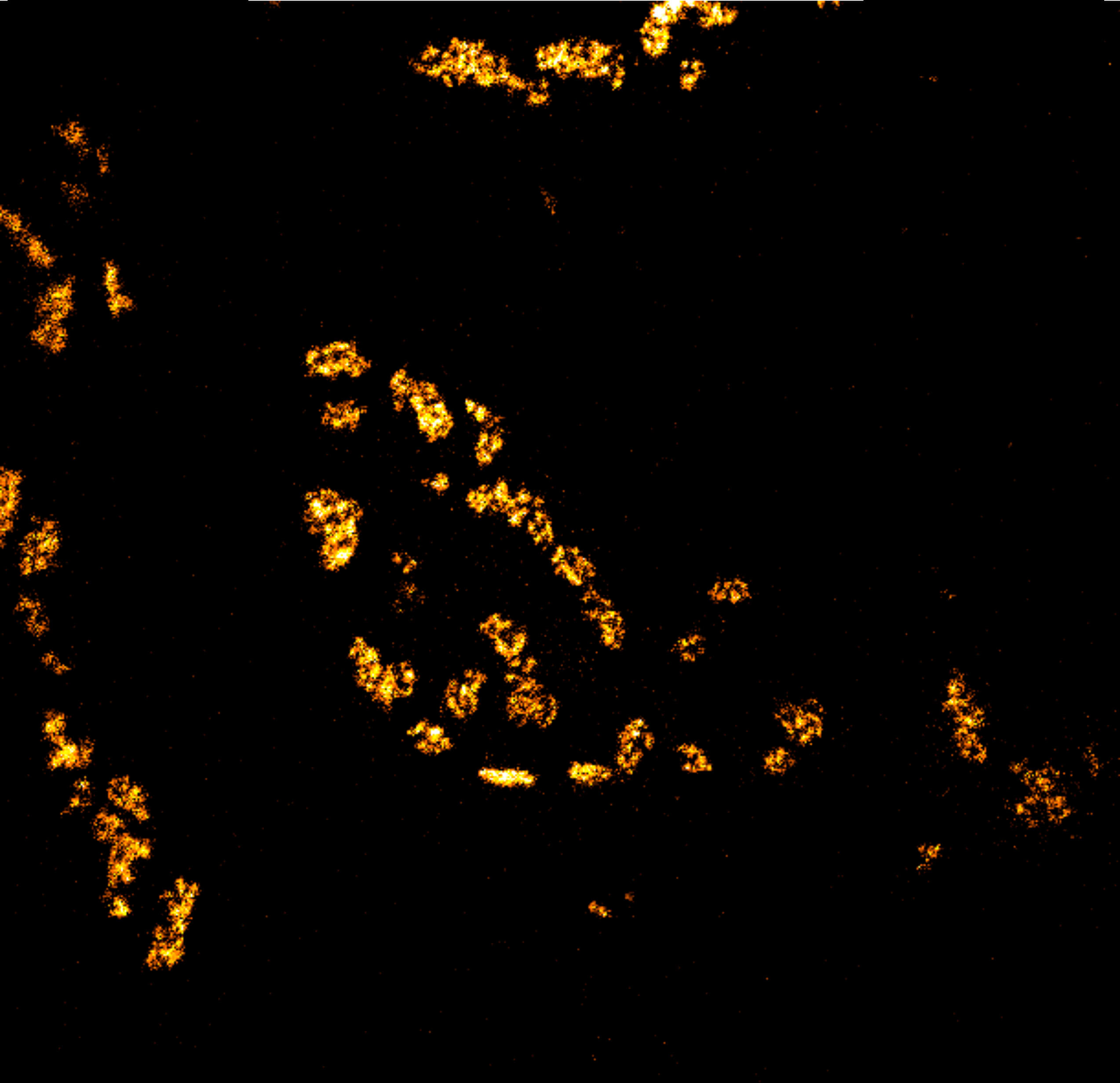
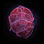
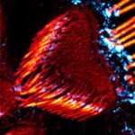
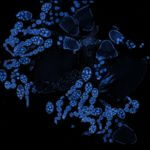
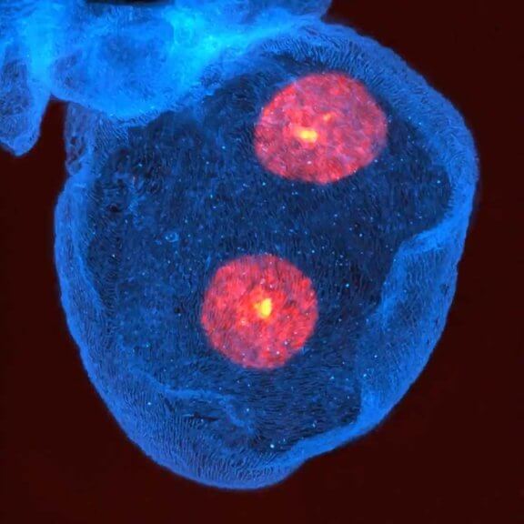
Drosophila ovariole stained for the synaptonemal complex protein C3G (cyan, abberior STAR RED), showing how the complex is formed and disassembled during oocyte development. DNA was stained with DAPI (purple), spectrin with abberior STAR ORANGE (green).

Description
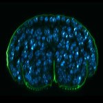
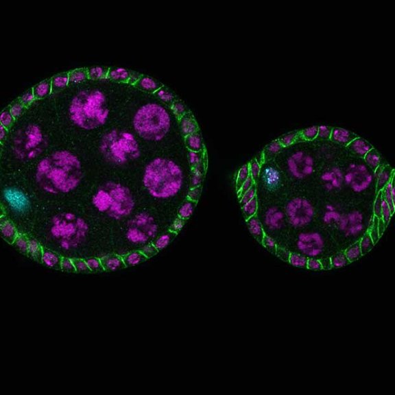
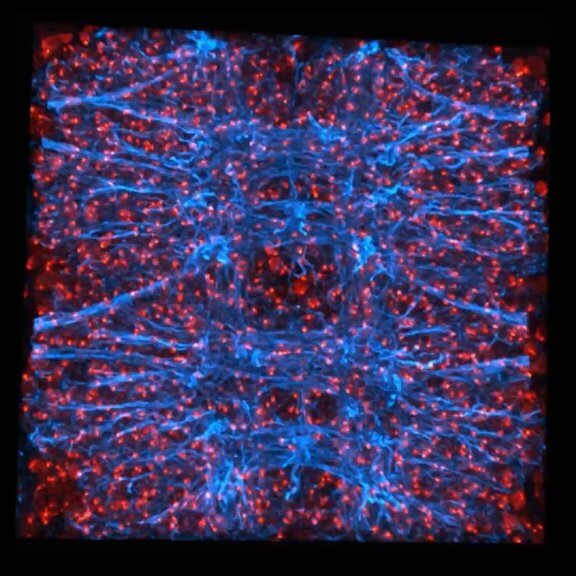
Drosophila egg in the ovary stained for tubulin (green, abberior STAR RED), actin (blue, abberior STAR GREEN), and DNA (red, abberior LIVE 590).
Description
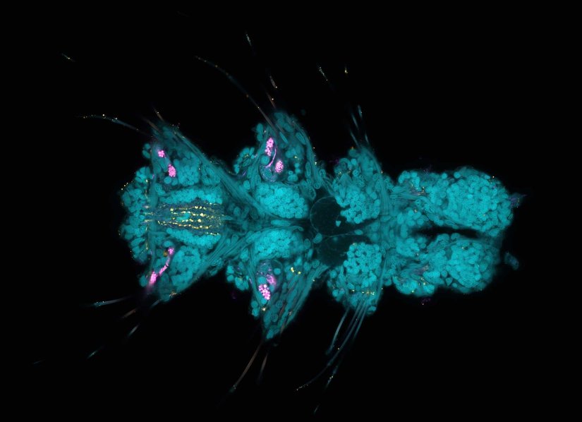
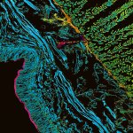
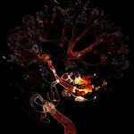
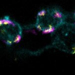
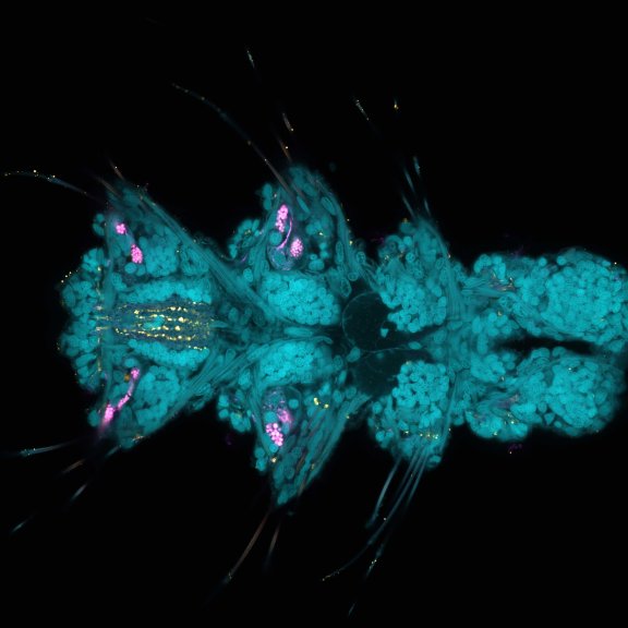
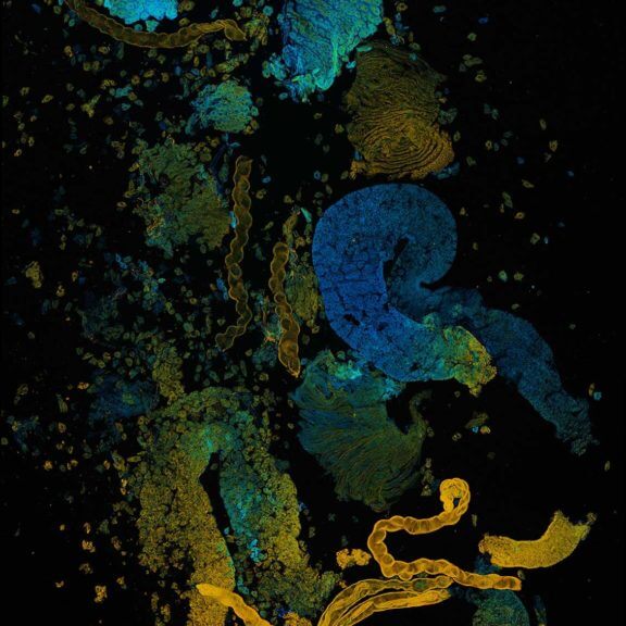
Larva of Platynereis dumerilii acquired in a confocal overview. Nuclei are shown in cyan (DAPI), tubulin in magenta, and serotonin-positive neurons in yellow.
Description
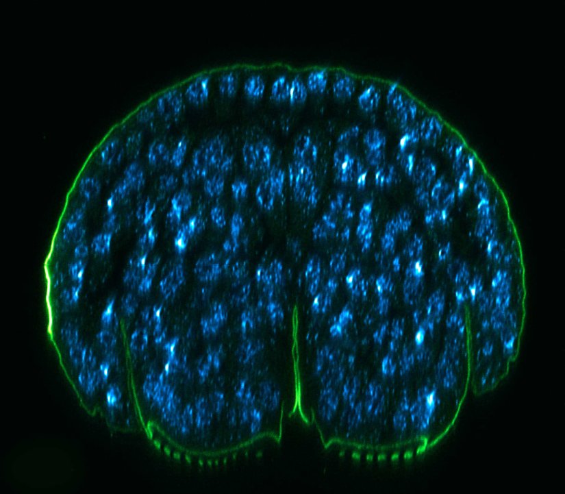
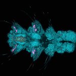
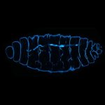
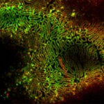
3 color confocal image of a Drosophila embryo stained for chitin (abberior STAR RED, cyan), tubulin (abberior STAR ORANGE, magenta), and DNA (PicoGreen, green).
Description
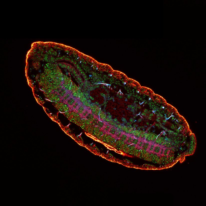
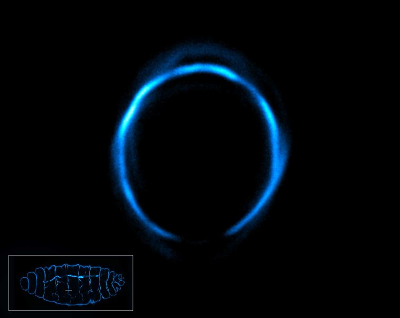
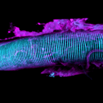
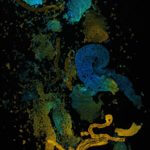
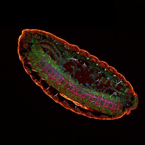
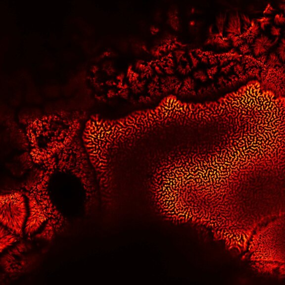
Confocal image of autofluorescence in a cross-section of the earthworm Lumbricus terrestris. TIMEBOW lifetime imaging detects differences in fluorescence lifetime, which depend on the type of autofluorescent molecule and its nano-environment, and visualizes them in distinct colors.
Modules:
Description
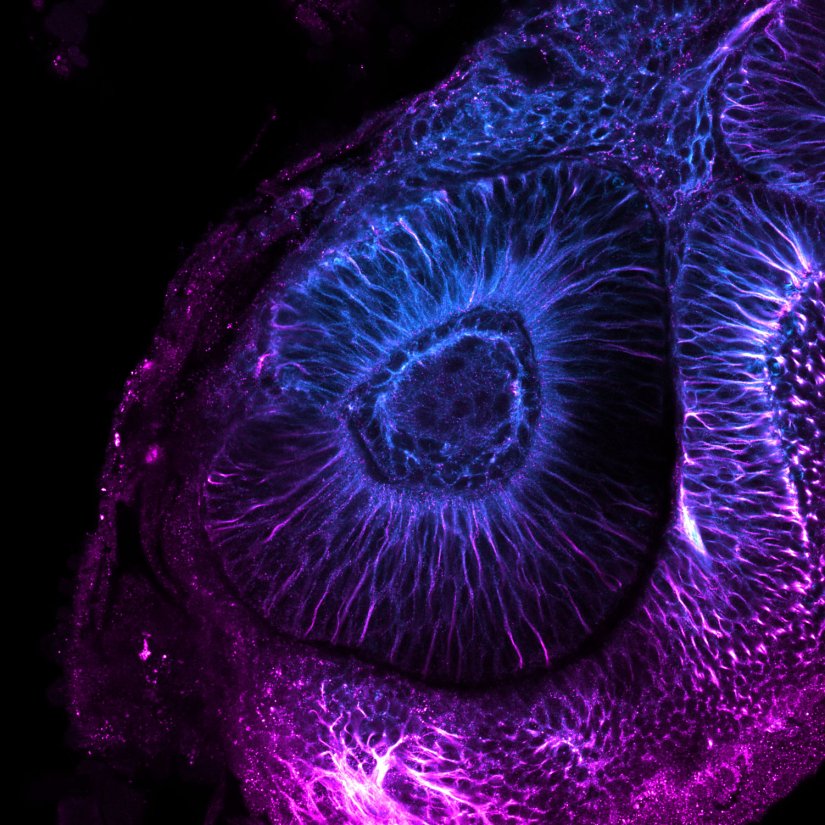
Confocal image of a developing zebrafish eye showing tubulin stained with abberior STAR RED and abberior STAR ORANGE.
Image courtesy of Graziamaria Paradisi, Marco Tartaglia, Antonella Lauri, Ospedale Pediatrico Bambino Gesù, Rome, Italy
Modules:
Description
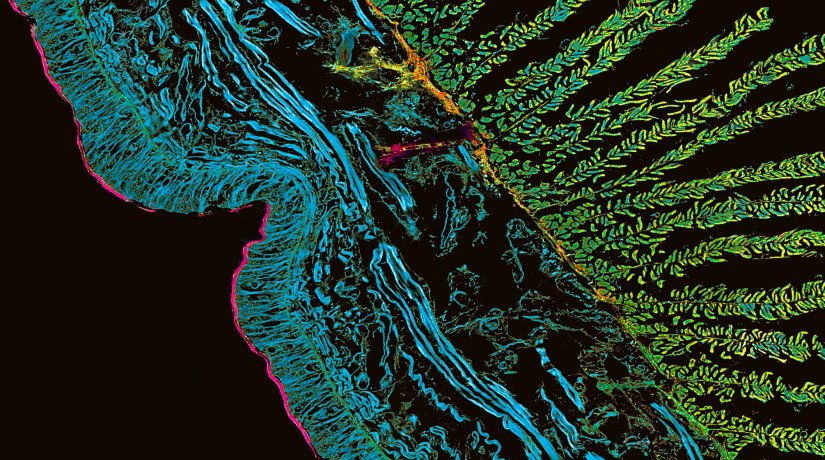
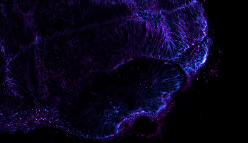
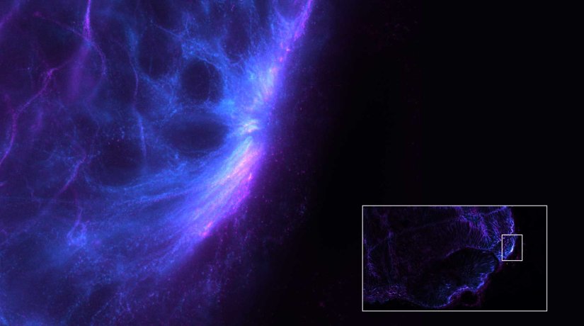
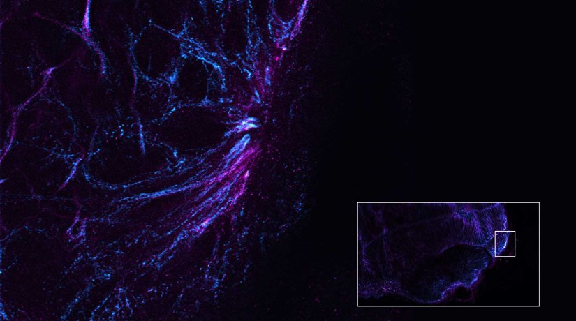
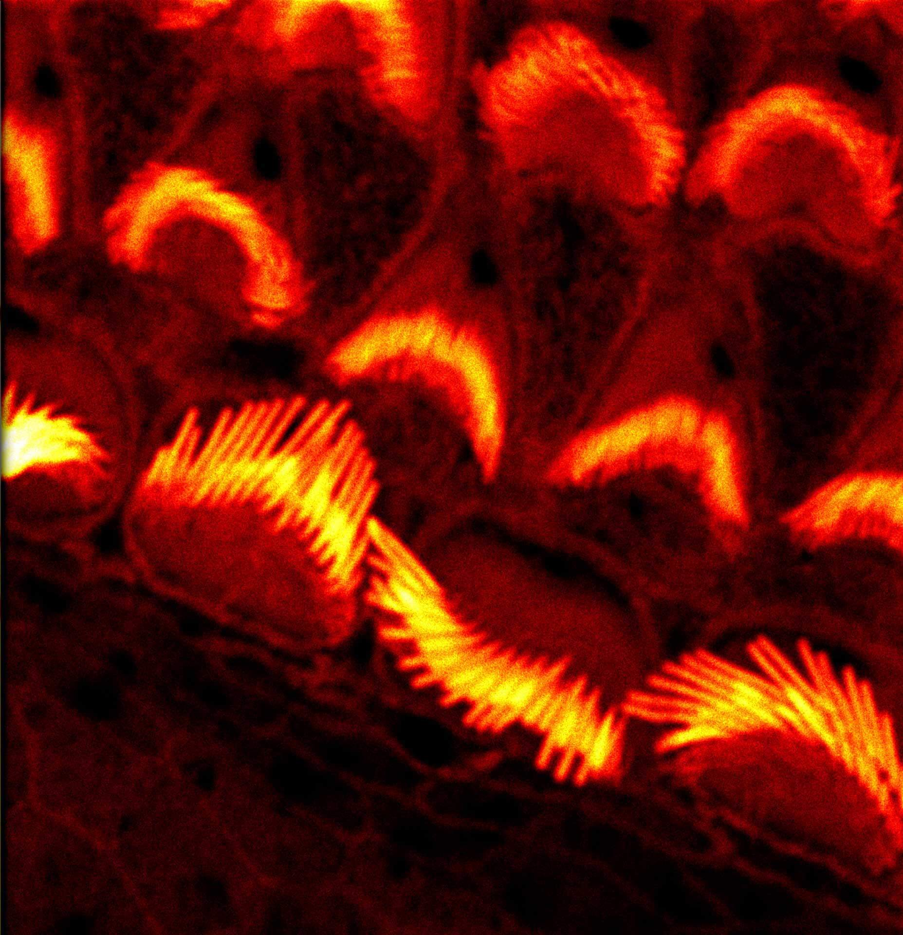
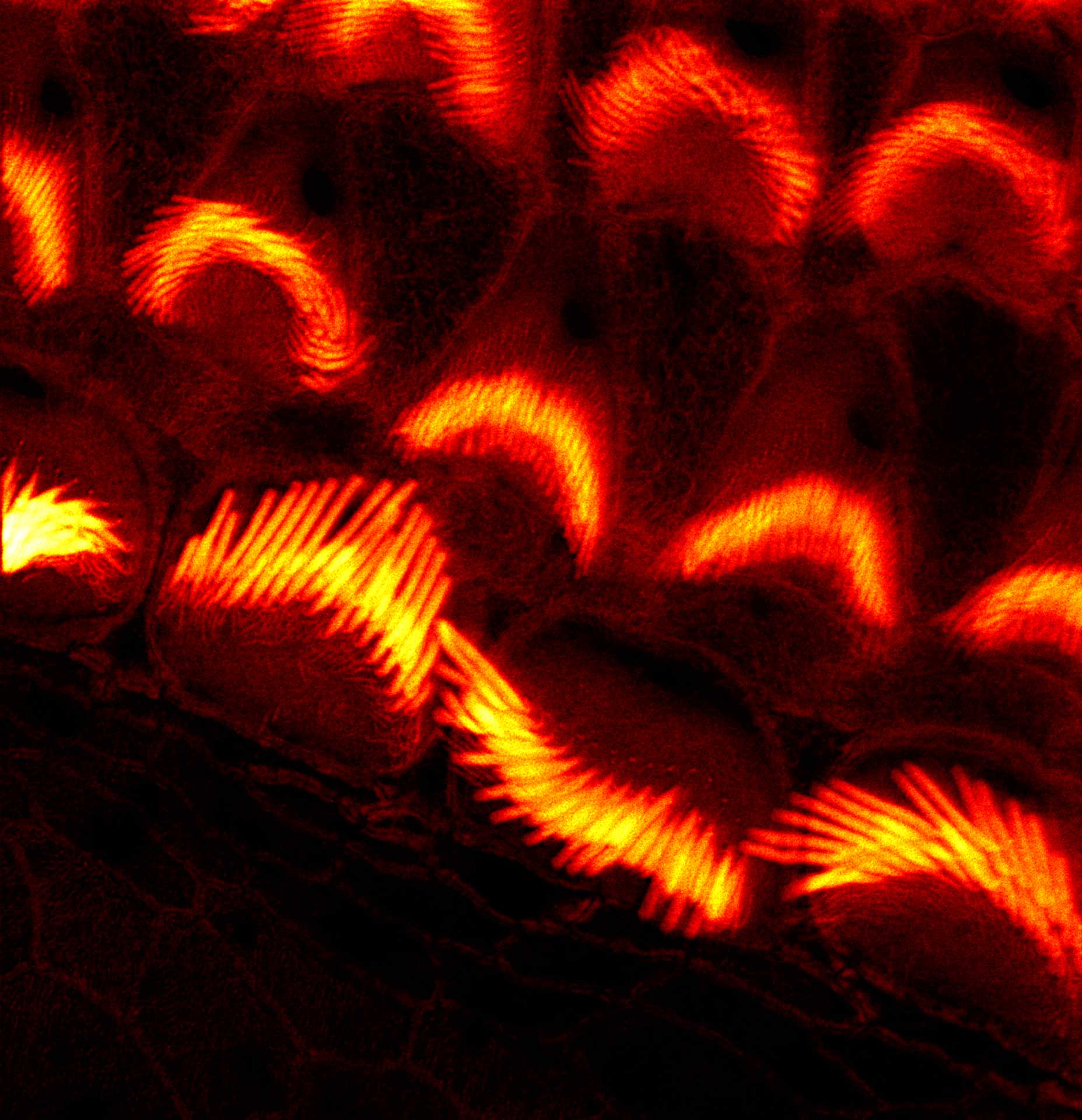
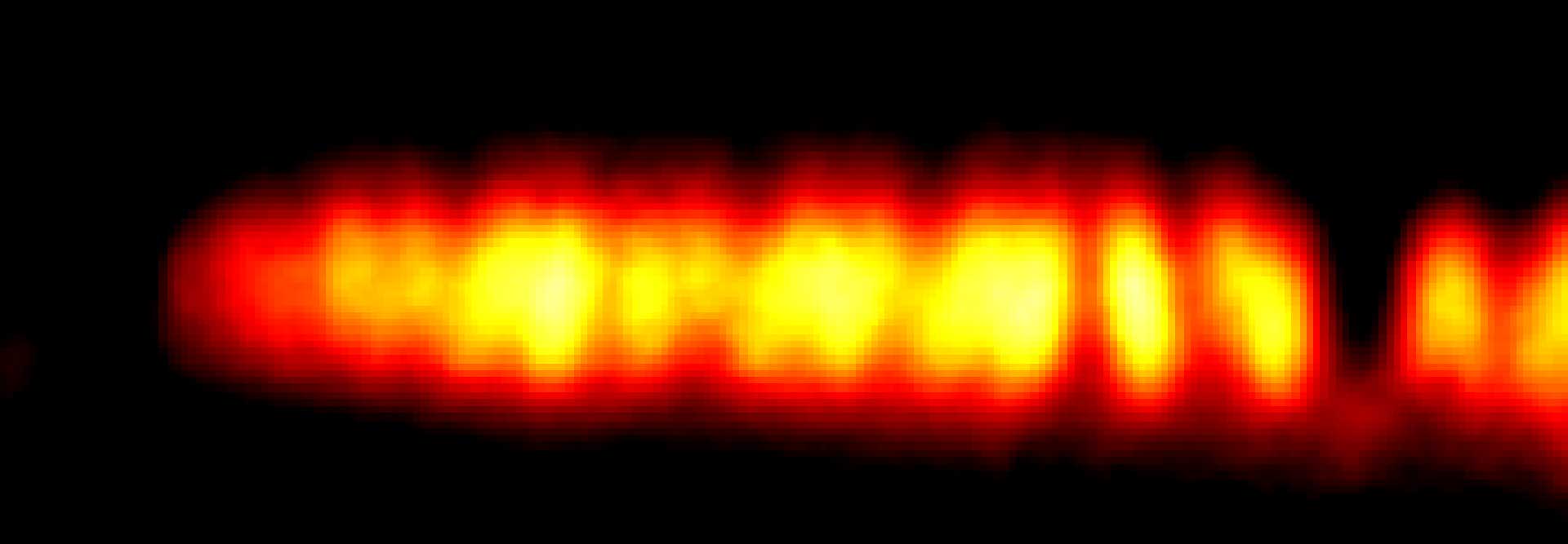
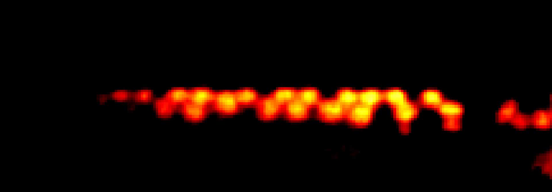
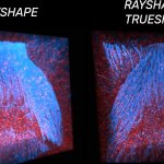
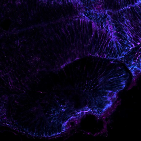
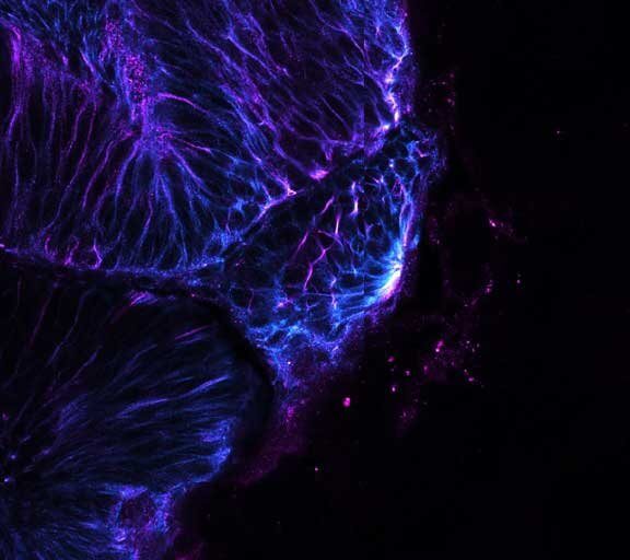
Gain in both signal-to-background ratio and resolution: MATRIX detection dramatically improves a conventional STED image of the zebrafish olfactory epithelium, resulting in a perfect and crystal-clear image revealing even more detail than STED alone can.
Modules:
Description
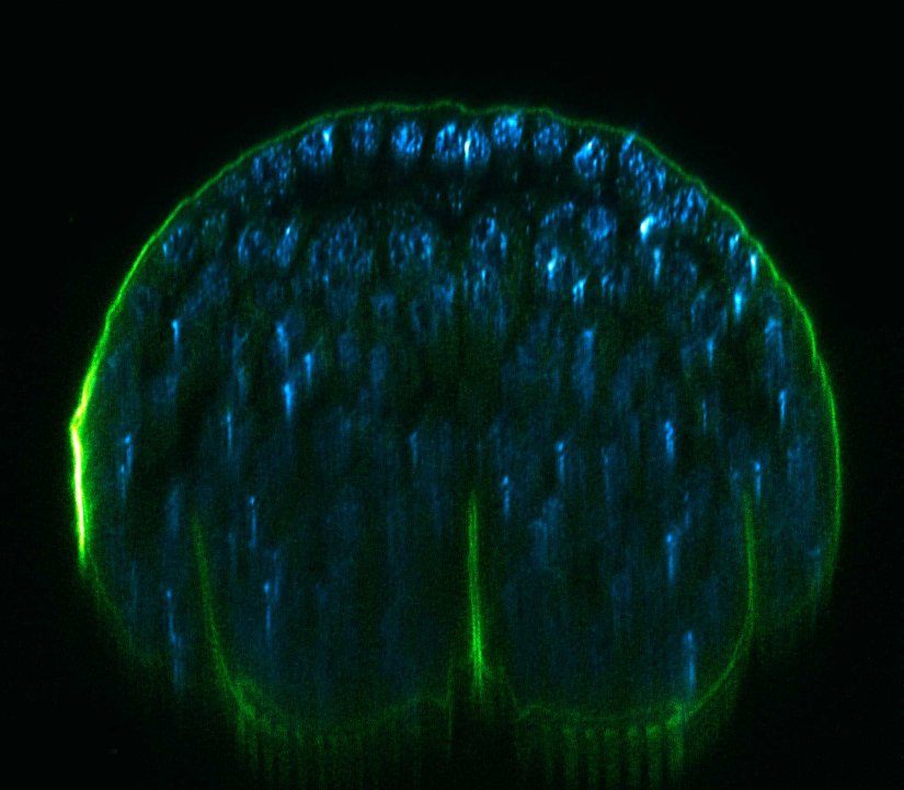
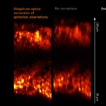
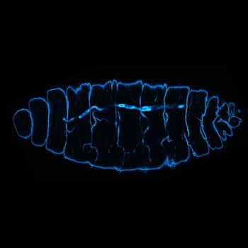
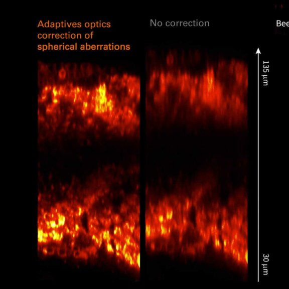
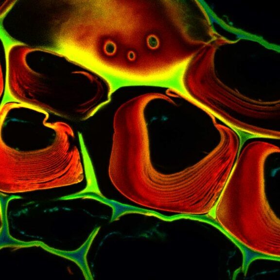
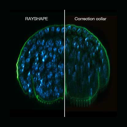
xz section of a stage 17 Drosophila embryo stained for chitin (abberior LIVE 610, green) and DNA (abberior LIVE 550, cyan).
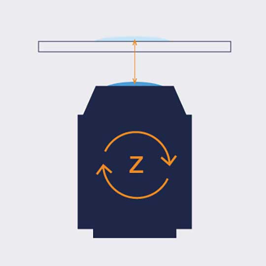
RAYSHAPE preserves resolution and brightness over the whole sample depth of about 200 µm by dynamically redirecting aberrated light to the right places.
In comparison, mechanical optics using a correction collar can only correct a limited z-range of approximately 20 µm
Modules:
Description
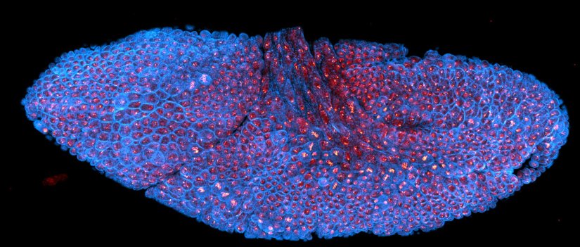
Drosophila stage 12 embryo, imaged with RAYSHAPE, stained for tubulin with abberior STAR RED and for DNA with abberior LIVE 550.
Modules:
Description
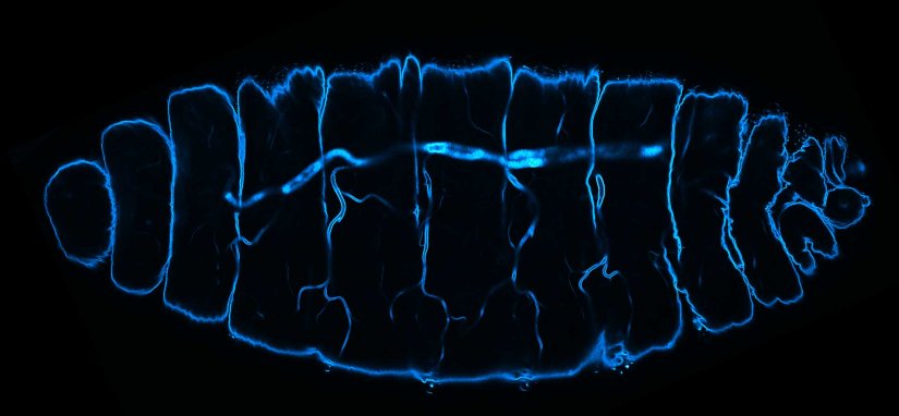
Deep tissue imaging with RAYSHAPE of a stage 17 Drosophila embryo stained for chitin with abberior LIVE 610.
Modules:
Description
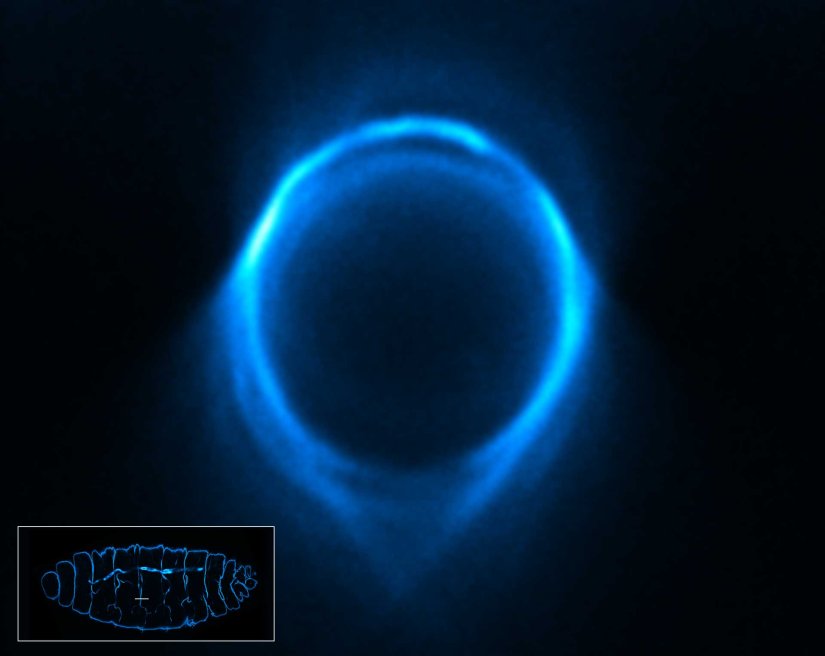
3D STED xz section of a Drosophila embryo trachea, imaged with RAYSHAPE and without abberation correction, depth 15 µm. Chitin was stained with abberior LIVE 610.
Modules:
Description
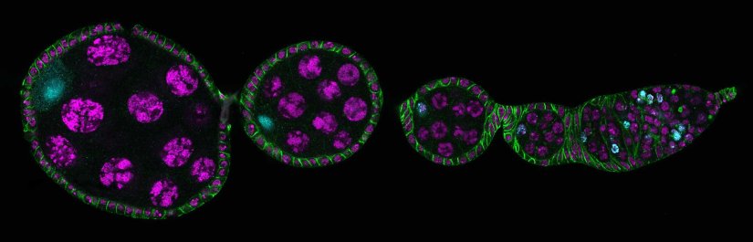
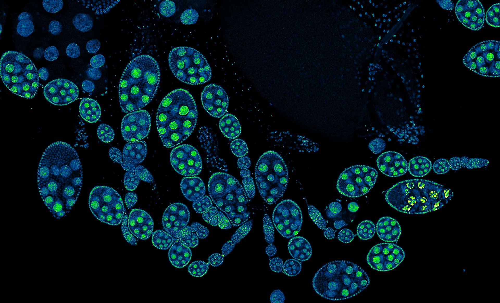
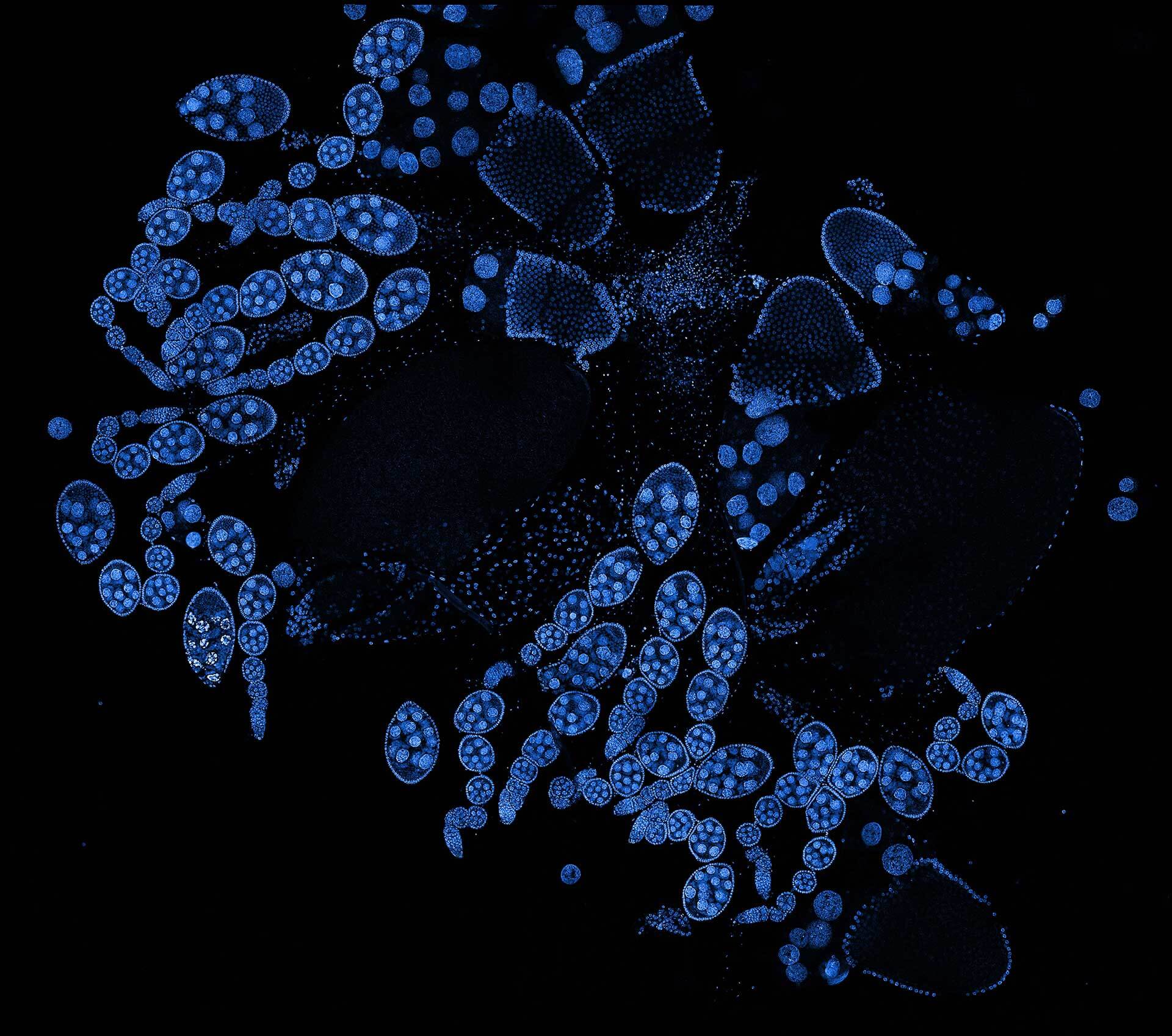
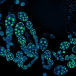
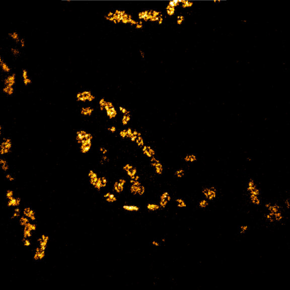
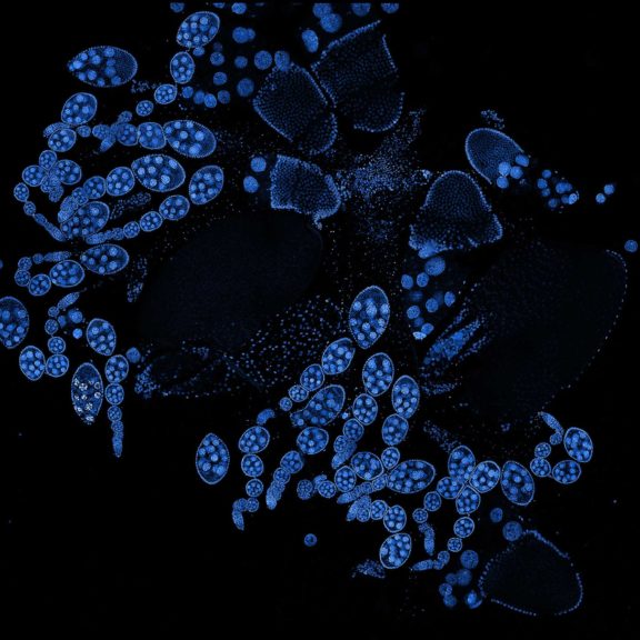
Drosophila ovariole stained with abberior LIVE 560 DNA showing nuclei in different cell types of the egg chamber. Ovaries were dissected from adult female fruit flies and were fixed prior to staining.
Image was acquired with the STEDYCON tiling feature and assembled with the SVI Huygens Stitcher.

Description
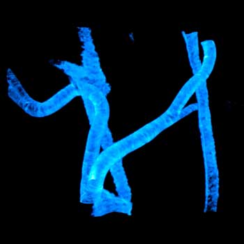
Drosophila spermatid tails were stained with abberior LIVE 610 tubulin. Testis were dissected from adult male fruit flies. Live cell imaging experiment was performed on a INFINITY microscope.

Description
Drosophila male accessory gland stained for F-actin using abberior STAR 580 phalloidin.
Sample was prepared in cooperation with Dr. H. R. Shcherbata at MPl for Biophysical Chemistry, Göttingen, Germany.
Description
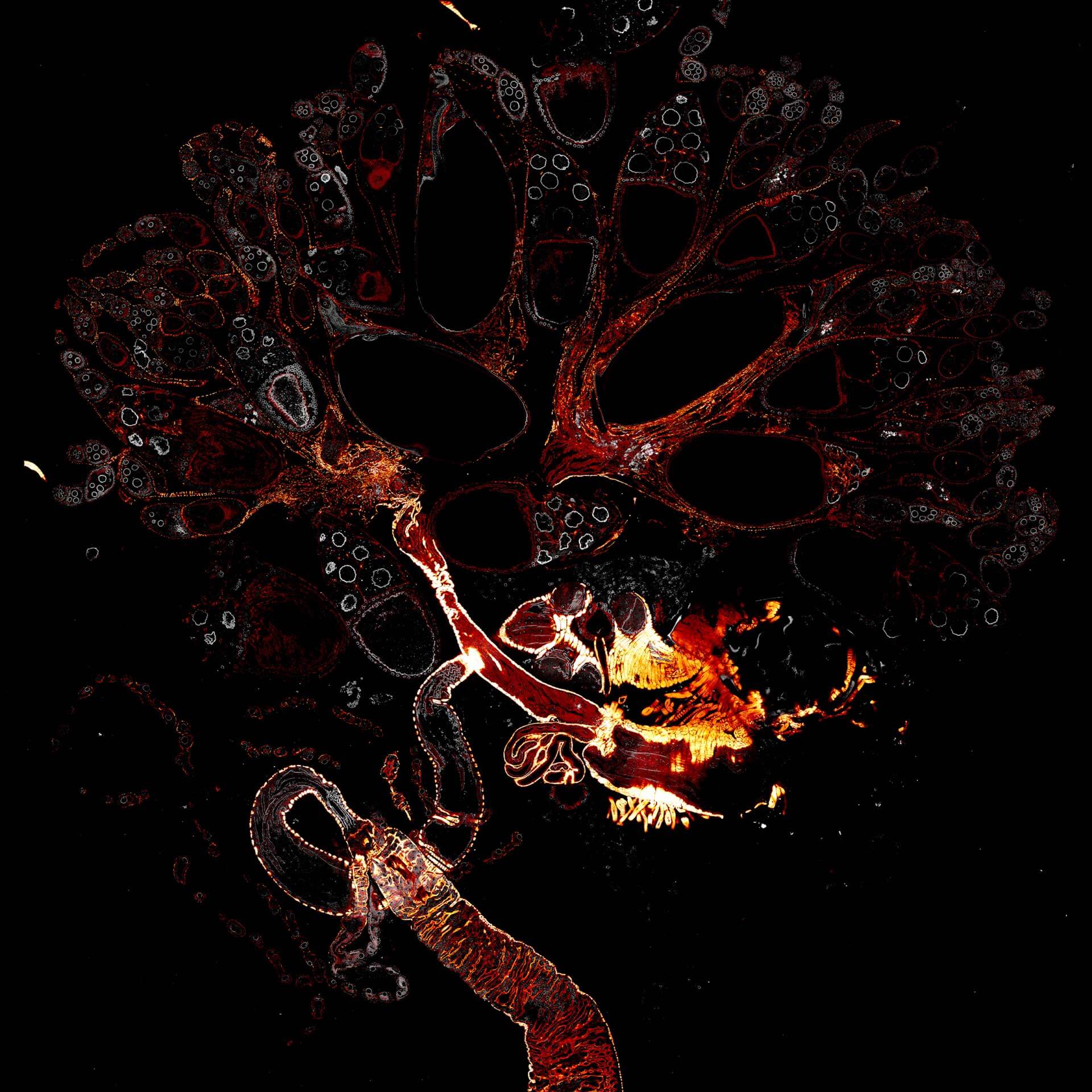
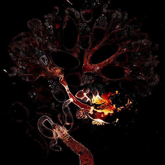
Drosophila female reproductive system stained for F-actin (red) with abberior STAR RED phalloidin. abberior STAR ORANGE is highlighting a nuclear pore protein (gray).
Image was acquired with the STEDYCON tiling feature and assembled with the SVI Huygens Stitcher.

Description
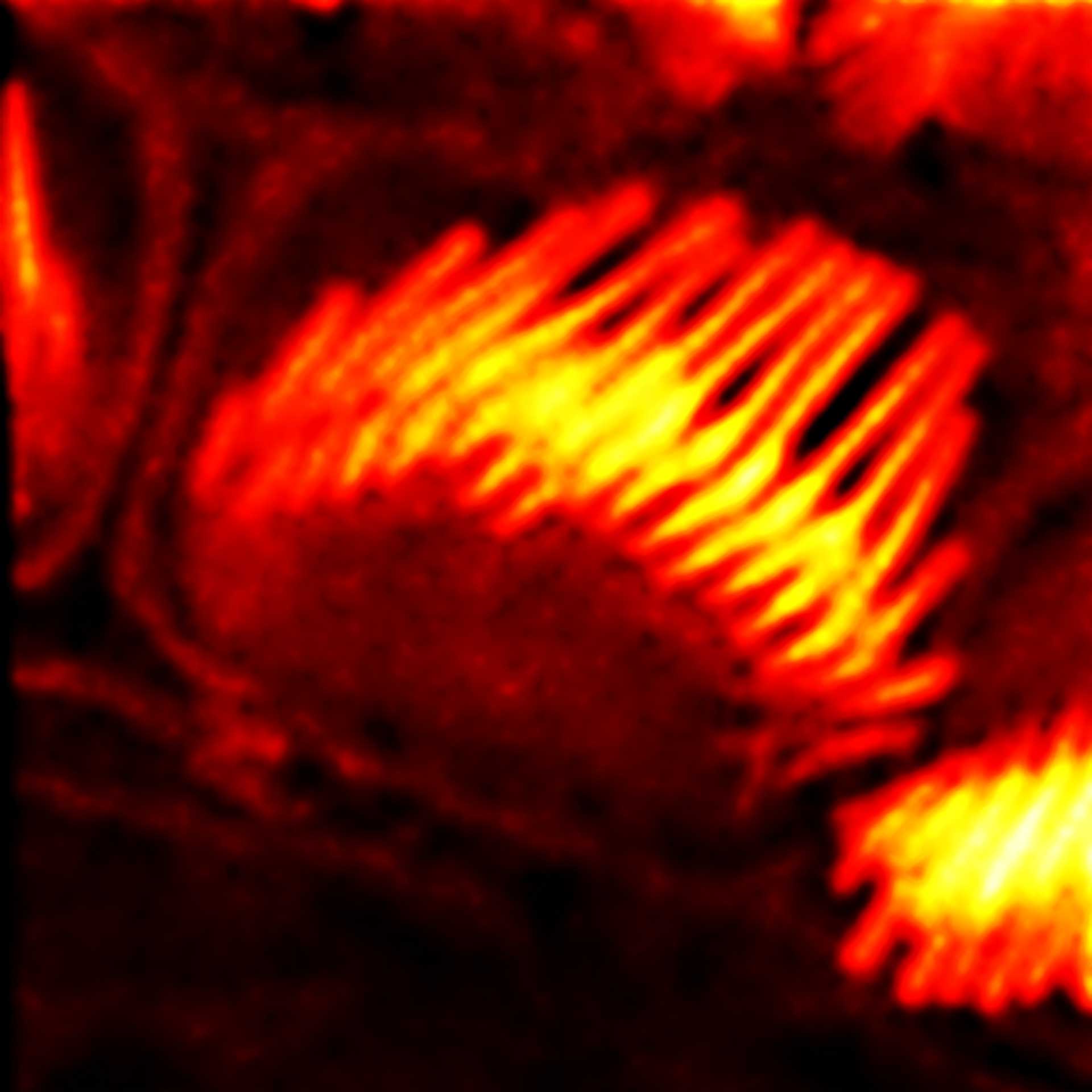
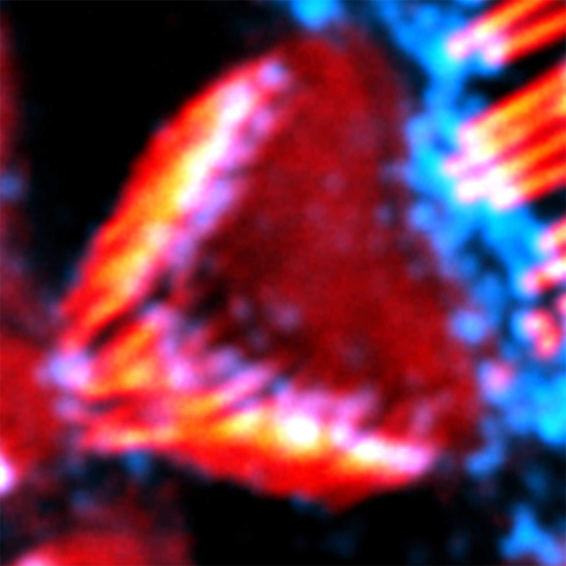
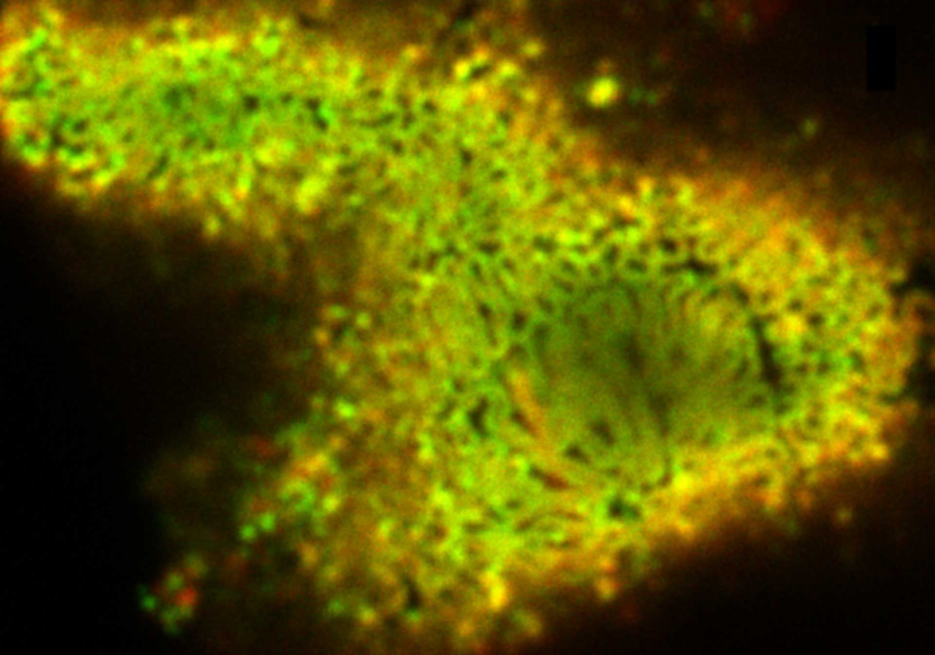
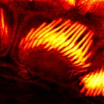
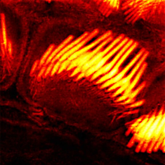
Actin stain of mouse inner ear hair cells using abberior STAR RED phalloidin.
Samples were prepared by Dr. Christian Vogl, InnerEarLab, UMG Göttingen, Germany.
Description
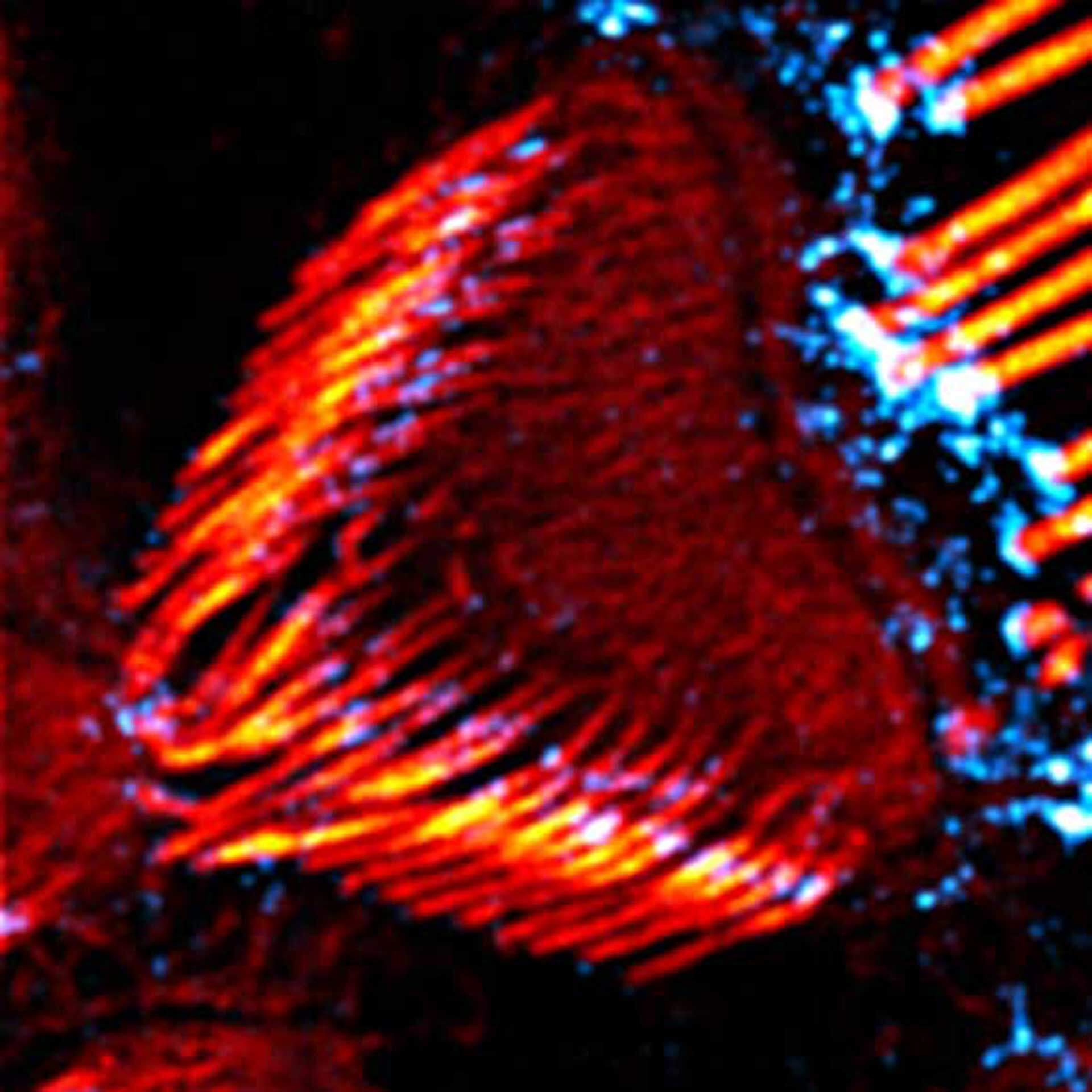
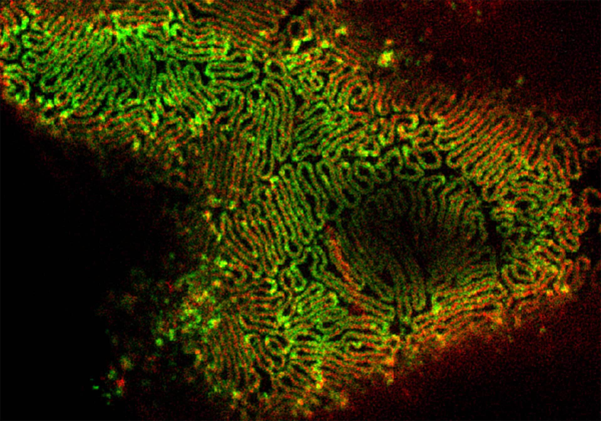
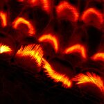
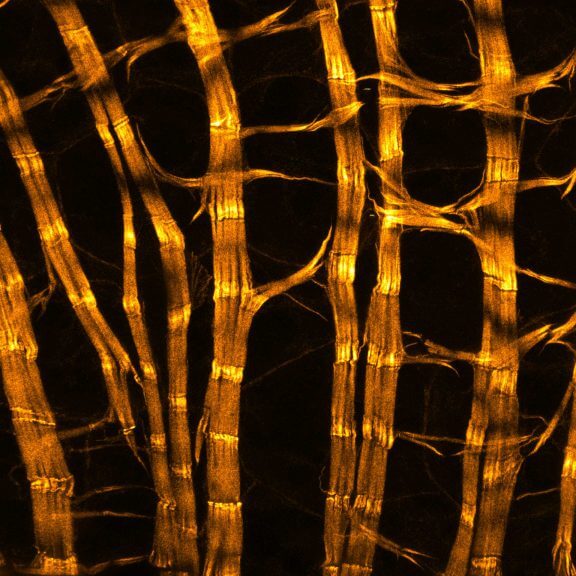
Actin stain of mouse inner ear hair cells using abberior STAR RED phalloidin. Zoom in for full effect.
Samples were prepared by Dr. Christian Vogl, InnerEarLab, UMG Göttingen, Germany.
Modules:
Description
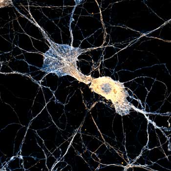
Body wall preparation of a 3rd instar Drosophila melanogaster larva. Synapses at the neuromuscular junction are visualized by immunostaining against Bruchpilot (Brp), an integral component of active zones in Drosophila. Images were acquired on a STEDYCON attached to an Olympus IX81. Images were kindly provided by Dr. Nadine Ehmann, Institute of Physiology, Neurophysiology, University of Würzburg.
Description
Actin stain of mouse inner ear hair cells using abberior STAR RED phalloidin.
Samples were prepared by Dr. Christian Vogl, InnerEarLab, UMG Göttingen, Germany.
Description
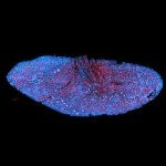
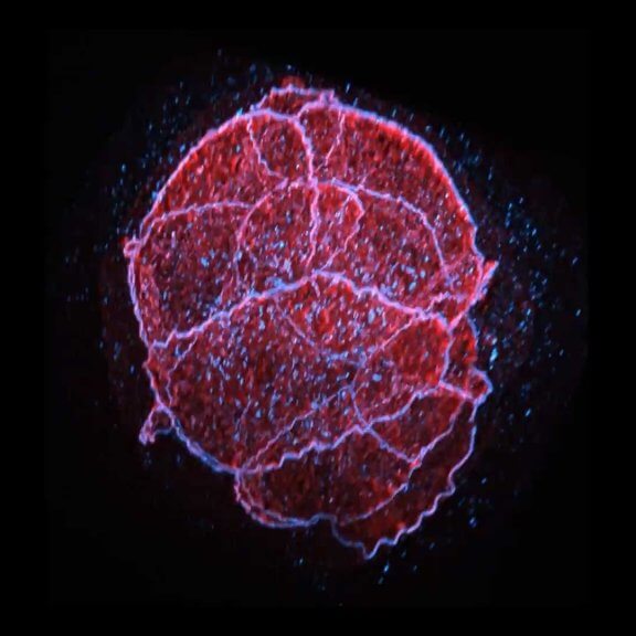
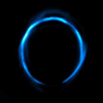
2-color 2D STED image of a cleared adult kidney sample of a rat. Shown is an image of a renal corpuscle showing Nephrin (red, abberior STAR 635P) structures inbetween the Podocin slits (green, AlexaFluor594).
Sample was prepared by D. Unnersjö Jess and H.G. Blom @ KTH Stockholm, Sweden.
Description
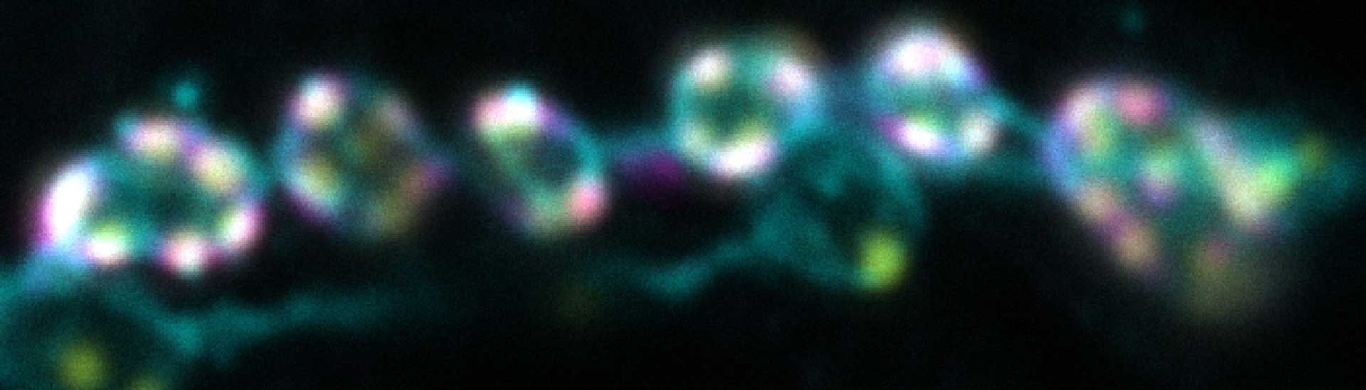
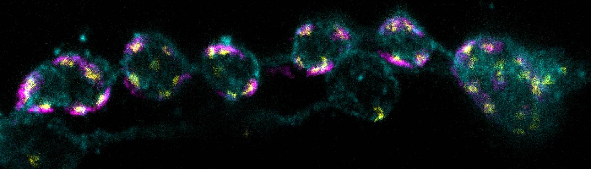
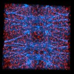
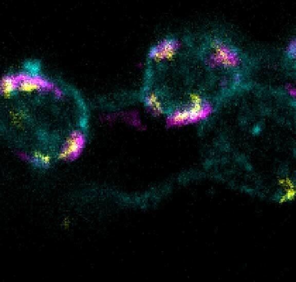
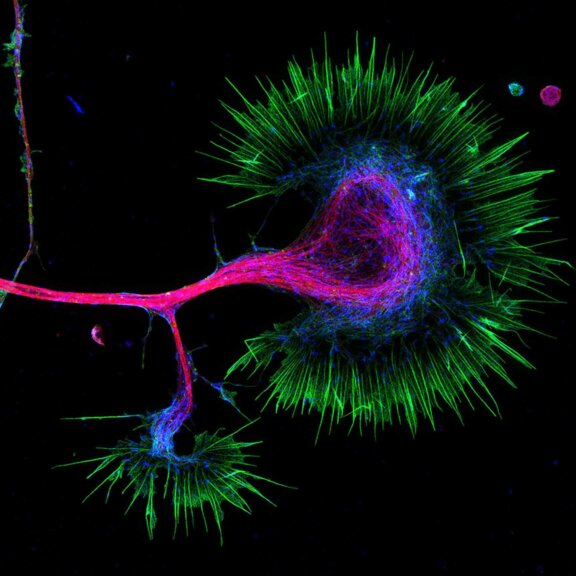
3-color STED imaging: active zones at the Drosophila larval neuromuscular junction immunostained for Bruchpilot and two other proteins.
Two superresolution channels (magenta, yellow) using a 775nm STED laser & one superresolution channel using a 595nm STED laser.
Samples by M. Lenz & M. Landgraf (University of Cambridge, UK).
Description
Drosophila ovariole stained with abberior LIVE 560 DNA showing nuclei in different cell types of the egg chamber. Ovaries were dissected from adult female fruit flies and were fixed prior to staining.
Image was acquired with the STEDYCON tiling feature and assembled with the SVI Huygens Stitcher.
Modules:


Discover the details –
with the must-have mic for zoology
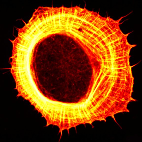
This tiled image of the Drosophila female reproductive system stained for F-actin (red, abberior STAR RED phalloidin), and a nuclear pore protein (gray, STAR ORANGE) was captured with the STEDYCON tiling feature and stitched with SVI Huygens. It is only one of many examples for the application of light microscopy in zoology. Generally, the demands on a light microscope are as diverse as the samples themselves. This logic applies to zoology in particular: your model organism may be mouse, Zebrafish, Xenopus, C. elegans or one of the many others. Then there is the question: Do you want to image entire animals, tissue sections, or individual cells? Do you want to produce a tiled image? Are you dealing with fixed samples or live cell imaging? How big or small are the structures to be resolved? And are the requirements in a particular lab always the same or do they vary depending on who is using the microscope? A variety of techniques and configurations come into question – and abberior is the right company to ask for a suitable instrument!
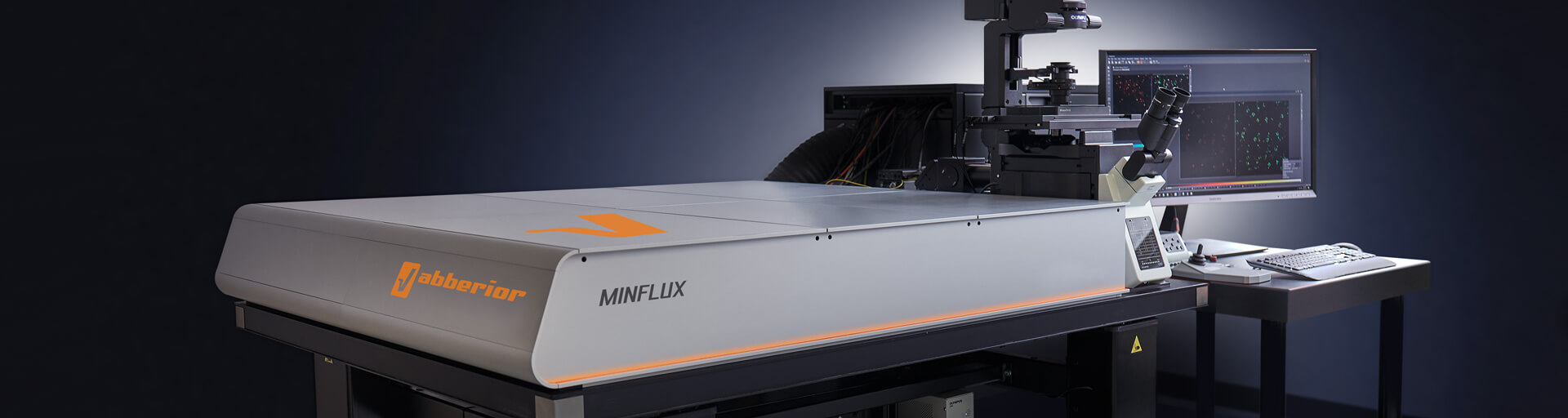
Get a demo >MINFLUX – unrivaled resolution and speed
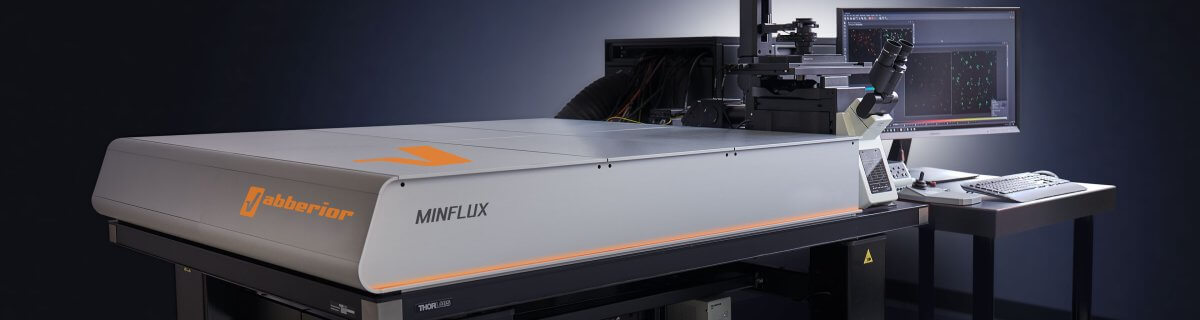
The MINFLUX platform offers an unprecedented array of imaging possibilities and allows you to resolve structures as small as a molecule, along all three dimensions. This unmatched resolution capability combined with unprecedented speed reveals sample details never seen before and helps to dissect fast and dynamic cellular processes in space and time. MINFLUX is the world’s most powerful fluorescence microscope. Details >
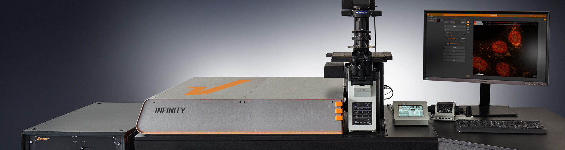
INFINITY – forever cutting edge
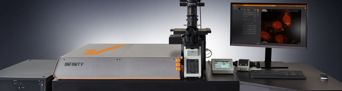
The INFINITY platform is the most customizable platform for all things microscopy and may be adapted to your particular demands depending on what type of sample you have and which question you want to address. INFINITY is always up to the challenge. Just tell us what you need and we will build you a customized, continuously upgradable system specialized for your research. Details >
MIRAVA POLYSCOPE – one for all and all for one

The MIRAVA® POLYSCOPE® combines every resolution – from millimeters down to 3 nm – in a singularly unique system. MIRAVA unites four microscopy technologies to cover an unprecedented resolution spectrum, extending over several orders of magnitude from diffraction-limited imaging all the way to true molecular resolution. Our LiGHTBOX software allows beginners to intuitively arrive at a top-notch image within three clicks, while also giving experts full control over the instrument. Analyze samples as diverse as whole organisms, tissues, single cells, or protein complexes with a single microscope! Details >
STEDYCON 2 – confocal, STED, lifetime… WOW!

The STEDYCON upgrades your existing widefield microscope to a confocal, STED, and lifetime machine with a resolution down to 30 nm. All that’s required is a free camera port and a good objective lens. With its super-intuitive user interface, the STEDYCON provides an intelligent microscope platform that enables everyone to acquire superb superresolution images after only minutes of training. Obtain premium confocal and STED images at the push of a button! Details >
A whole zoo
of modules for zoology
Every one of our modules adds another superpower to your microscope. In this xz-section of a stage 17 Drosophila embryo stained for chitin (green, abberior LIVE 610) and DNA (blue, abberior LIVE 550), RAYSHAPE was used to dynamically correct for aberrations over the entire z-range, restoring signal and focus. The result is a crisp and clear image from top to bottom.
Ask for detailed information >Choose a superpower for your experiment – our modules
Every MINFLUX, INFINITY, and FACILITY microscope can be upgraded with modules to overcome the specific imaging challenges in your research. Do you focus on stem cells and differentiation? mRNAs and their role in development and morphogenesis? Are you interested in cell migration? Or something entirely different? Whatever it is, your imaging will profit from one or more of our modules. Apply abberior’s unique RAYSHAPE aberration correction using a deformable mirror for aberration-free imaging in deep tissue. Improve optical sectioning and signal-to-background ratio with the MATRIX detector. Use TIMEBOW to separate signals according to their fluorescence lifetime and expand your labeling options, image resolution, and much more. And we have even more in store! If you can’t find what you need on our website, get in touch with us, and we’ll develop it for you.
MATRIX Detector
Many eyes see more than one. The MATRIX detector drastically improves signal-to-background ratio, resolution, and dynamic range.
TIMEBOW Imaging
TIMEBOW lifetime imaging for stunning results at confocal and STED super-resolution.
FLEXPOSURE Illumination
Brings down the light dose on your sample and lables dramatically. Key ingredient for volume and live-cell superresolution.

RAYSHAPE Mirror
Dynamic aberration correction with a deformable mirror over about 200 µm z-range. 140 digital actuators adjust the mirror surface within milliseconds.


Custom Solutions
We offer solutions for even the most challenging applications. Everything that can be done, we will do.
Broaden your knowledge
of microscopes, dyes, and superresolution
Confocal microscopy generates stunning images like this one of a Platynereis dumerlii larva 72 hours after fertilization stained for tubulin (cyan, abberior STAR RED), synapsin (STAR ORANGE, orange), and DNA (gray, DAPI). But how does confocal microscopy work? And what does STED do differently to reach resolution beyond the diffraction limit? What is resolution in the first place? How do aberrations arise and how can we get rid of them? Our knowledge base is your chance to close any gaps in fluorescence microscopy expertise you might have!
Tell me more >
Confocal microscopy offers superior optical sectioning. But what is that exactly? And what about other ways to get rid of the background, such as array-based detectors like the MATRIX? Details >
Deep and clear: where confocal beats out wide-field microscopy

Confocal microscopes were designed to get rid of background signal. How do they work? And when do you know it’s time to use one? The answer is in the pinhole. Details >

Fluorescent labeling strategies have become more and more sophisticated and offer ever-new options to improve microscopic imaging. Among the latest are exchangeable HaloTag ligands that put an end to photobleaching for good. Details >
How to correct for aberrations in light microscopy

Aberrations can give microscopists a hard time. They belong to microscopy like pathogens belong to life. There are ways to bring diverted rays back on track, but some are better than others. The question is: deformable mirror or correction collar? Details >
Why do superresolution microscopists love alpacas?

It is a very simple yet very important fact: the localization precision of any superresolution microscope can only be as good as the size of the fluorescent staining allows. In other words, when your fluorescent dye is too big or too far away from the protein you want to label, you will never be able to reach a resolution that is higher than this offset. The good news is: there are ways to reduce the offset between target protein and fluorescent label. And one of these are nanobodies. Details >
STEDYCON: ease-of-use in a shoebox

A sleek, black-and-orange box transforms your widefield microscope into a confocal and a superresolution STED instrument and your exploration of subcellular structures into a seamless, discovery-rich experience. Carefully designed with masterly engineering, STEDYCON breaks the stereotype of the finicky, hard-to-use scope. It opens new possibilities at the press of a button for any user and almost any location. How does it do it? The secret’s in the box. Details >
Additional information and articles
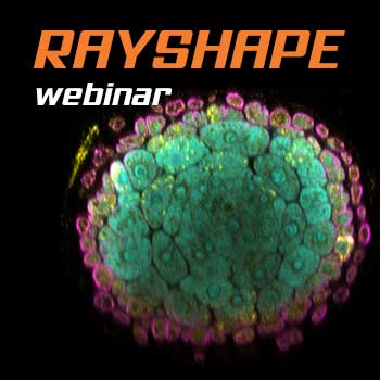
RAYSHAPE webinar – dynamic aberration correction for deep tissue imaging
Hello RAYSHAPE! Bye-bye, aberrations!
RAYSHAPE special
RAYSHAPE dynamic aberration correction with a deformable mirror preserves resolution and increases brightness. 140 actuators adjust mirror surface within milliseconds for over 200 µm z-range.

TIMEBOW Lifetime Imaging – gives time color
Explore abberior’s TIMEBOW lifetime toolbox with our application experts Julia Menzel and Jan-Gero Schloetel.
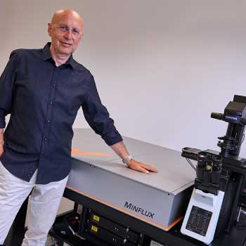
MINFLUX
The world’s most powerful fluorescence microscope MINFLUX, based on Stefan Hell’s invention, allows to resolve structures as small as a molecule in 3D.
Why is MINFLUX the best tool for single particle tracking?
The sampling rate of MINFLUX is 100 times higher than that of camera-based techniques. With only a few photons, we achieve a precision in the single-digit nanometer range.
MINFLUX webinar – Tracking with unprecedented spatio-temporal resolution!
Thanks MINFLUX it’s possible to track the movement of individual molecules in the cell, e.g. kinesin. See what EMBL scientists have achieved.