


From synapses to systems – Neurobiology


FAQ Video 37

“How does TIMEBOW lifetime imaging work and why is MATRIX array detection a perfect match?”
FAQ Video 38

MINFLUX allows an everyday 3D resolution of 2 – 3 nm and offers about 100 times faster tracking than with a camera-based system.
Neurobiology unites a highly diverse set of disciplines like (electro-)physiology, anatomy, molecular and developmental biology, or optogenetics, to name a few.
Many of them hinge on the imaging of cultured cells, tissues, or whole organs, both fixed and alive. Nowadays, complex model systems like organoids are investigated, as well. Light microscopy is a valuable tool in neurobiology, since it allows the analysis of the structure, function, and interaction of neuronal cells and systems in an intact and even living state deep within tissue. It may even be used for calcium imaging to monitor physiological dynamics within cells. As many structural details of vesicles, synapses, spines, and other neuronal components are frequently beyond the resolution limit of conventional light microscopy, superresolution methods play a critical role in neuroscience.
We at abberior provide microscopes for both confocal and STED imaging with superior performance as well as add-on modules addressing the particular challenges of neurobiological microscopy like deep tissue imaging.
Furthermore, we facilitate unprecedented spatial and temporal resolution with MINFLUX, which has been applied, for example, to map the presynaptic active zone of rod photoreceptors and dissect the walking of kinesin-1 along microtubules in vivo.
Our product portfolio is completed by a comprehensive selection of fluorescent dyes and labels suitable for various applications.
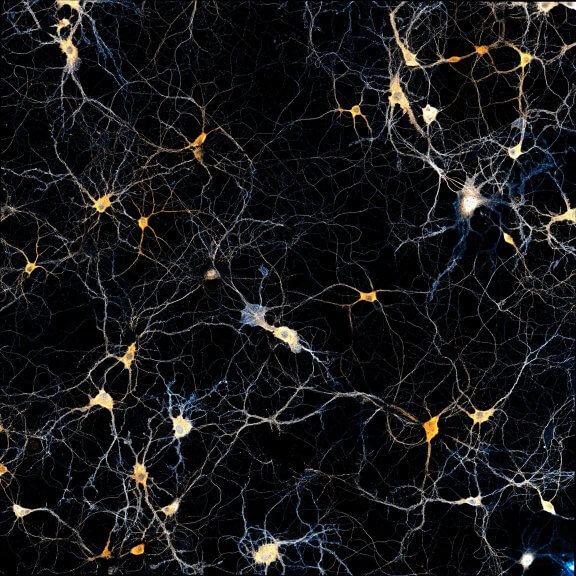
Talk to a scientist >Untangling the maze within
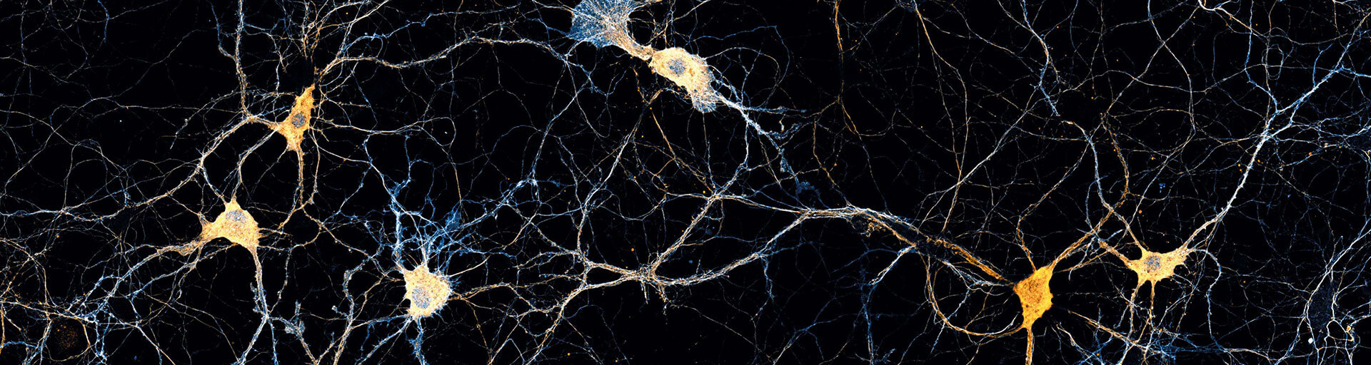
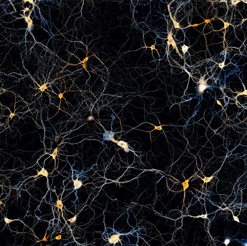
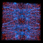
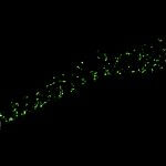
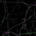
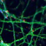
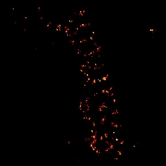
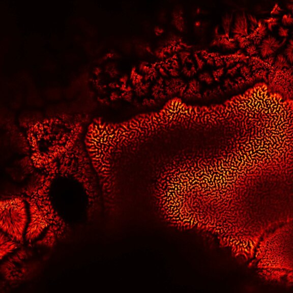
Imaging neurobiological samples, while often challenging, generates images that are not only scientifically valuable but often also highly aesthetic. The one you see here, bearing resemblance to a starry sky, stems from a brain section stained for spectrin and adducin (abberior STAR RED and STAR ORANGE).
Test your sample >The bright side of the brain
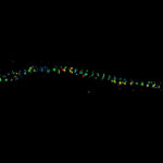
Fluorescence microscopy of neurons, synapses, glia cells, or whole nervous tissue sections provides us with unique insights into the structure and organizational details of the nervous system. Superresolution microscopy adds a level of detail unavailable to light microscopy until recently. Flip through our gallery to see the characteristic banding pattern of spectrin along axons becoming discernable with STED, or admire the beauty of an axonal growth cone imaged in multicolor superresolution.
Description
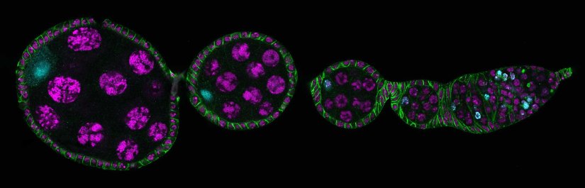
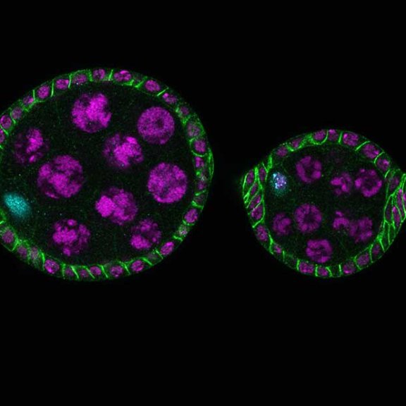
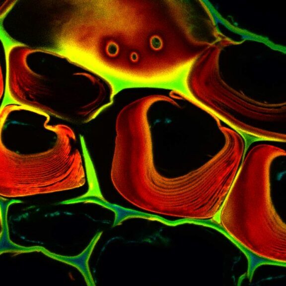
Drosophila ovariole stained for the synaptonemal complex protein C3G (cyan, abberior STAR RED), showing how the complex is formed and disassembled during oocyte development. DNA was stained with DAPI (purple), spectrin with abberior STAR ORANGE (green).
Description
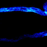
Dendrite with spines of a neuron grown on a coverslip, stained for βII spectrin and imaged in confocal mode and with the MINFLUX module of the MIRAVA POLYSCOPE.
Modules:

Description
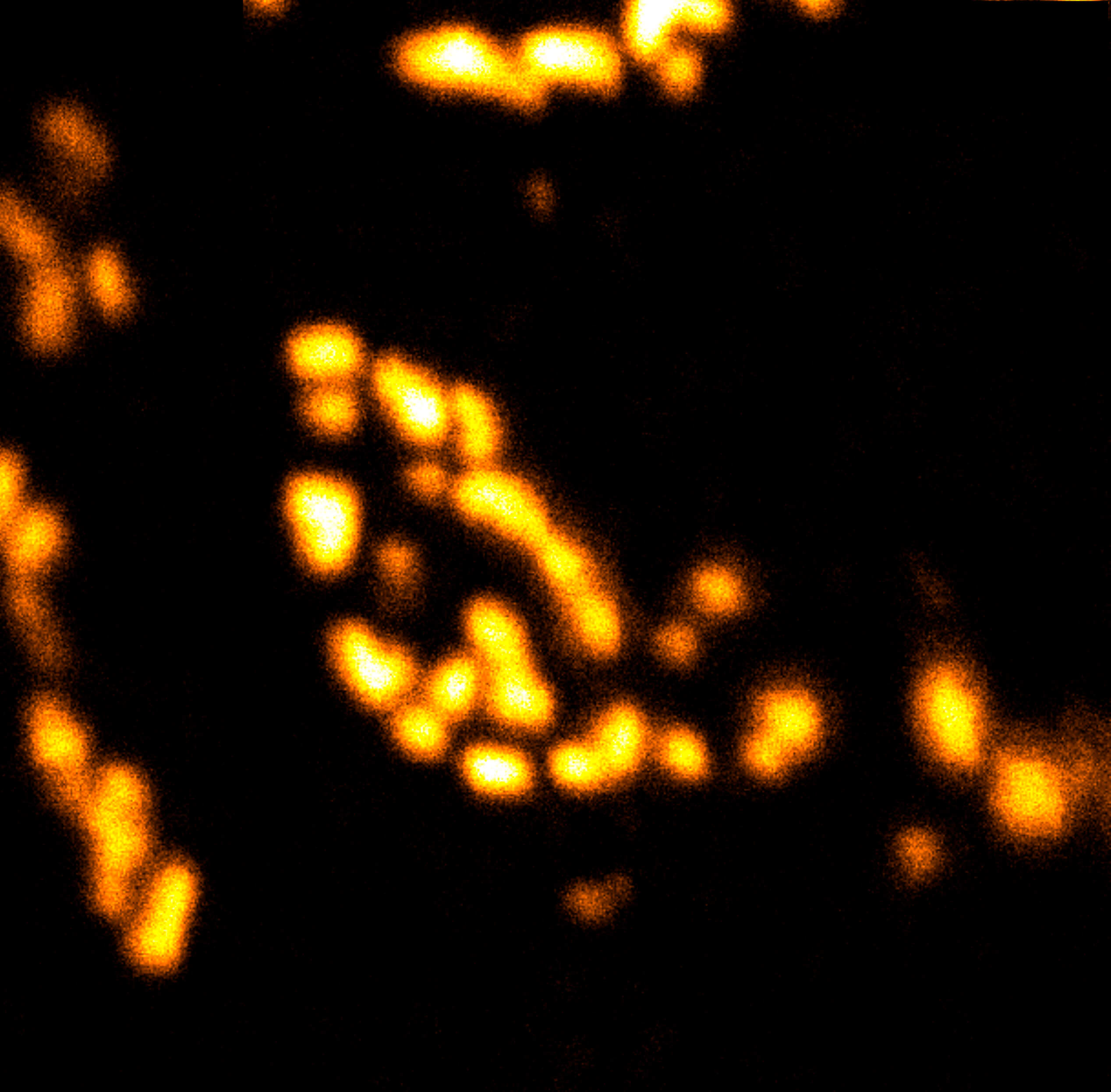
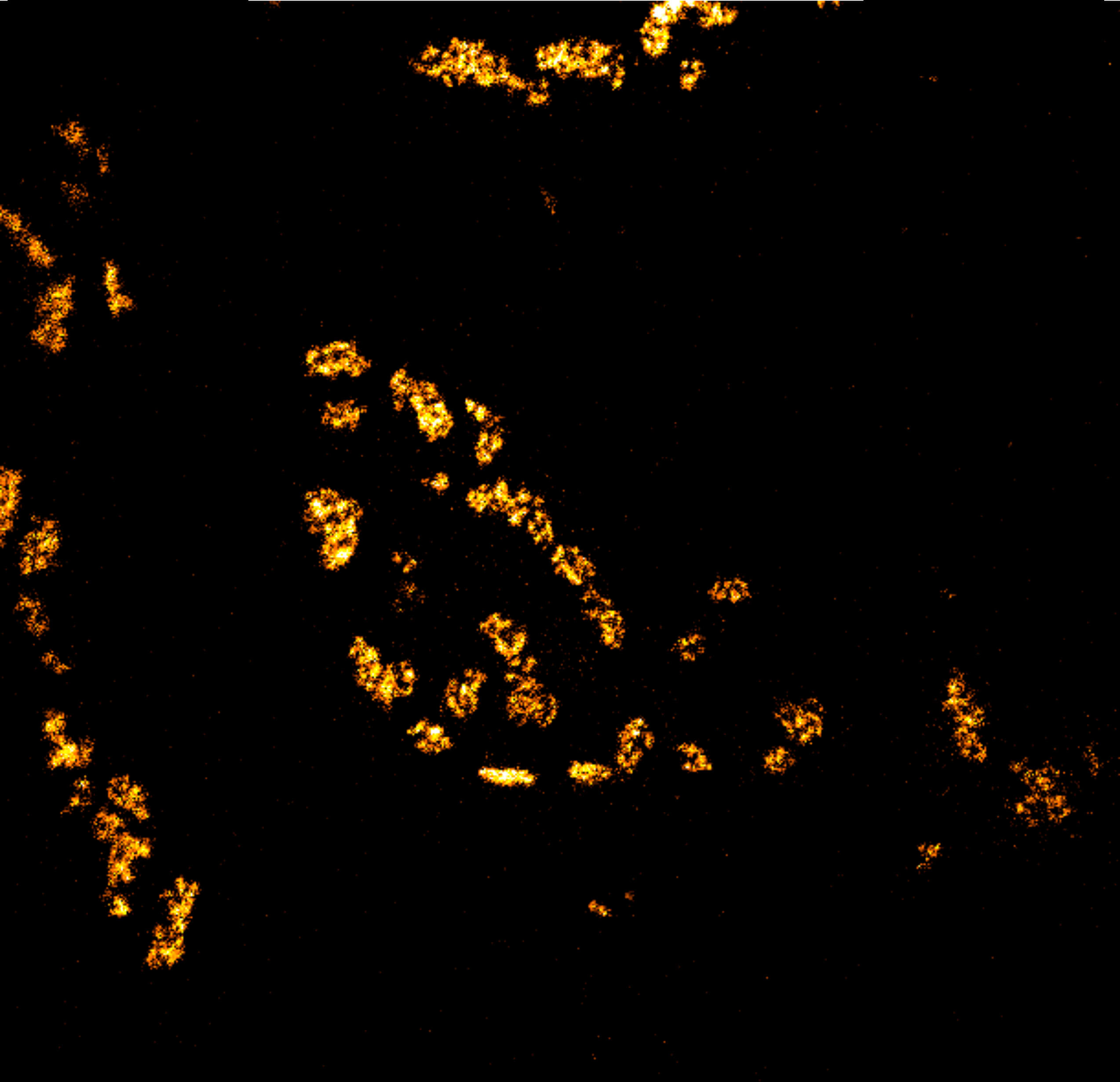
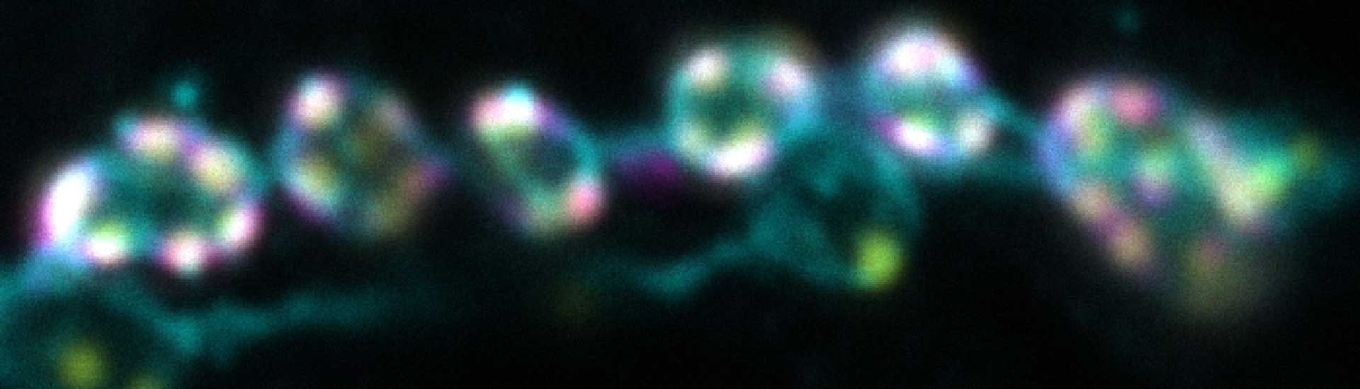
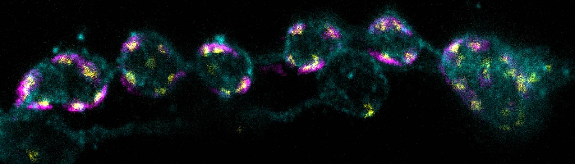
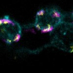
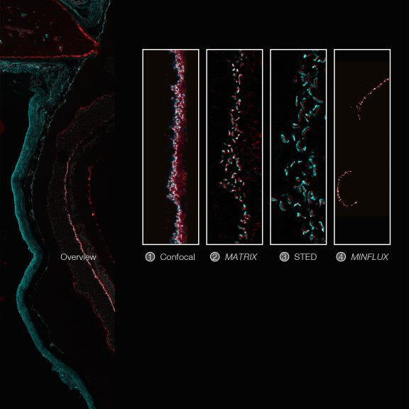
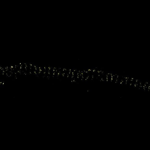
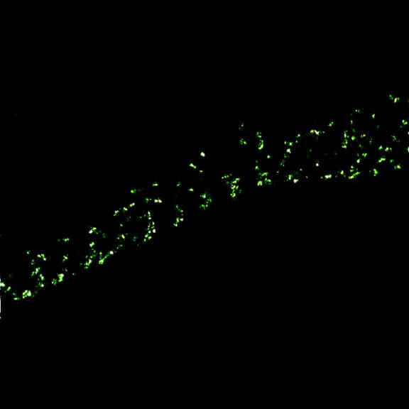
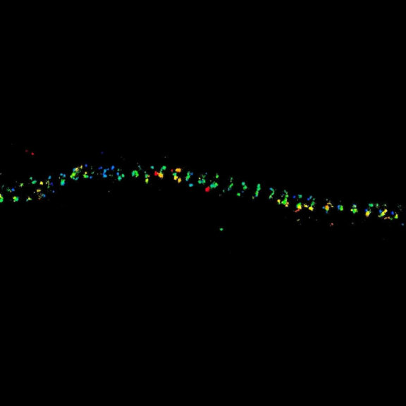
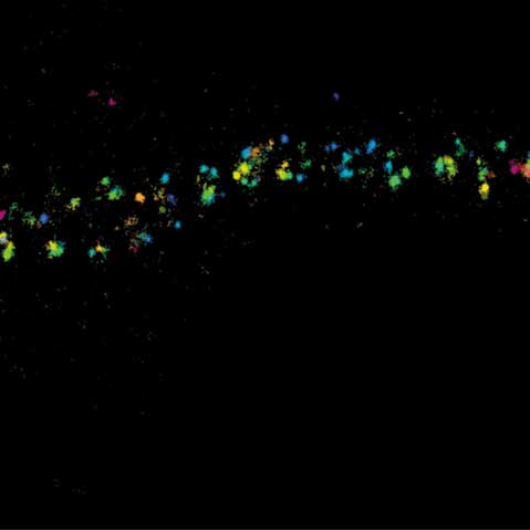
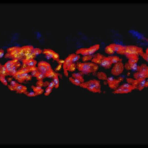
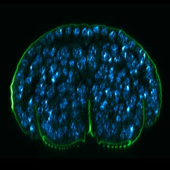
Imaging across scales from diffraction-limited to molecular resolution: ribbon synapses in fixed mouse retina tissue stained for VAMP1 (abberior STAR RED) and CtBP2 (abberior STAR ORANGE) (confocal, MATRIX, STED) or for Bassoon (MINFLUX).
Sample courtesy: Arlene Hirano, PhD, University of California Los Angeles, Los Angeles, CA, USA (confocal, MATRIX, STED), and by Dr. Chad Grabner and Prof. Dr. Tobias Moser, Max Planck Institute for Multidisciplinary Sciences, Göttingen, Germany (MINFLUX).
Modules:
Description
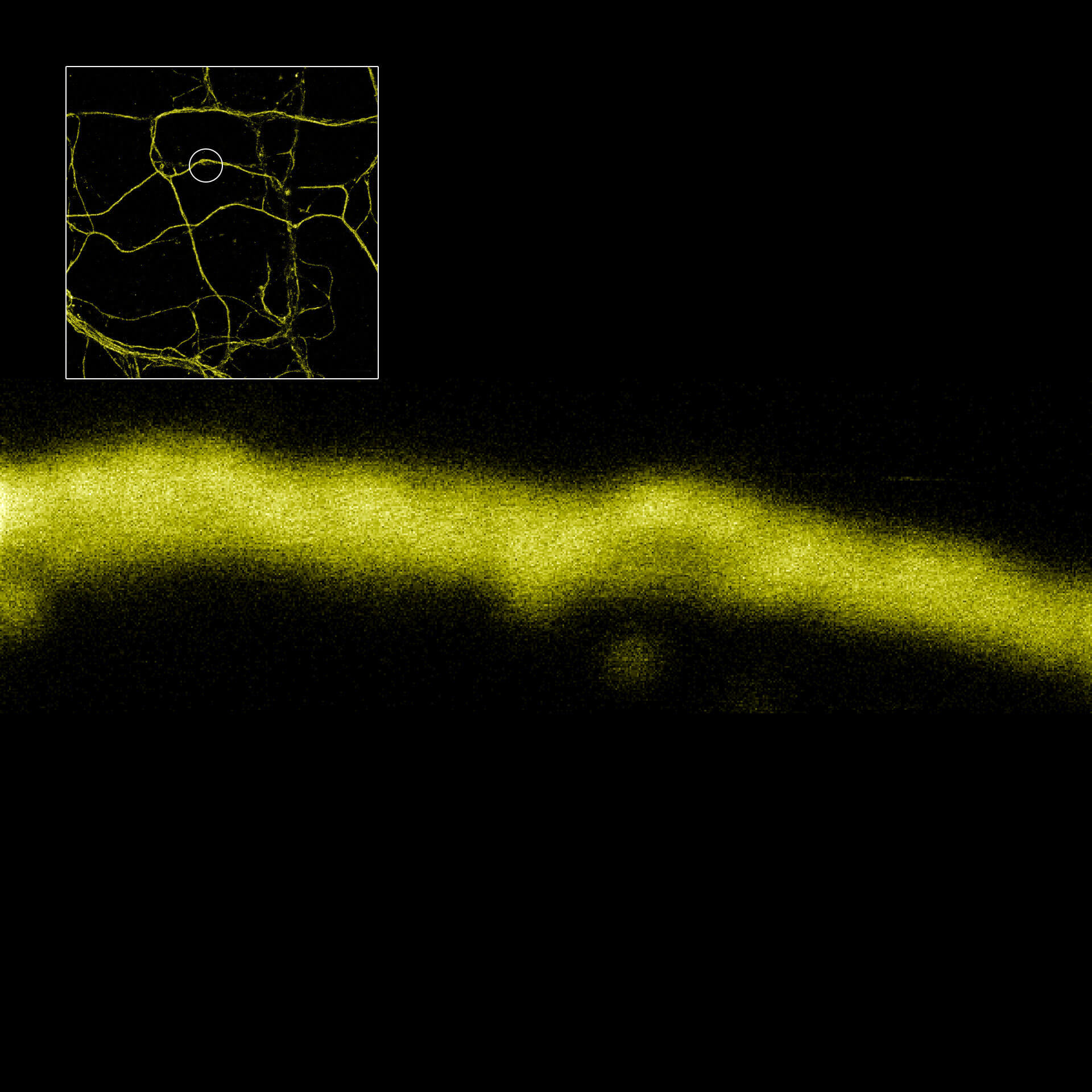
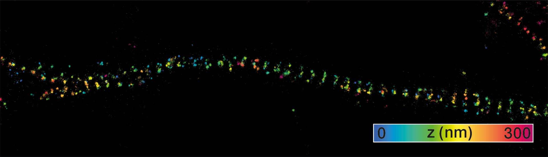
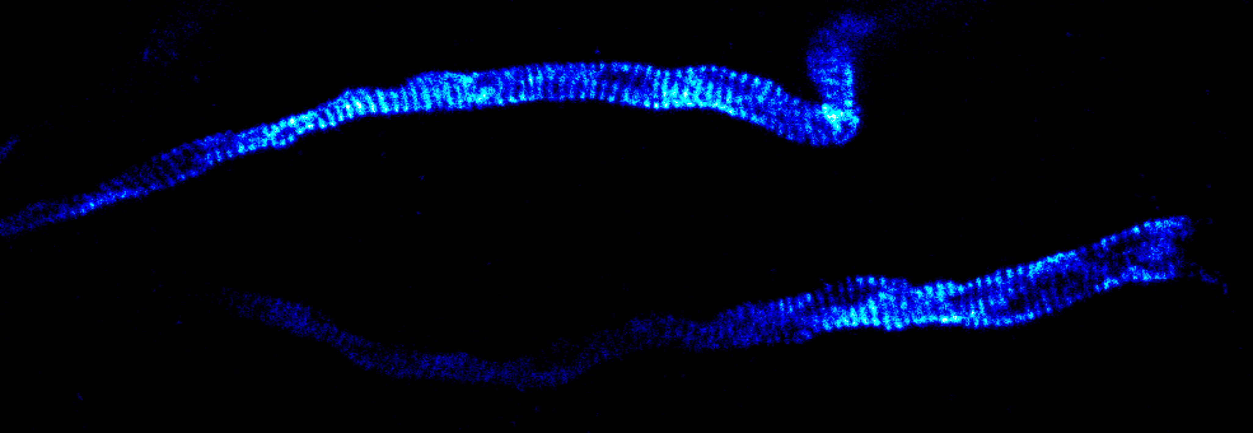
MINFLUX imaging of βII spectrin in a primary hippocampal neuron labeled with abberior FLUX 680 by indirect immunofluorescence. Please note the periodic arrangement of spectrin along the axon.
Description
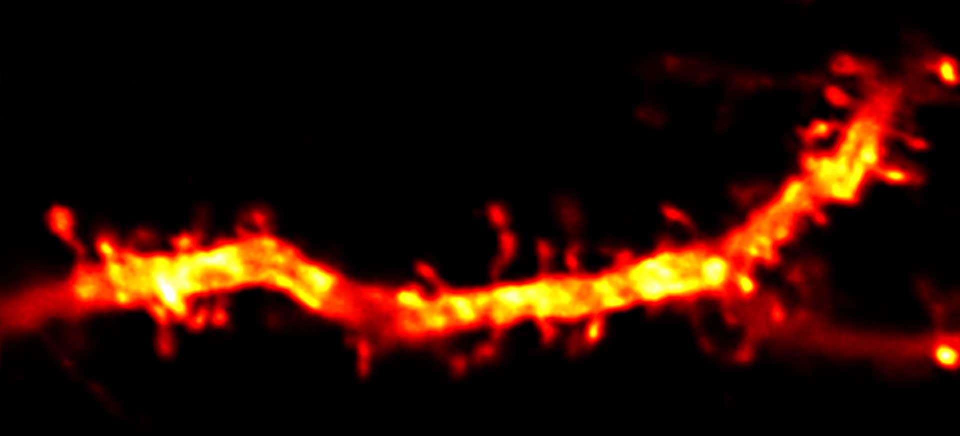
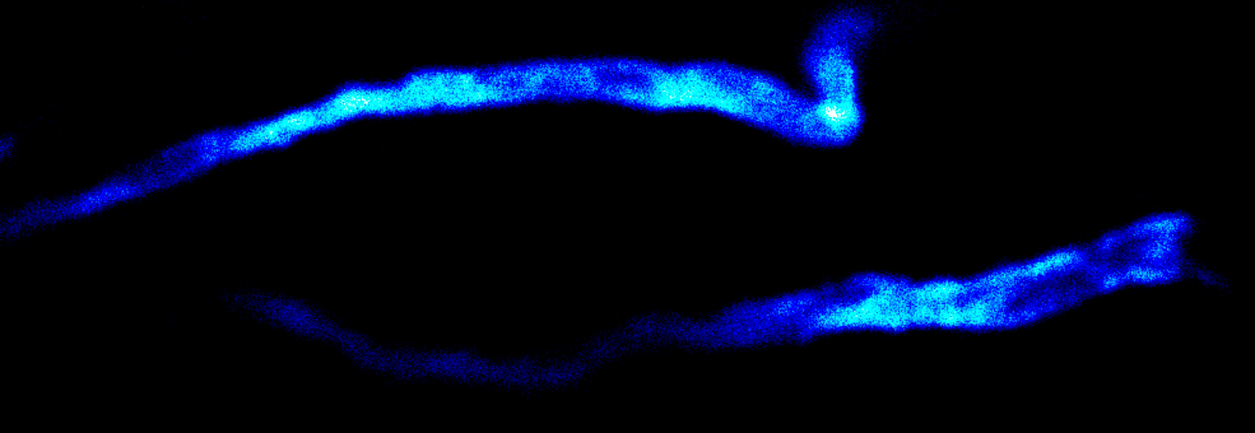
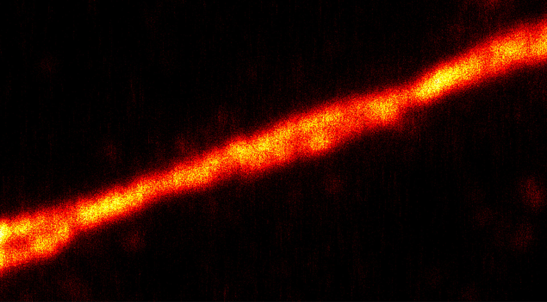
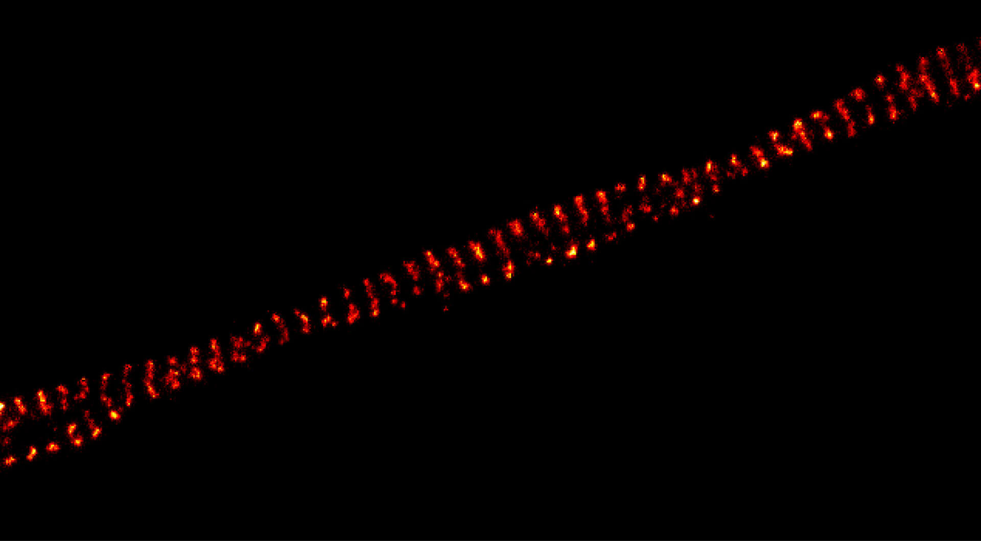
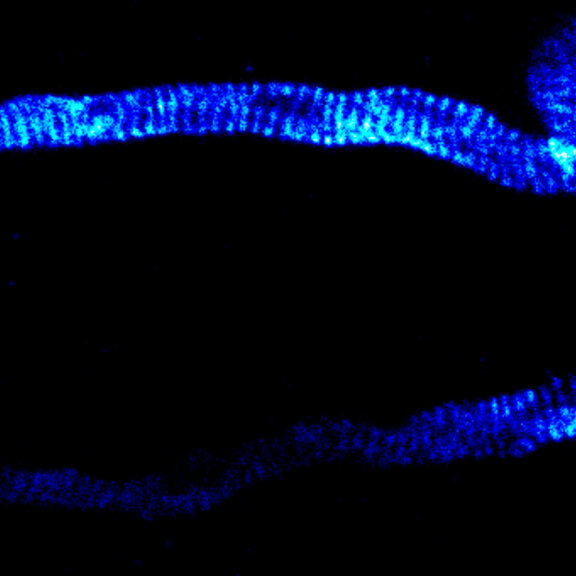
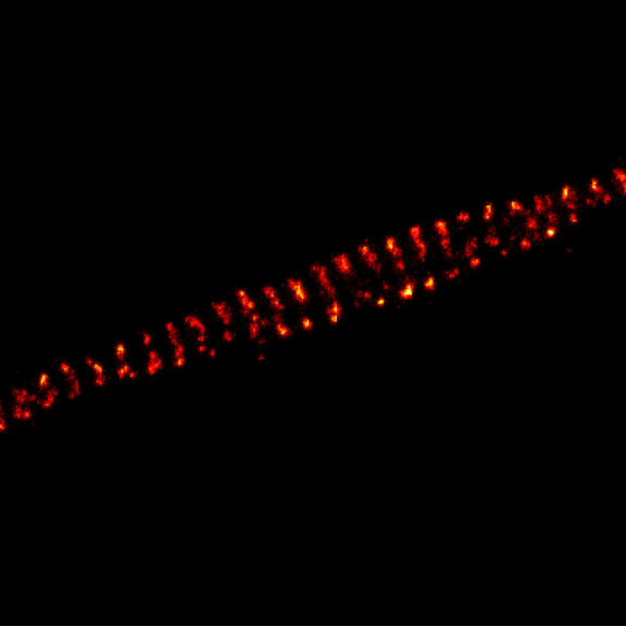
MINFLUX image of axonal βII spectrin labeled with abberior FLUX 660 in primary hippocampal neurons. Note the periodic arrangement of spectrin along the axon, and the absence of any details in the confocal counterpart image.
Description
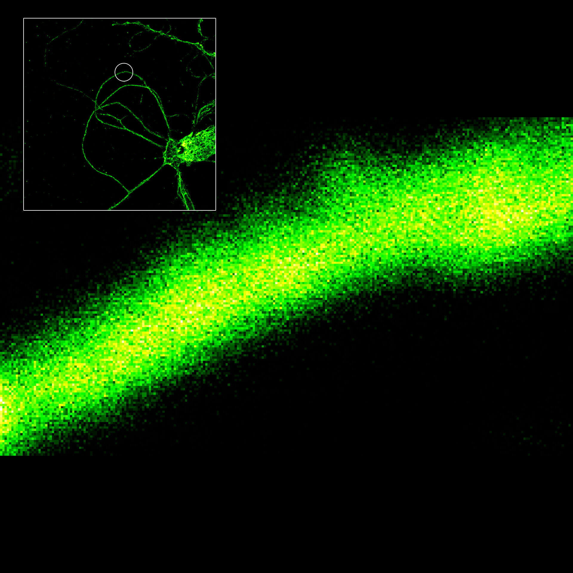
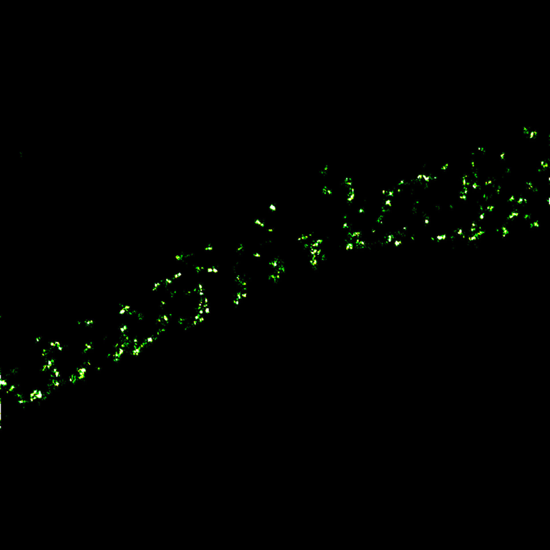
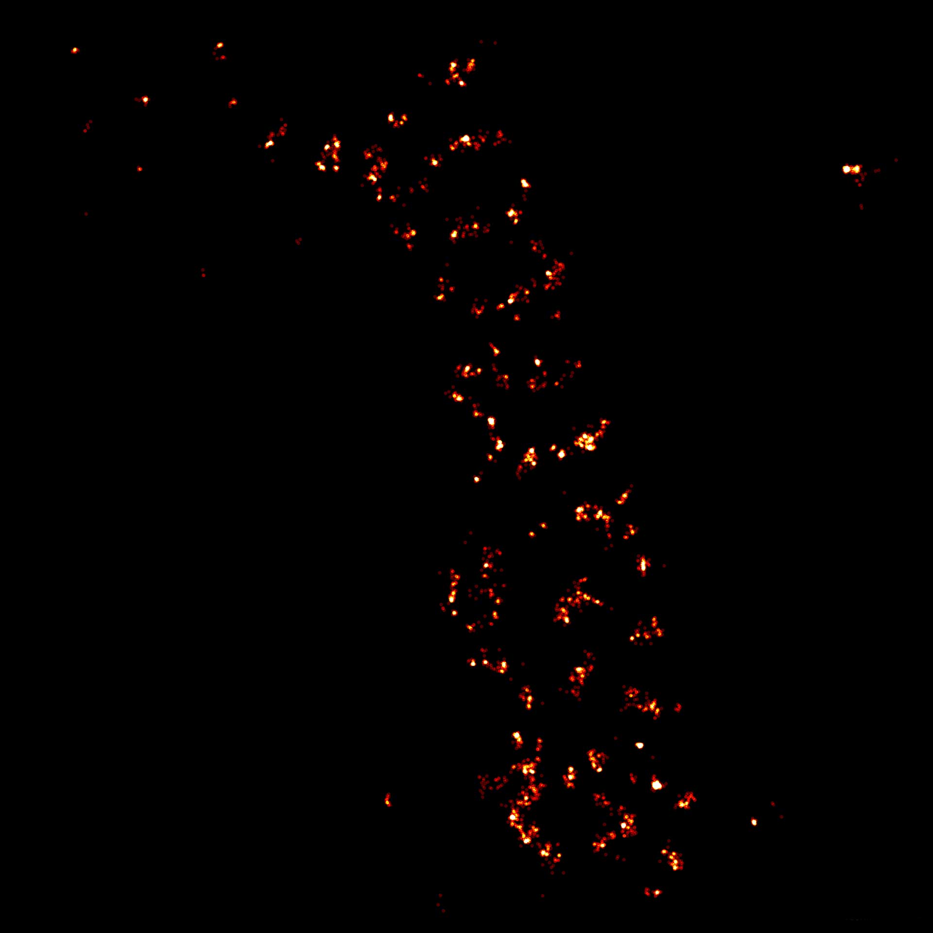
3D MINFLUX imaging of βII spectrin in a primary hippocampal neuron labeled with Alexa Fluor 647 by indirect immunofluorescence
Description
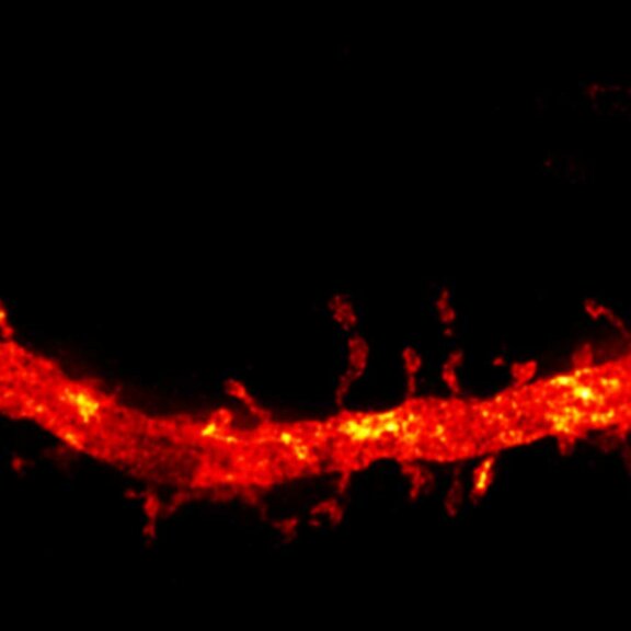
Confocal and 2D STED imaging of dendrites in brain slices. GFP-tagged proteins were expressed and immunolabelled using primary antibodies against GFP and secondary antibodies coupled to abberior STAR 635P.
Sample was prepared by O. Kaplan and H. Kawabe @ MPI of Experimental Medicine, Göttingen, Germany.
Description
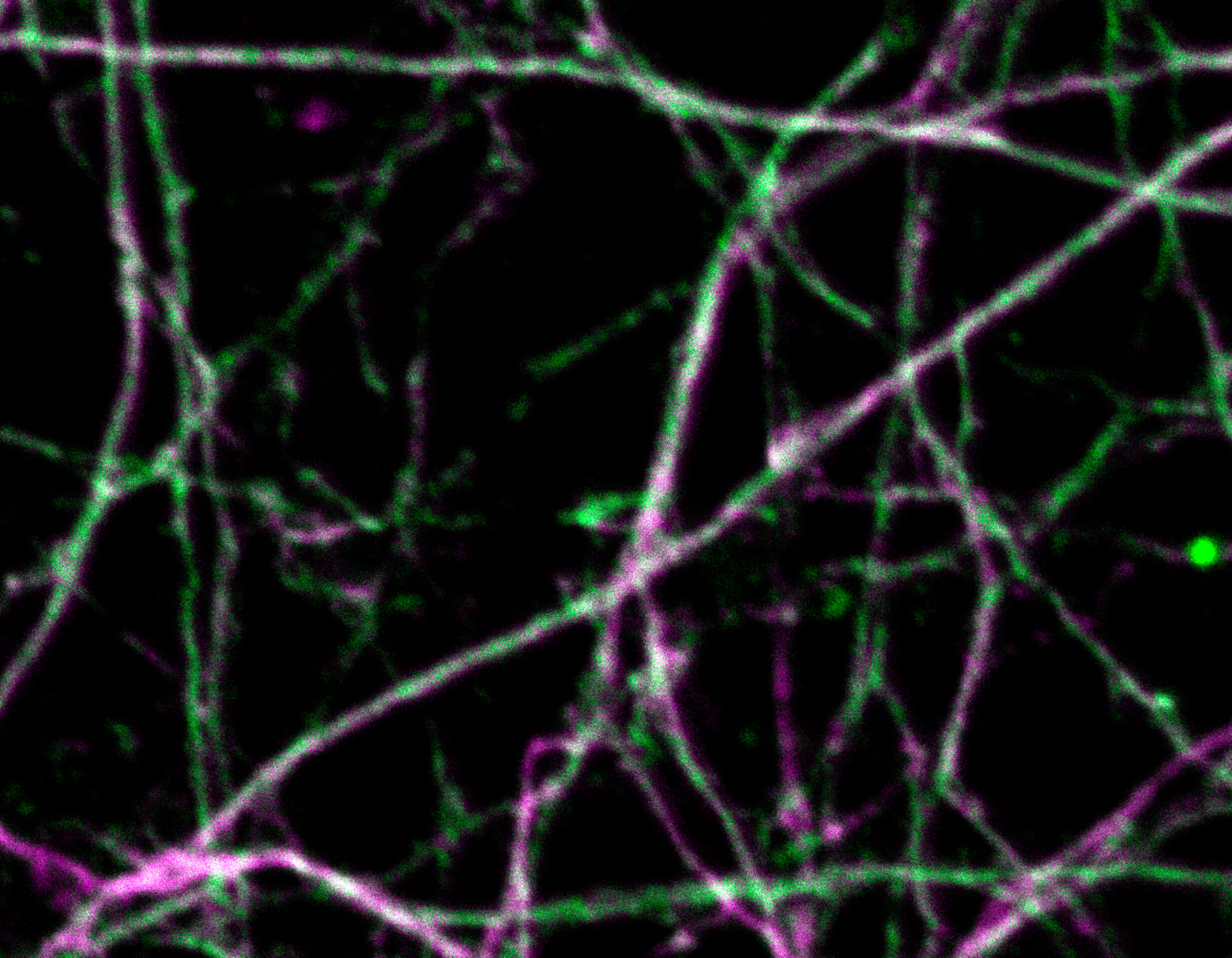
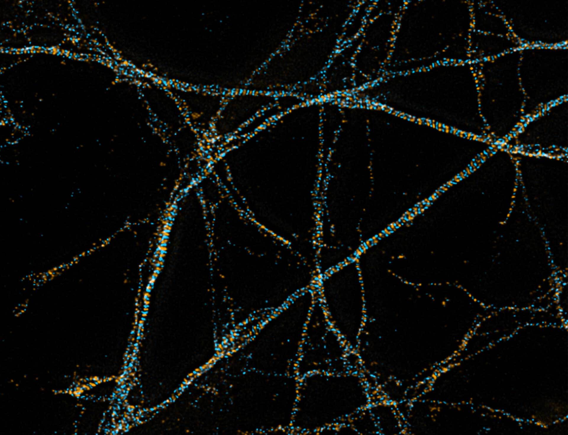
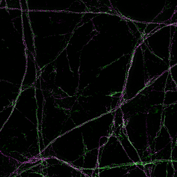
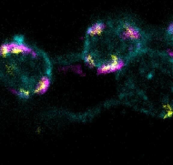
Primary hippocampal neurons with cytoskeleton proteins labeled (magenta, alpha-Adducin, abberior STAR 635P and green, ßII spectrin, Alexa 594). Imaged with abberior Instruments’ STEDYCON and deconvolved with SVI Huygens optimized for STEDYCON.
Description
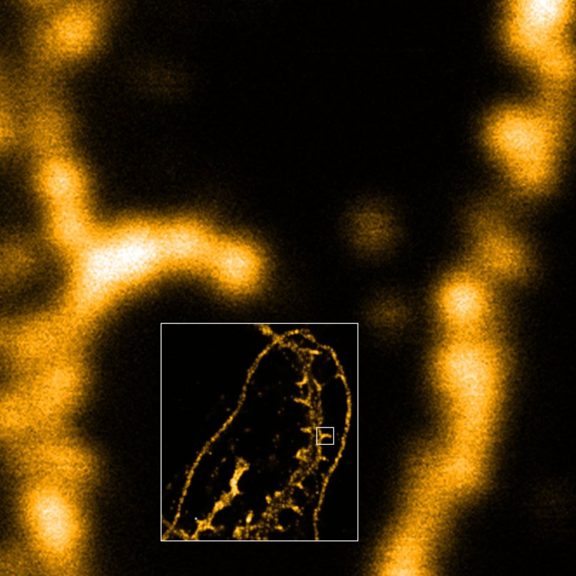
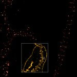
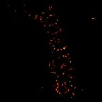
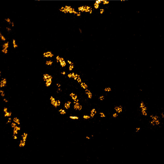
Body wall preparation of a 3rd instar Drosophila melanogaster larva. Synapses at the neuromuscular junction are visualized by immunostaining against Bruchpilot (Brp), an integral component of active zones in Drosophila. Images were acquired on a STEDYCON attached to an Olympus IX81. Images were kindly provided by Dr. Nadine Ehmann, Institute of Physiology, Neurophysiology, University of Würzburg.
Description
Immunolabelling of βIV-spectrin, a scaffolding protein of the axon initial segment, using a self-made primary antibody and Alexa 594 secondary antibody. Cryo-section of mouse neocortex cut at 20 µm. Images were acquired using a STEDYCON attached to a Zeiss AxioImager Z2. The sample was kindly provided by Dr. Maren Engelhardt, Institute of Neuroanatomy, Medical Faculty Mannheim, Heidelberg University.

Description
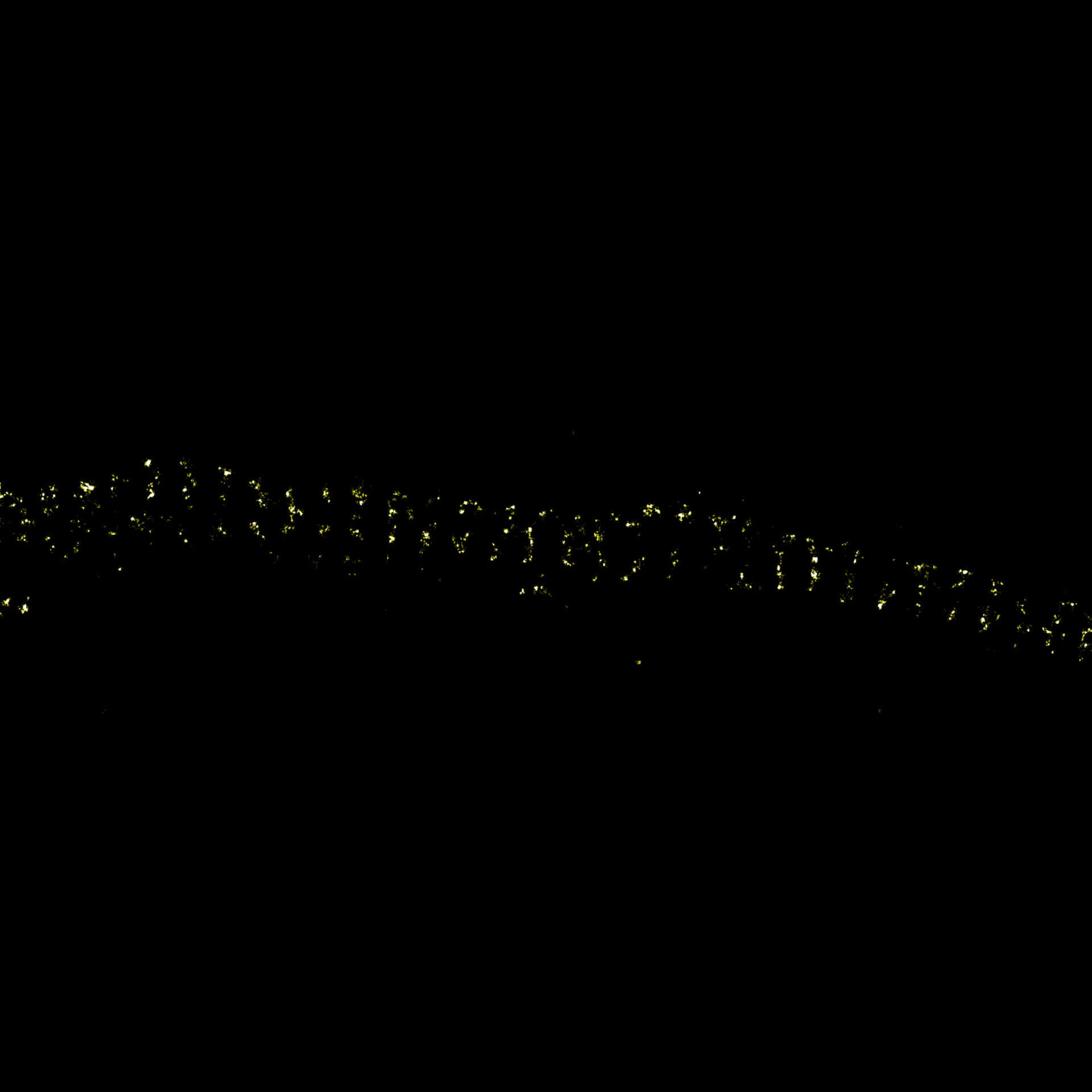
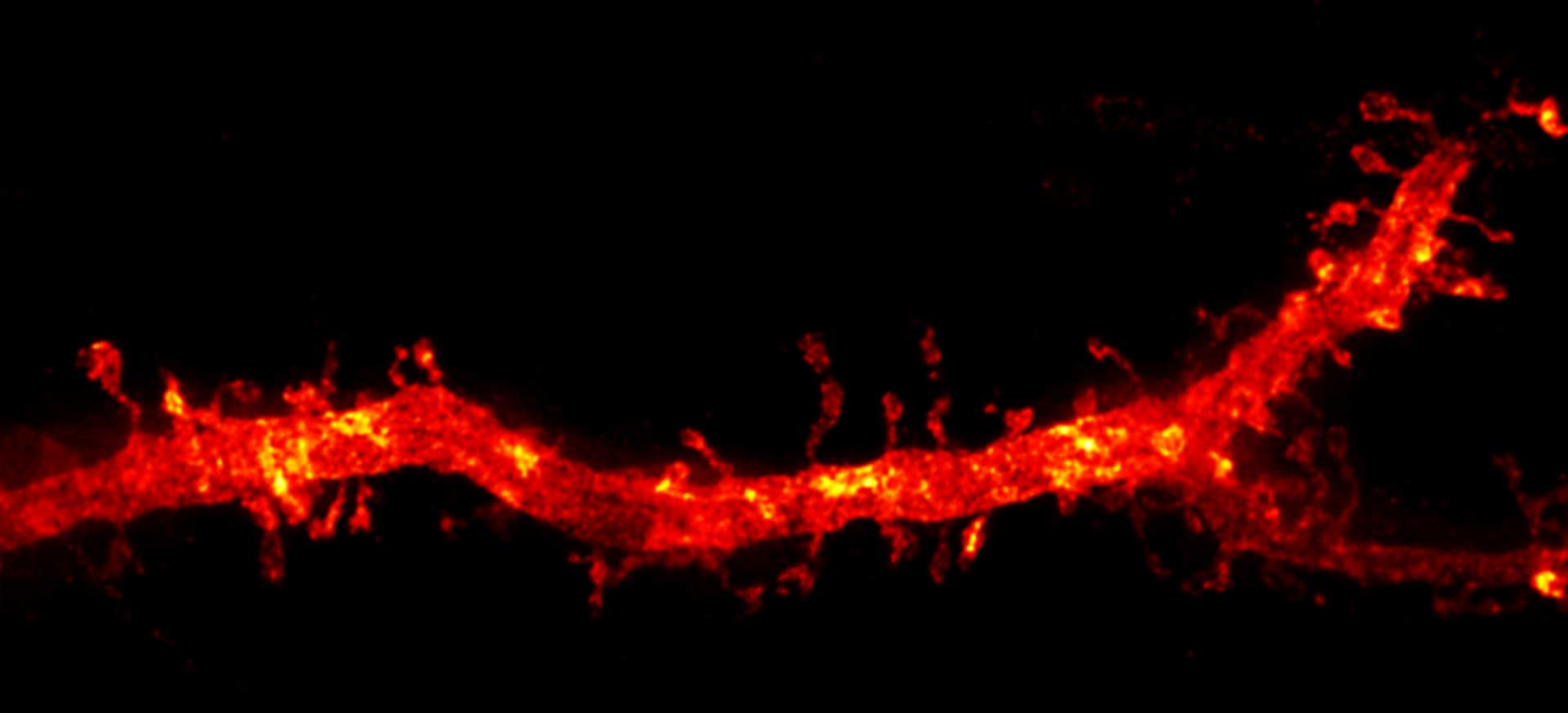
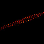
MINFLUX image of axonal bII spectrin in primary hippocampal neurons with < 2 nm resolution. Note the periodic arrangement of spectrin along the axon, and the absence of any details in the confocal counterpart image.
Description
Modules:
Description
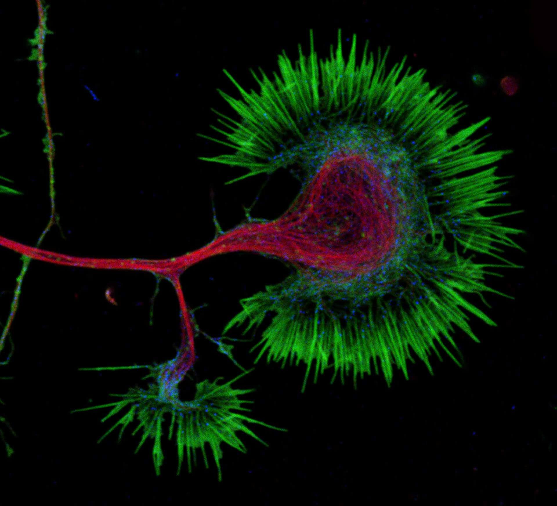
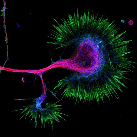
Growth cone at the tip of the axon of a primary hippocampal neuron at 1 day in vitro. Microtubules (Tuj1, abberior STAR 580, red) are bundled in the central-domain suggesting a pausing state. The molecular motor myosin IIB (confocal, Alexa488, blue) is enriched at the transition-zone, along the F-actin arcs. In the peripheral domain actin forms bundles in the filopodia (Phalloidin, abberior STAR 635, green). Sample courtesy: Elisa D’Este, Max Planck Institute for Biophysical Chemistry, Göttingen, Germany.
Description
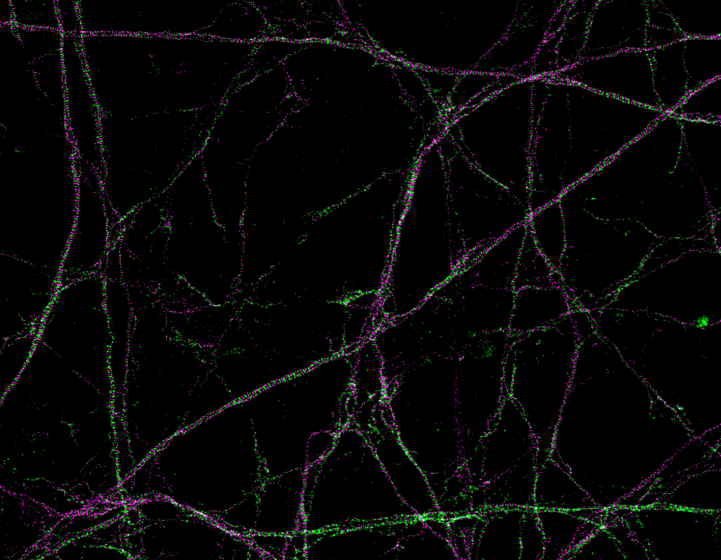
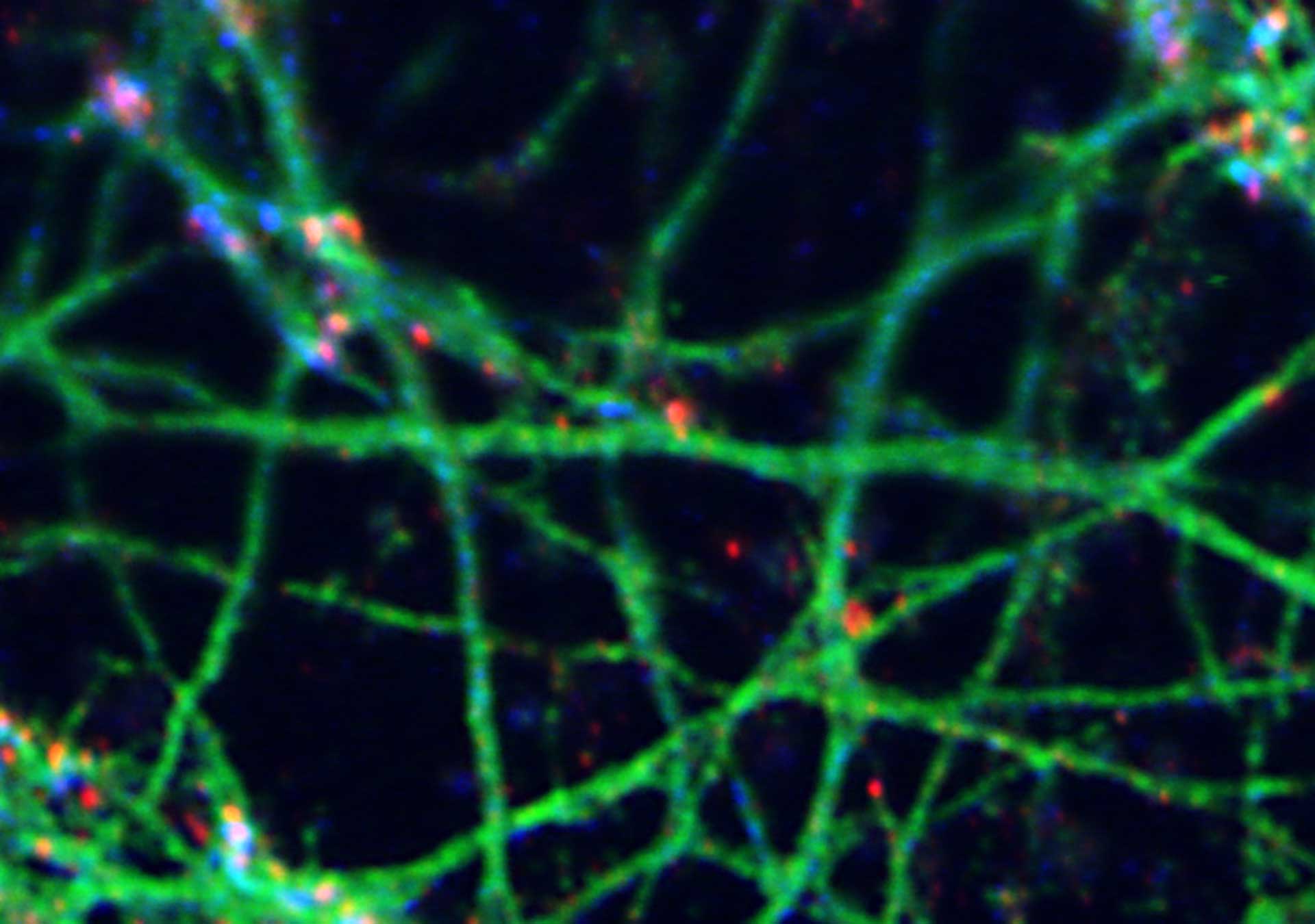
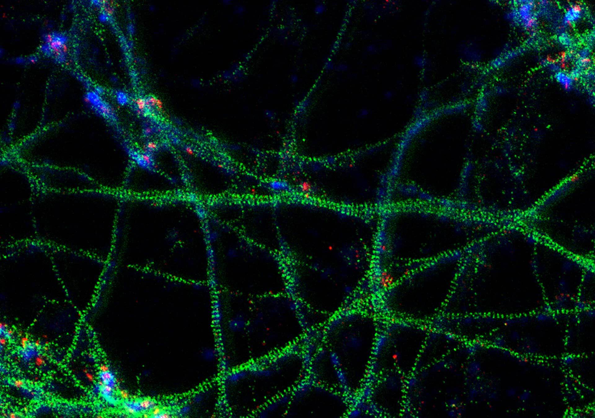
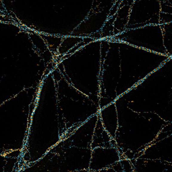
3-color STED image of primary hippocampal neurons. Please note the characteristic ~190 nm beta II spectrin periodicity along distal axons (green) which is only visible in the STED image. Labelled structures: beta II spectrin (green, abberior STAR 635P), Bassoon (red, abberior STAR 580), Actin cytoskeleton (blue, phalloidin, Oregon Green 488). Imaged with abberior Expert Line with 595nm and 775nm STED laser. Sample was prepared by Elisa D’Este @ MPIBPC, Göttingen.
Modules:
Description
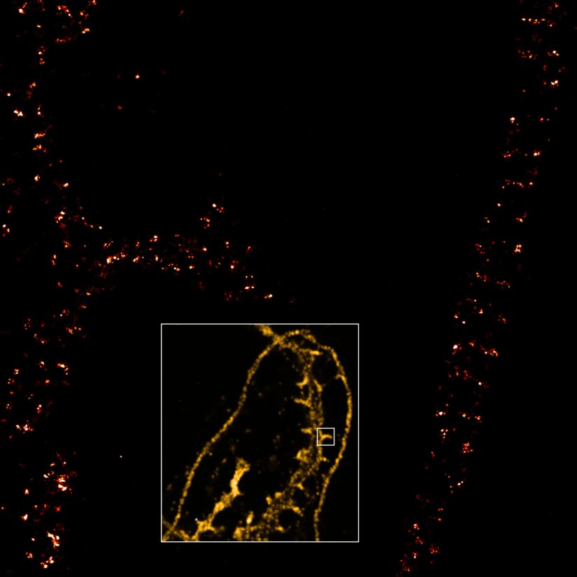
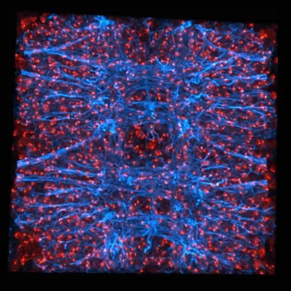
3-color STED imaging: active zones at the Drosophila larval neuromuscular junction immunostained for Bruchpilot and two other proteins.
Two superresolution channels (magenta, yellow) using a 775nm STED laser & one superresolution channel using a 595nm STED laser.
Samples by M. Lenz & M. Landgraf (University of Cambridge, UK).


Imaging beyond the synapse –
the best microscopes for neurobiology
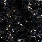
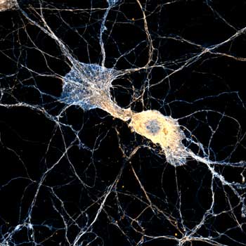
The STEDYCON is a confocal and STED microscope in a shoebox. In spite of its small size, there is nothing modest about its imaging performance. This STEDYCON live cell image of a neuron stained with abberior LIVE 590 for tubulin, LIVE 460L for actin, and STAR RED for membranes, imaged with a single STED laser, is beautiful proof. Our microscopes’ optics, developed for the high demands of superresolution STED, also bring confocal images to a new level. On top of confocal and STED microscopes, we are the only commercial provider of MINFLUX, and we also offer a comprehensive array of dyes for high-end fluorescence microscopy.
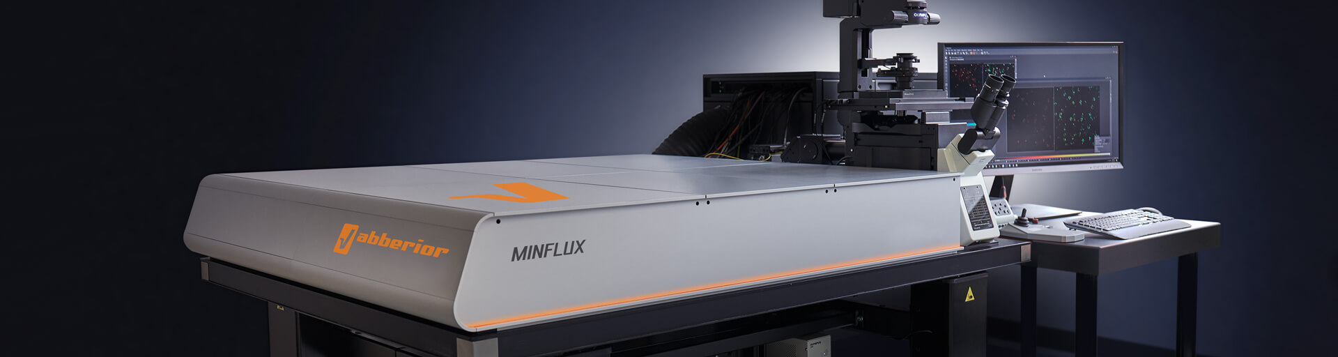
Get a demo >MINFLUX – unrivaled resolution and speed
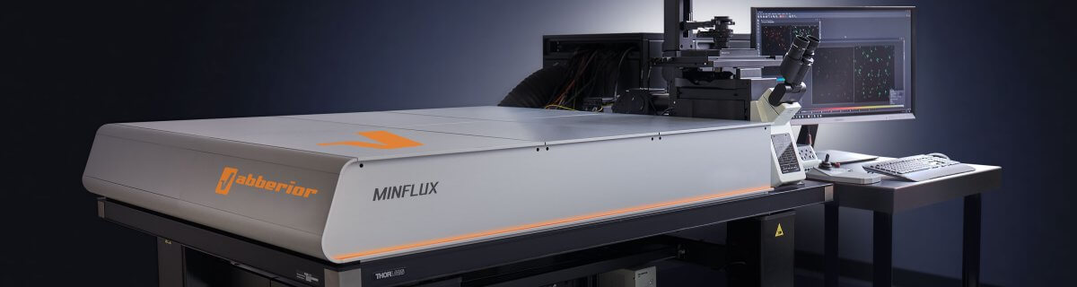
The MINFLUX platform offers an unprecedented array of imaging possibilities and allows you to resolve structures as small as a molecule, along all three dimensions. This unmatched resolution capability combined with unprecedented speed reveals sample details never seen before and helps to dissect fast and dynamic cellular processes in space and time. MINFLUX has been used to e.g. resolve the molecular architecture of the photoreceptor’s presynaptic active zone. It is the world’s most powerful fluorescence microscope. Details >
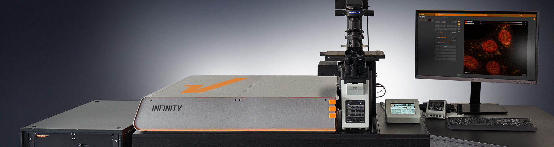
INFINITY – forever cutting edge
The INFINITY platform is the most customizable platform for all things microscopy and may be adapted to your particular demands in neurobiological imaging. INFINITY is always up to the challenge. Just tell us what you need and we will build you a customized, continuously upgradable system specialized for your research. Details >
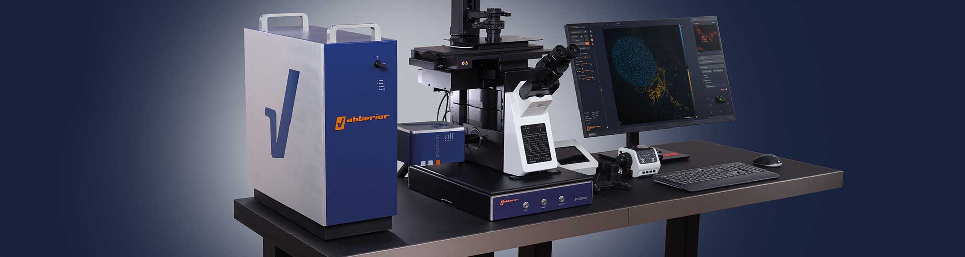
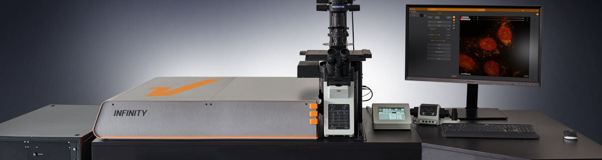
MIRAVA POLYSCOPE – one for all and all for one

The MIRAVA® POLYSCOPE® combines every resolution – from millimeters down to 3 nm – in a singularly unique system. MIRAVA unites four microscopy technologies to cover an unprecedented resolution spectrum, extending over several orders of magnitude from diffraction-limited imaging all the way to true molecular resolution. Our LiGHTBOX software allows beginners to intuitively arrive at a top-notch image within three clicks, while also giving experts full control over the instrument. Details >
STEDYCON 2 – confocal, STED, lifetime… WOW!

The STEDYCON upgrades your existing widefield microscope to a confocal, STED, and lifetime machine with a resolution down to 30 nm. All that’s required is a free camera port and a good objective lens. With its super-intuitive user interface, the STEDYCON provides an intelligent microscope platform that enables everyone to acquire superb superresolution images after only minutes of training. Details >
Cutting-edge tools
to resolve the mind
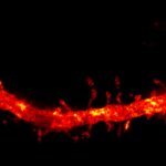
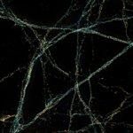
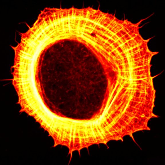
Every one of our modules adds another superpower to your microscope. Pulsed STED, for example, enhances resolution while reducing the light burden on your sample. It contributes to STED images like the one you see here, revealing the characteristic banding pattern of the cytoskeletal proteins α adducin (cyan, abberior STAR 635P) and βII spectrin (yellow, Alexa 594) along axons and dendrites in primary hippocampal neurons.
Ask for detailed information >Choose a superpower for your experiment – our modules
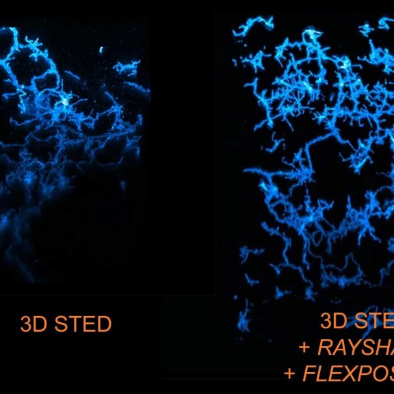
Every MINFLUX, INFINITY, and FACILITY microscope from abberior can be equipped with different modules, powerful functional units that expand the system’s core functionality to reach new imaging heights. You may, for instance, use TIMEBOW lifetime imaging to separate and remove the autofluorescence of lipofuscin from images. Your deep tissue images will be crisp and clear from top to bottom thanks to RAYSHAPE aberration correction with a deformable mirror. FLEXPOSURE adaptive illumination puts light only where it has an effect and may reduce the light burden by orders of magnitude, facilitating extended live cell imaging and volume scans without bleaching or phototoxic effects. Our modules can do all of this and even more! And if you can’t find what you need on our website, get in touch with us, and we’ll develop it for you.
MATRIX Detector
Many eyes see more than one. The MATRIX detector drastically improves signal-to-background ratio, resolution, and dynamic range.
TIMEBOW Imaging
TIMEBOW lifetime imaging for stunning results at confocal and STED super-resolution.
FLEXPOSURE Illumination
Brings down the light dose on your sample and lables dramatically. Key ingredient for volume and live-cell superresolution.
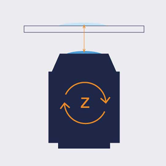
RAYSHAPE Mirror
Dynamic aberration correction with a deformable mirror over about 200 µm z-range. 140 digital actuators adjust the mirror surface within milliseconds.


Custom Solutions
We offer solutions for even the most challenging applications. Everything that can be done, we will do.
Broaden your knowledge
of microscopes, dyes, and superresolution
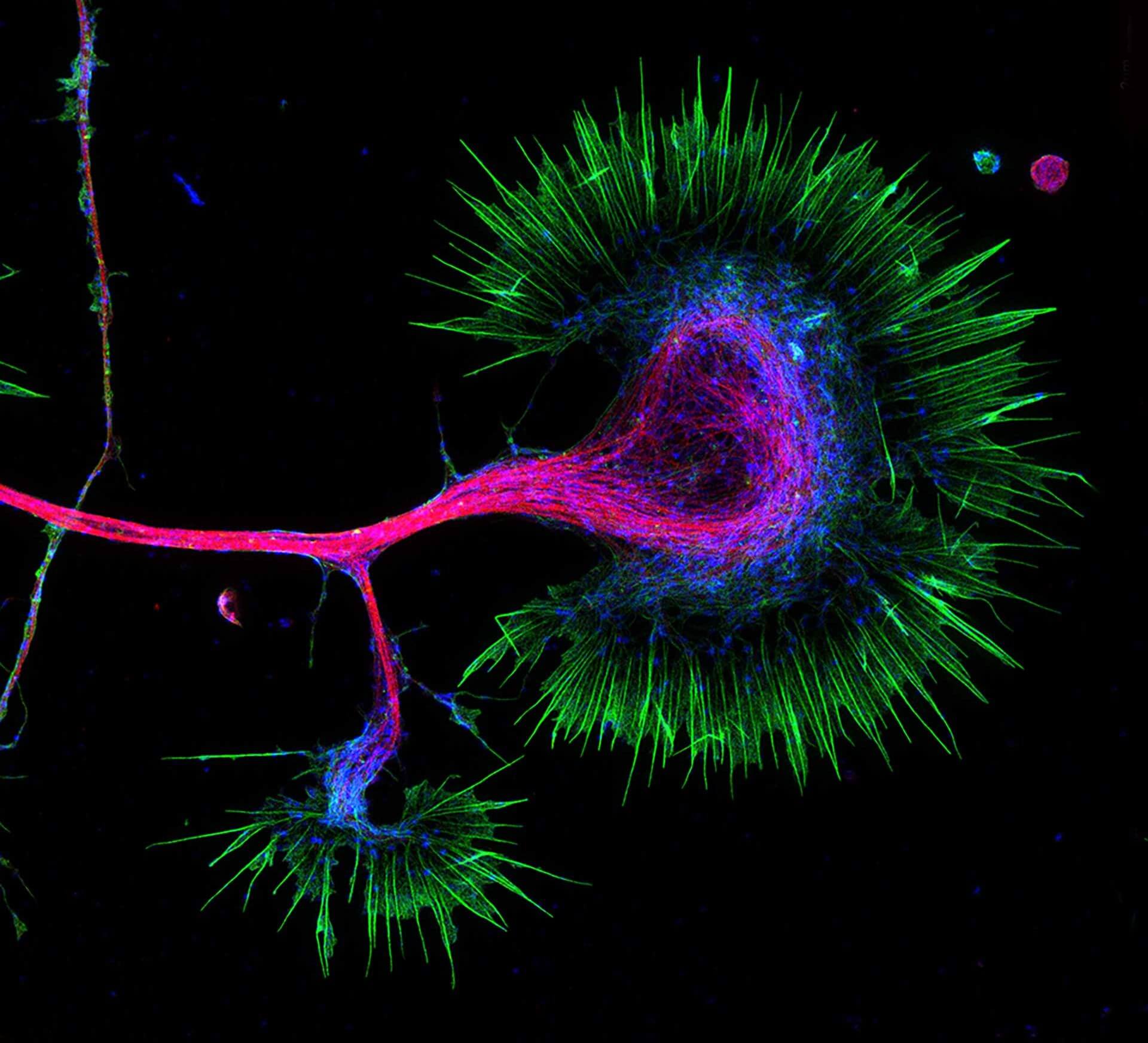
Artwork created by life in the petri dish: axonal growth cone of a primary hippocampal neuron in vitro at day 1, stained for microtubules (magenta, Tuj1, abberior STAR 580), myosin IIB (blue, Alexa488), and actin (green, abberior STAR 635 phalloidin). If you want to make your theoretical expertise on light microscopy grow, check out our knowledge base! It covers topics from the very basics (for example: “What is resolution?”) to high-end challenges (“How to correct for aberrations?”) and the many questions that lie in between.
Tell me more >How to correct for aberrations in light microscopy

Aberrations can give microscopists a hard time. They belong to microscopy like pathogens belong to life. There are ways to bring diverted rays back on track, but some are better than others. The question is: deformable mirror or correction collar? Details >

Every technique that allows to observe cells is more or less invasive and fluorescence microscopy is no exception. Many imaging situations profit from a reduction in light dose as provided by FLEXPOSURE adaptive illumination. Details >
Let the cells shine with immunofluorescence labeling

The most versatile and therefore most common strategy to bring the dye to the sample is immunofluorescence. In case you always wanted to know how immunofluorescence works and which properties of antibodies make it so powerful and at the same time define its limits! Details >
Superresolution for biology: when size, time, and context matter

The spatial resolution achievable with today’s light microscopes has unveiled life at the scale of individual molecules. Size is no longer a barrier to seeing biology at the most fundamental level. But life is not static. It emerges from movement and change. How do superresolution technologies hold up to the challenges of documenting dynamic biological mechanisms? Details >

For all the talk about criteria and definitions, measuring the resolution of a microscope is more nuanced than you’d think. The scales at which microscopes operate today are subject to noise and background that obscure and distort signals. What you use for the measurement can make a big difference. The second article in our “Resolution” series. Details >
STEDYCON: ease-of-use in a shoebox

A sleek, black-and-orange box transforms your widefield microscope into a confocal and a superresolution STED instrument and your exploration of subcellular structures into a seamless, discovery-rich experience. Carefully designed with masterly engineering, STEDYCON breaks the stereotype of the finicky, hard-to-use scope. It opens new possibilities at the press of a button for any user and almost any location. How does it do it? The secret’s in the box. Details >
Additional information and articles
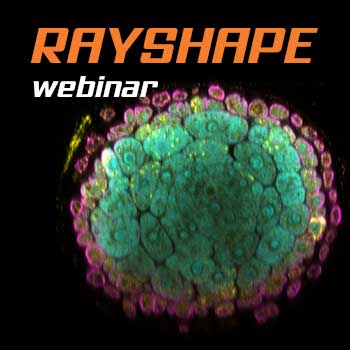
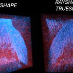
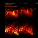
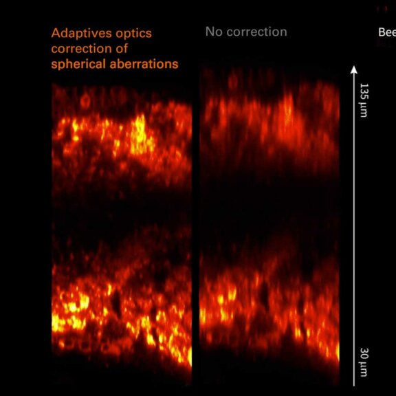
RAYSHAPE webinar – dynamic aberration correction for deep tissue imaging
Hello RAYSHAPE! Bye-bye, aberrations!
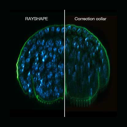
RAYSHAPE special
RAYSHAPE dynamic aberration correction with a deformable mirror preserves resolution and increases brightness. 140 actuators adjust mirror surface within milliseconds for over 200 µm z-range.

TIMEBOW Lifetime Imaging – gives time color
Explore abberior’s TIMEBOW lifetime toolbox with our application experts Julia Menzel and Jan-Gero Schloetel.
FLEXPOSURE special
Fluorescence requires light. Light inevitably bleaches fluorophores. What a dilemma. abberior has the solution: FLEXPOSURE! It brings down the light dose by up to two orders of magnitude, without compromising…
MINFLUX webinar – Tracking with unprecedented spatio-temporal resolution!
Thanks MINFLUX it’s possible to track the movement of individual molecules in the cell, e.g. kinesin. See what EMBL scientists have achieved.

Summer Symposia: Adaptive Optics
All about aberration correction in STED and how the outstanding capabilities of abberior’s Adaptive Optics helps overcome the challenges of complex samples.

Adaptive STED in Neuroscience Research
Prof. Hiroshi Kawabe presents his research with STED for neuroscience applications and Martin Meschkat explains the benefits MATRIX array detection.

Insights into Synaptic Plasticity using STED Microscopy
Dion Dickmann talks about his work on synaptic organization and plasticity. Joachim Fischer presents the theory of STED and the shoebox-sized STEDYCON

Summer Symposia: STED-PAINT
Mike Heilemann highlights transient labeling with exchangeable fluorophores in superresolution imaging. Jan-Gero Schloetel images DNA and cell walls.