


Zooming in on cellular life – Cell Biology


FAQ Video 37

“How does TIMEBOW lifetime imaging work and why is MATRIX array detection a perfect match?”

Cell biology is one of the broadest research areas within molecular biology. Correspondingly diverse are the requirements for light microscopy as one of its pivotal experimental techniques. Cell cycle, cellular architecture and organization, communication and signal transduction, transport processes – every field poses special demands on staining and imaging.
For some purposes in cell biology, the resolution of a confocal microscope is sufficient, but special modules to reduce background signal or to analyze fluorescent lifetime may be required. In other cases, you will need superresolution microscopy like STED to capture relevant details beyond the diffraction limit, or MINFLUX to reach single-digit nanometer resolution and to track the movement of individual molecules in a living cell.
Whatever the need, abberior has the solution! We offer confocal and STED microscopes with superior performance as well as add-on modules tailored to your particular imaging demands. And we are the only manufacturer of MINFLUX, the fluorescent microscope with unsurpassed spatiotemporal resolution. Our product portfolio is completed by a comprehensive selection of fluorescent dyes and labels suitable for various applications.
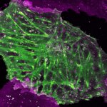
Talk to a scientist >Cel(l)ebrities in focus
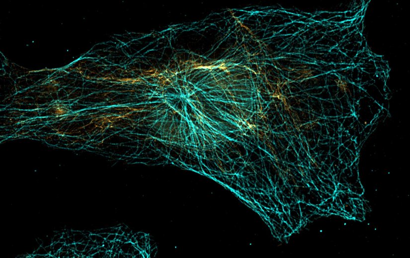
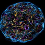
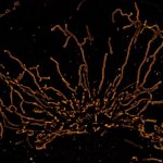
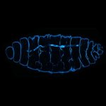
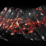
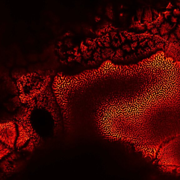
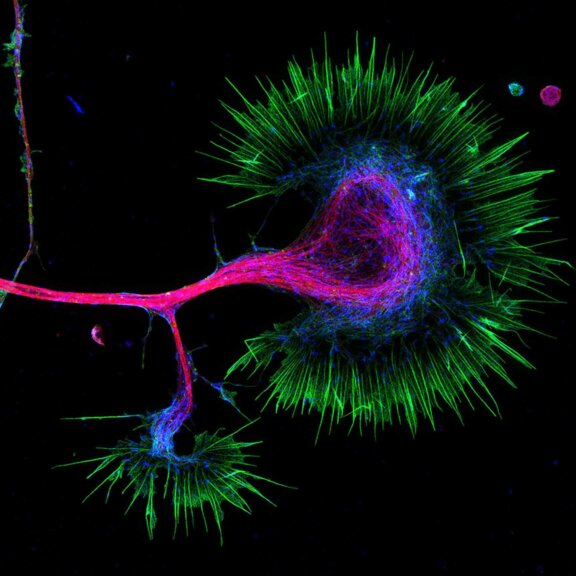
It’s virtually impossible to overestimate the complexity of what’s going on inside a cell. Multicolor staining is a way to visualize at least some of the countless subcellular structures. This four-color STED image shows Vero cells stained for F-actin (red, abberior STAR RED phalloidin), vimentin (cyan, STAR 460L), nuclear pore complexes (green, STAR ORANGE), and golgi (yellow, CF594). STAR ORANGE and CF594 were separated by their fluorescence lifetime with TIMEBOW imaging.
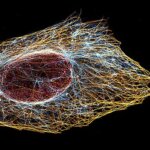
Test your sample >Cellfies for science
Fluorescence microscopy reveals the diversity of cellular life and landscapes. Organelles like mitochondria, peroxisomes, golgi, nucleus, or the ER, and structures such as the cytoskeleton, chromatin, membranes, vesicles – countless components may today be visualized under the microscope. And imaging beyond the diffraction limit with STED and MINFLUX reveals details never seen before. Our gallery takes you on a colorful journey through life in the microscopic world of cells.

Description
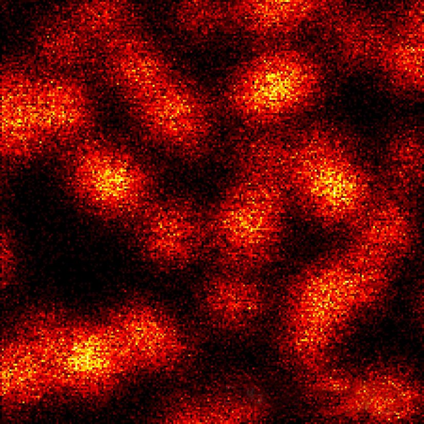
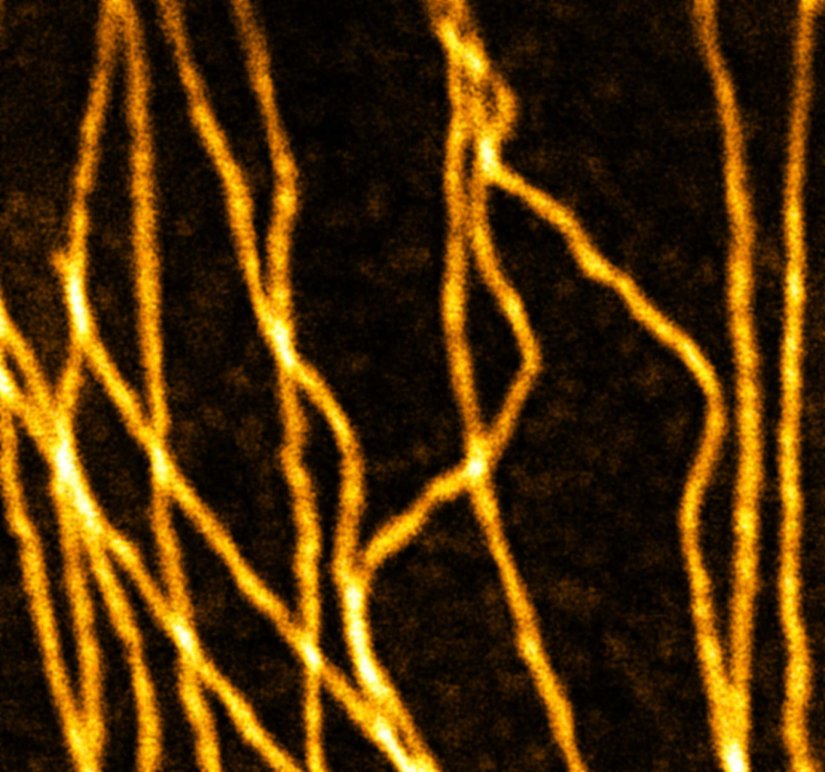
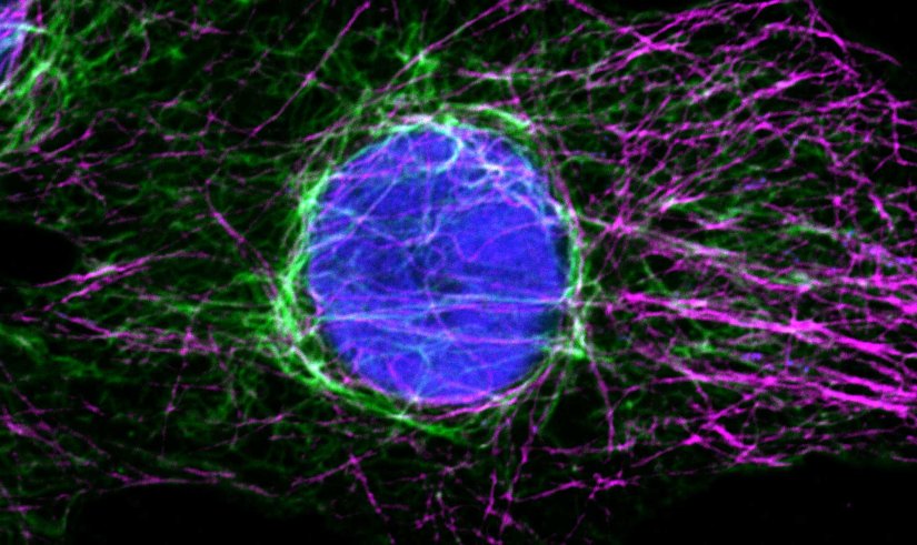
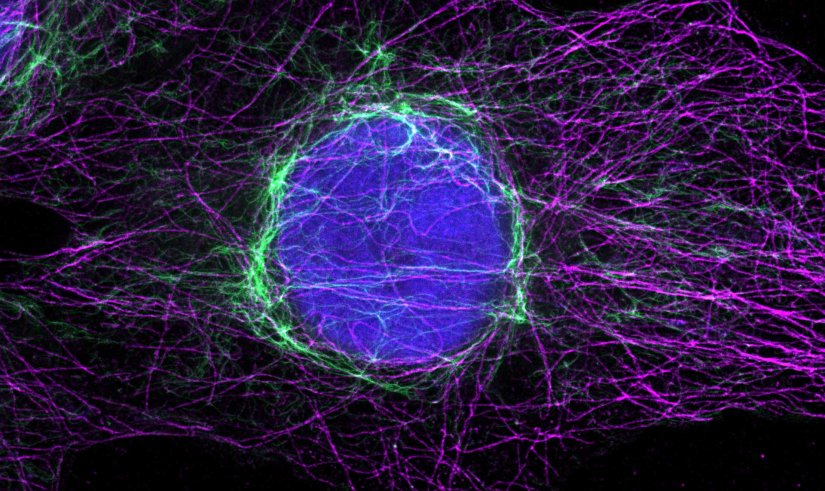
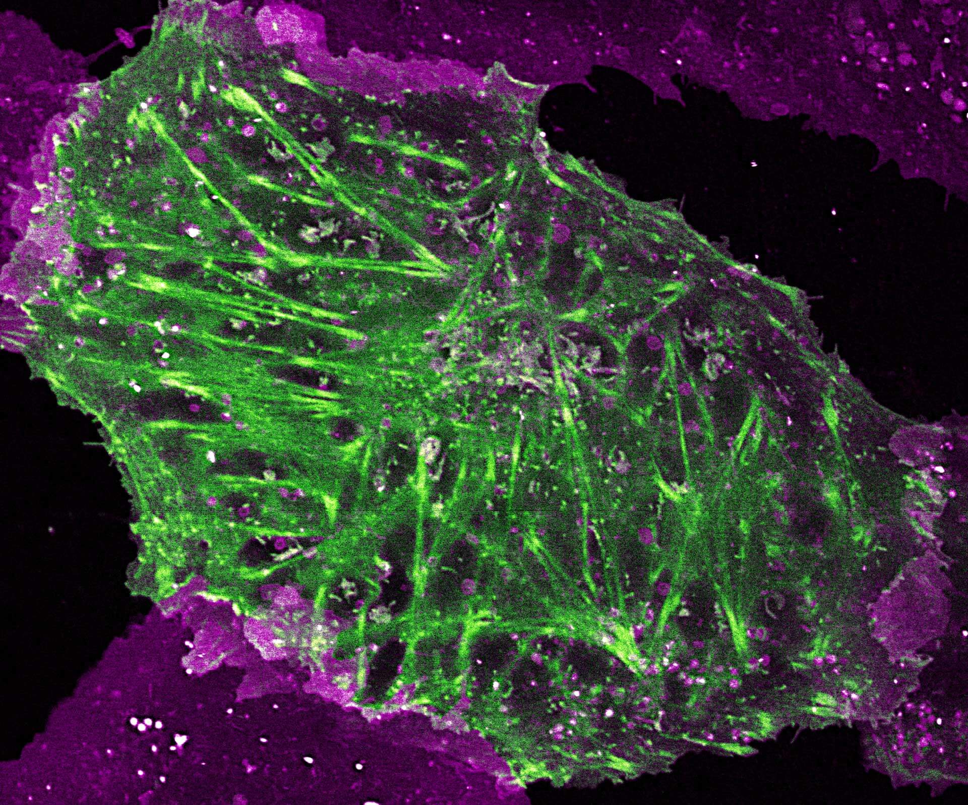
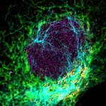
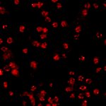
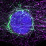
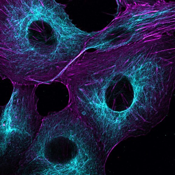
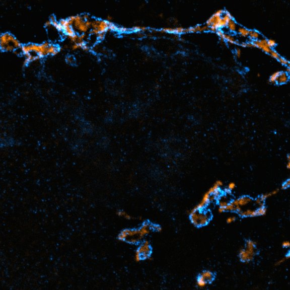
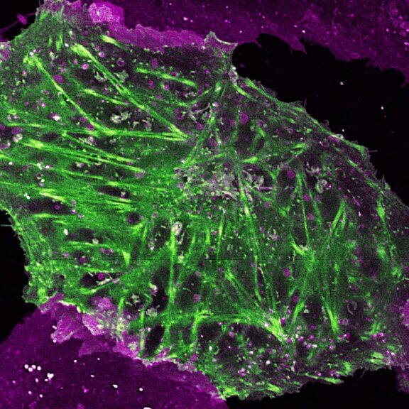
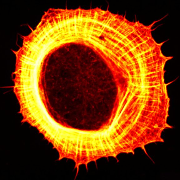
Mammalian cells stained for actin (purple, abberior STAR RED) and vimentin (cyan, abberior LIVE 460L). Differential detection with the MATRIX array detector was used to improve resolution, optical sectioning, and signal-to-background-ratio.
Description
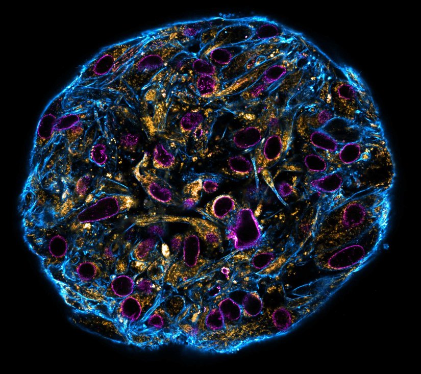
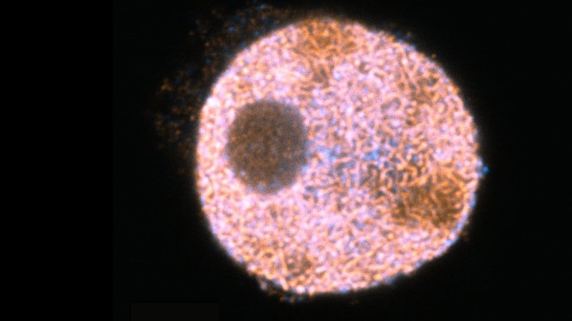
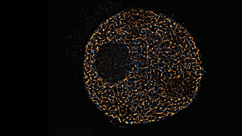
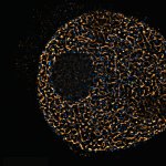
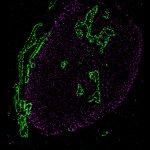
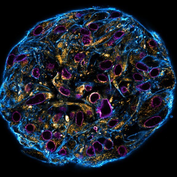
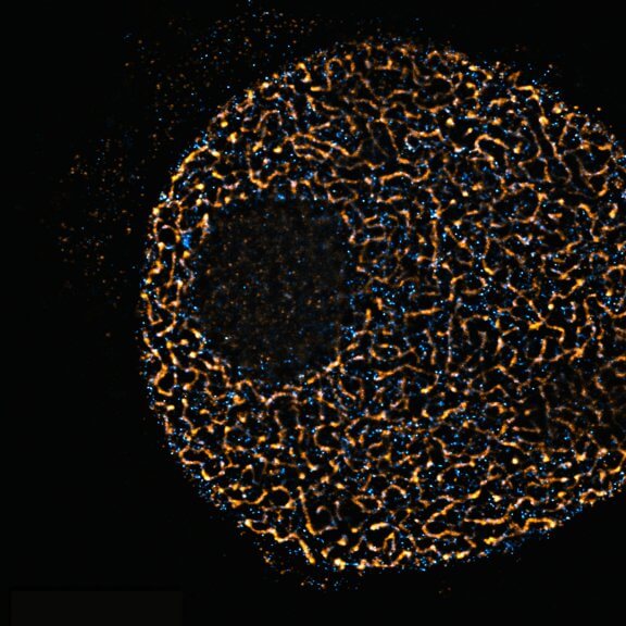
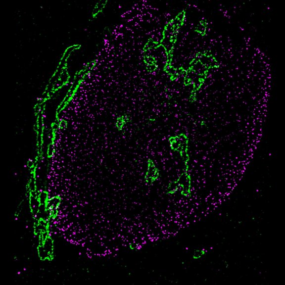
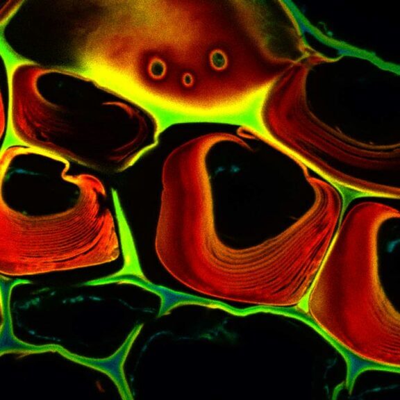
Confocal stitched image of a spheroid stained for nuclear pores (magenta), actin cytoskeleton (blue), and mitochondria (orange).
NIH-313 spheroid, fixed with 4% PFA, kindly provided by ibidi and prepared on µ-Slide VI 0.4 µ-Pattern ibiTreat.

Description
PFA-fixed mammalian cell stained by using Smart Secondaries® from NanoTag Biotechnologies. Stained structures are intermediate filaments (cyan, abberior STAR 460L), mitochondria (green, abberior STAR GREEN), golgi (orange, abberior STAR ORANGE) and nuclear pore complex (magenta, abberior STAR RED).

Description
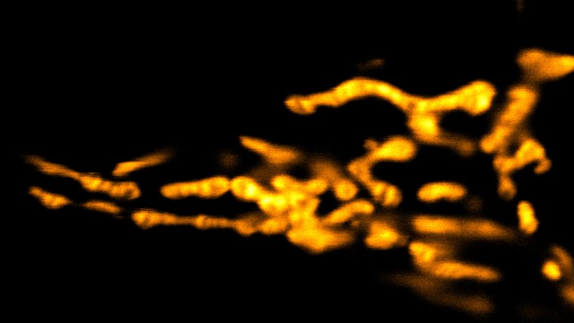
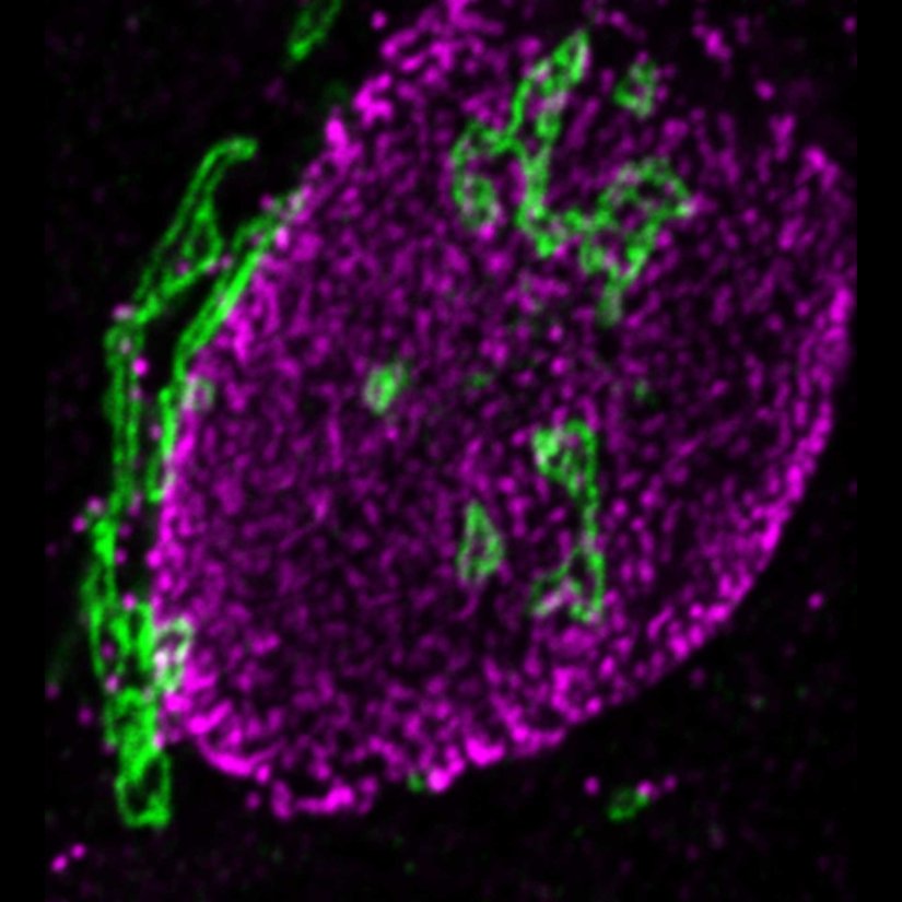
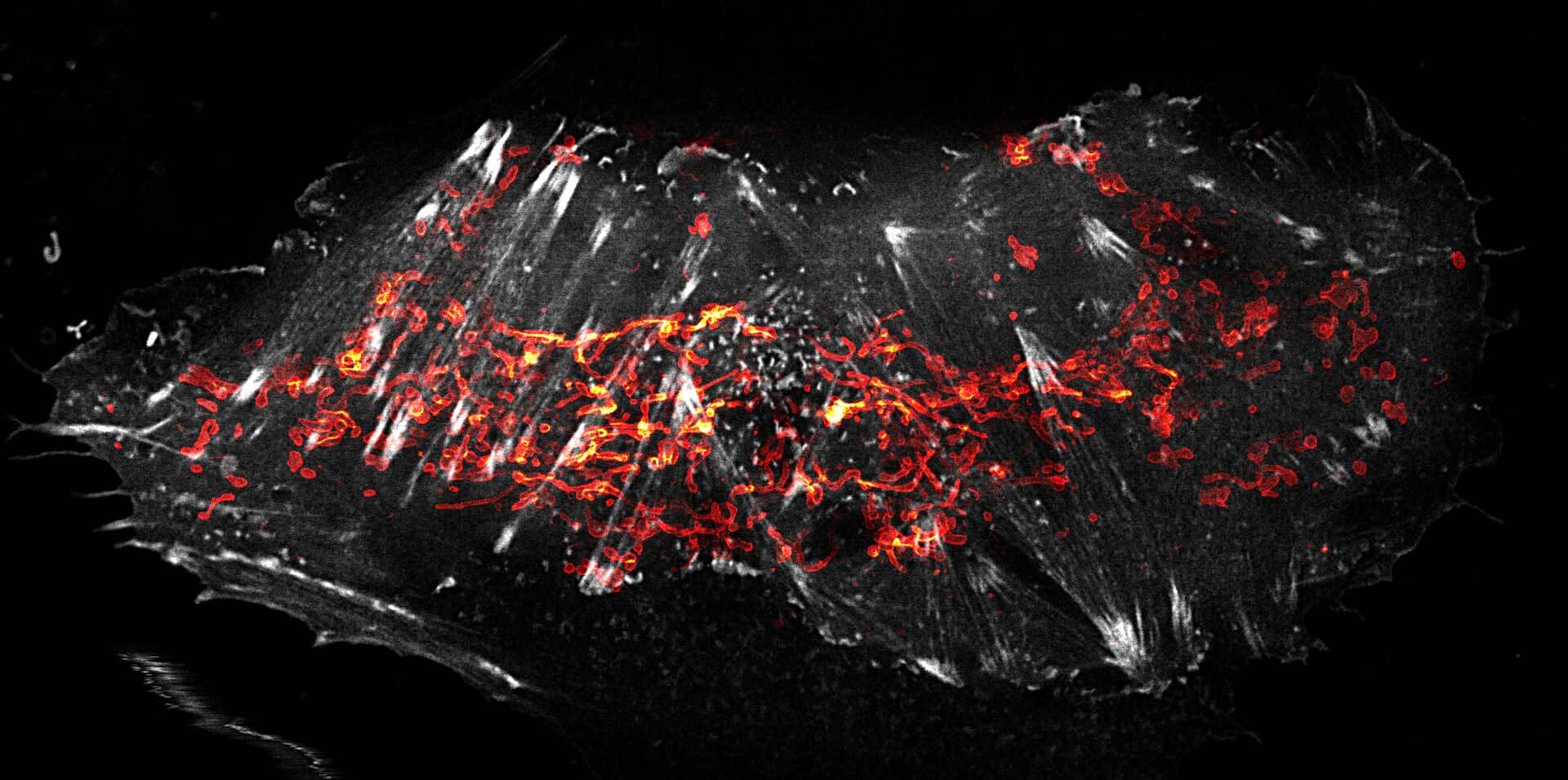
Mitochondria labeled for the outer membrane protein TOMM20, imaged in confocal mode and with the MINFLUX module of the MIRAVA POLYSCOPE.
Modules:
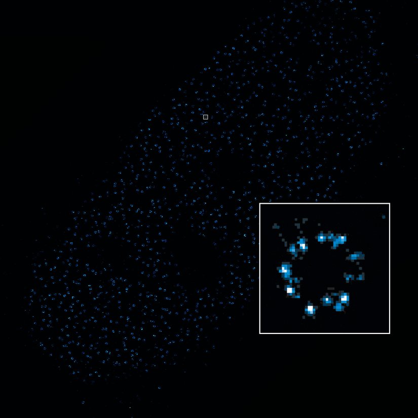
Description
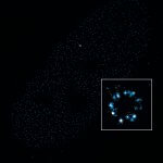
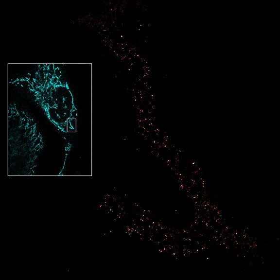
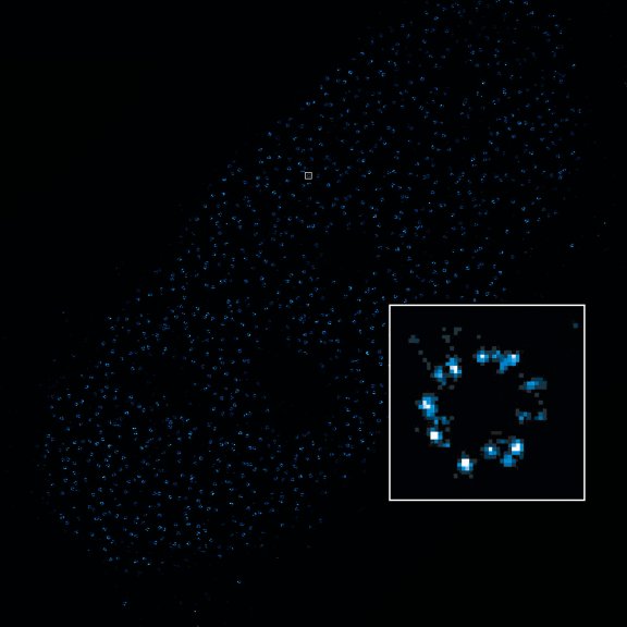
2D MINFLUX image of nuclear pore complex subunits, imaged in fixed mammalian cells expressing GFP-tagged NUP96 stained with abberior DNA-PAINT 660. The molecular resolution of the MINFLUX module of the MIRAVA POLYSCOPE allows visualizing the shape and arrangement of individual subunits of the nuclear pore complex.
Modules:

Description
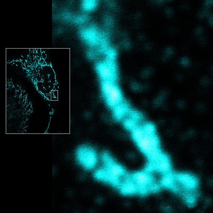
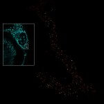
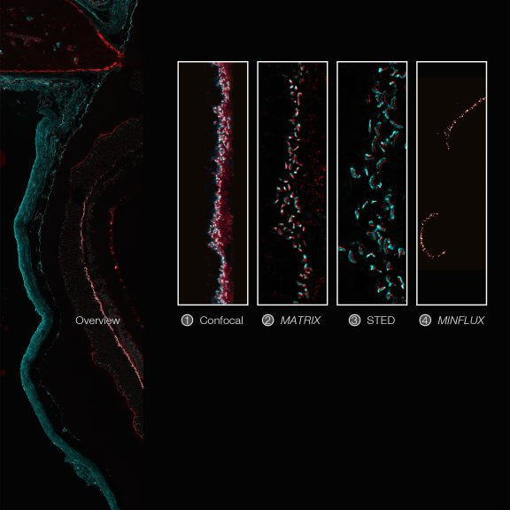
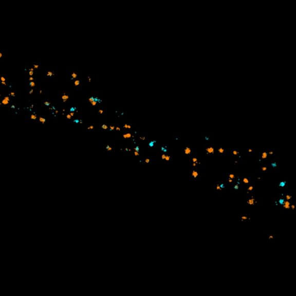
Imaging across scales from diffraction-limited to molecular resolution: ribbon synapses in fixed mouse retina tissue stained for VAMP1 (abberior STAR RED) and CtBP2 (abberior STAR ORANGE) (confocal, MATRIX, STED) or for Bassoon (MINFLUX).
Sample courtesy: Arlene Hirano, PhD, University of California Los Angeles, Los Angeles, CA, USA (confocal, MATRIX, STED), and by Dr. Chad Grabner and Prof. Dr. Tobias Moser, Max Planck Institute for Multidisciplinary Sciences, Göttingen, Germany (MINFLUX).
Modules:
Description
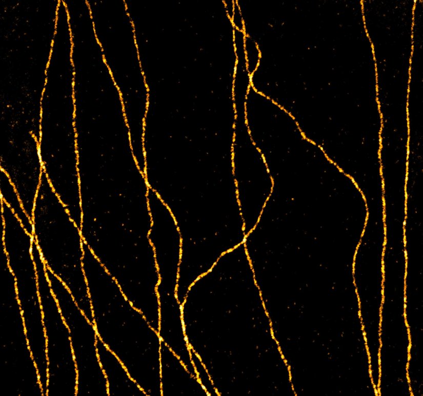
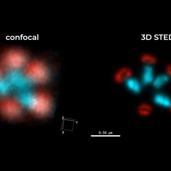
A confocal image compared to a MATRIX + TRUESHARP STED image of nuclear pore complexes in mammalian cells stained for NUP96 with abberior STAR RED.
Modules:
Description
Living HeLa cells stained with the mitochondrial membrane marker abberior LIVE ORANGE mito, visualizing both outer and inner membranes. Confocal and STED images where deconvolved with TRUESHARP image boosting.
Modules:
Description
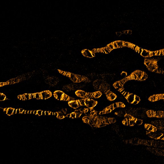
Confocal and STED image of a meiotic cell of Chinese spring wheat stained for two synaptonemal complex components.
Sample courtesy: Sepsi Adél, HUN-REN, Centre for Agricultural Research, Martonvásár, Hungary.
Modules:

Description
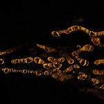
STED image of the golgi apparatus in a fixed mammalian cell, recorded with MATRIX array detection and deconvolved with TRUESHARP image boosting. Stained are the golgi proteins GM130 (cyan, abberior STAR RED) and giantin (orange, abberior STAR ORANGE).
Modules:
Description
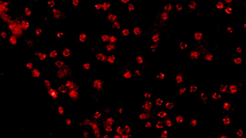
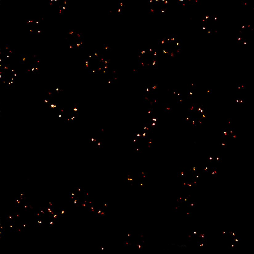
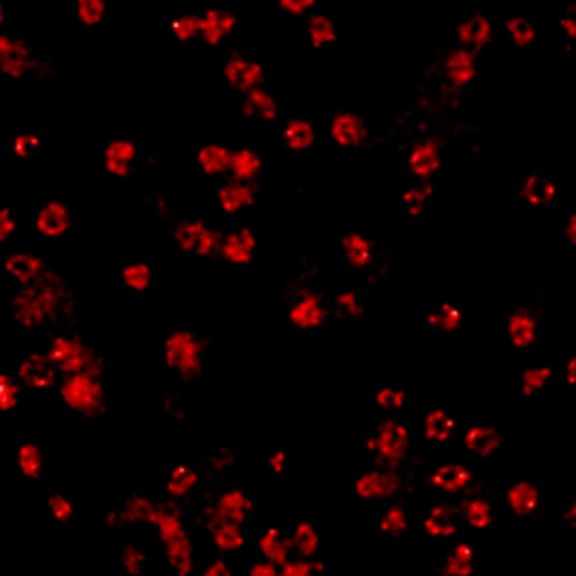
2D MINFLUX nanoscopy of the nuclear pore complex subunits, labeled with abberior FLUX 647 conjugated to JIR AffiniPure-VHH Fragment antibodies (secondary nanobodies). In contrast to confocal microscopy, 2D MINFLUX allows visualization of the shape and arrangement of individual nuclear pore complex subunits. Here, we reach localization precisions of ~ 2 nm in raw localization data.
Description
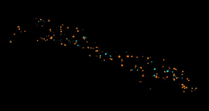
Two-color 3D MINFLUX revealing an inner and outer mitochondrial membrane marker. Cultured mammalian cells labeled with indirect immunofluorescence using JIR AffiniPure-VHH Fragment antibodies (secondary nanobodies) coupled to abberior FLUX 640 (orange) and FLUX 680 (cyan). MINFLUX enables the visualization and separation of both structures.
Description
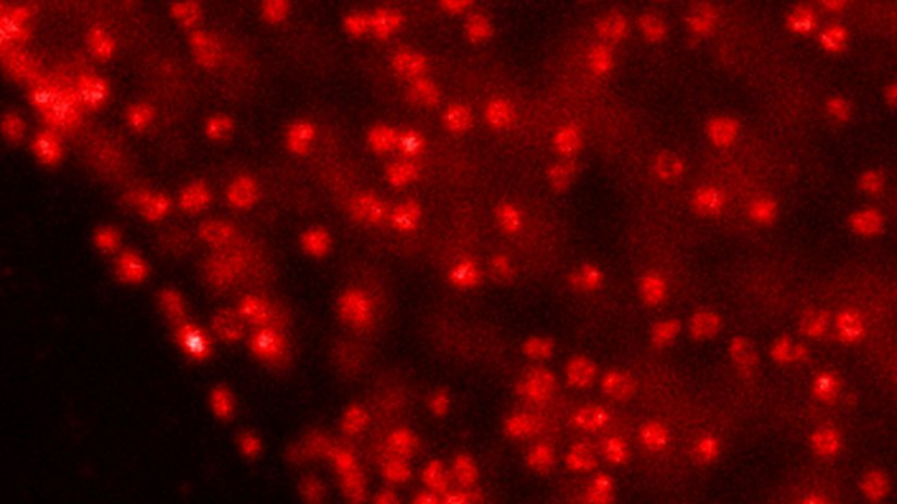
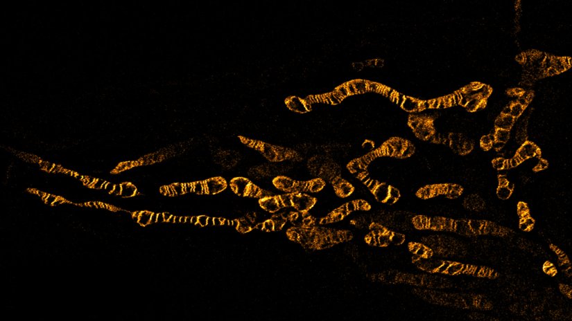
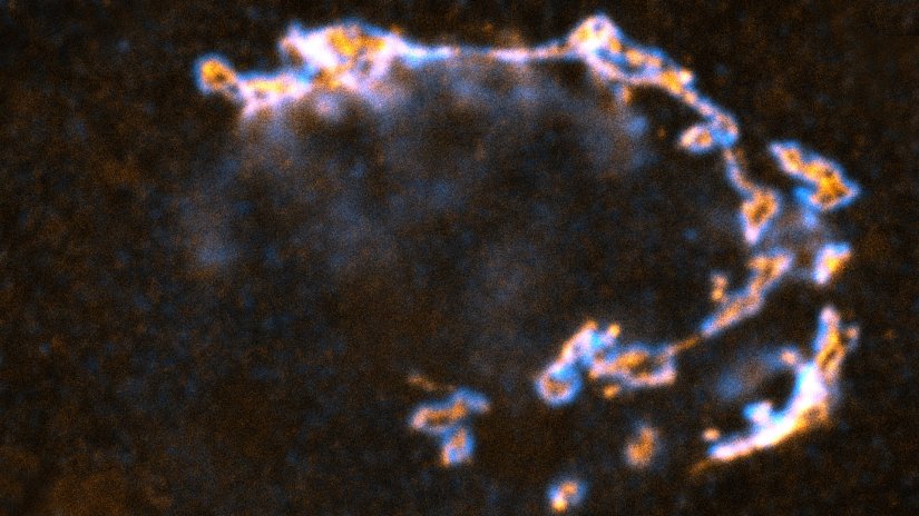
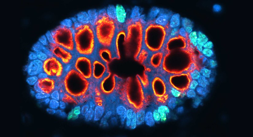
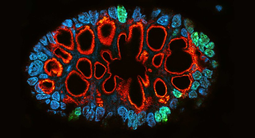
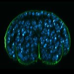
Paraffin section of gut biopsy stained for Ki67 (abberior STAR ORANGE), Muc2 (abberior STAR RED), and DAPI.
Modules:
Description
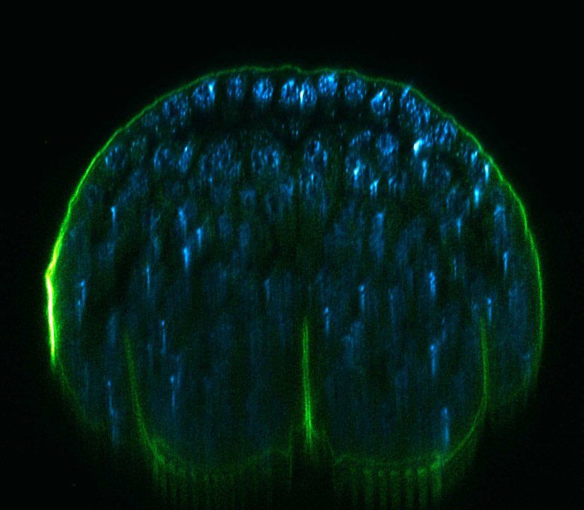
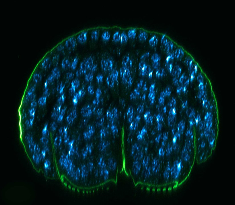
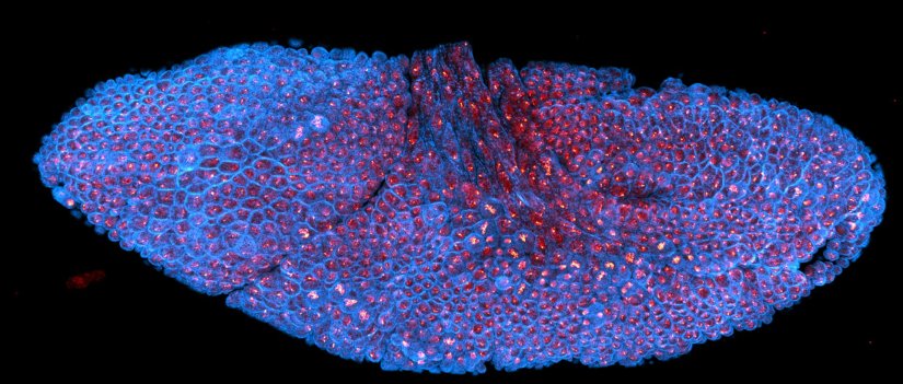
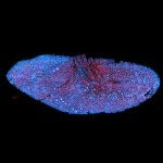
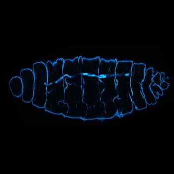
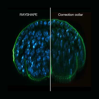
xz section of a stage 17 Drosophila embryo stained for chitin (abberior LIVE 610, green) and DNA (abberior LIVE 550, cyan).
RAYSHAPE preserves resolution and brightness over the whole sample depth of about 200 µm by dynamically redirecting aberrated light to the right places.
In comparison, mechanical optics using a correction collar can only correct a limited z-range of approximately 20 µm
Modules:
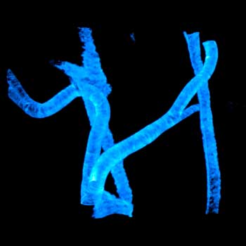
Description
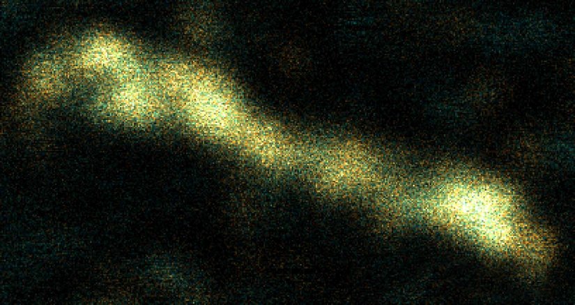
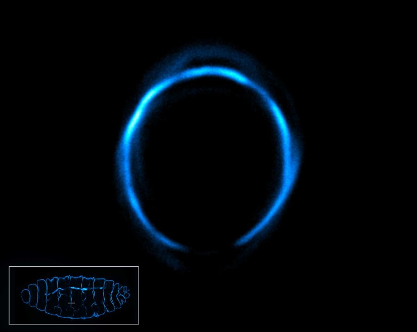
Drosophila stage 12 embryo, imaged with RAYSHAPE, stained for tubulin with abberior STAR RED and for DNA with abberior LIVE 550.
Modules:
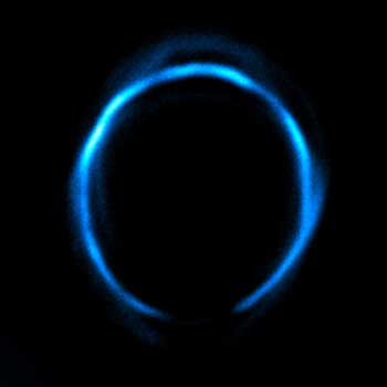
Description
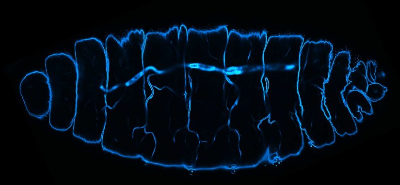
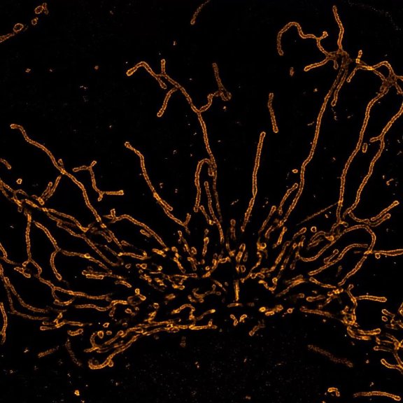
Deep tissue imaging with RAYSHAPE of a stage 17 Drosophila embryo stained for chitin with abberior LIVE 610.
Modules:
Description
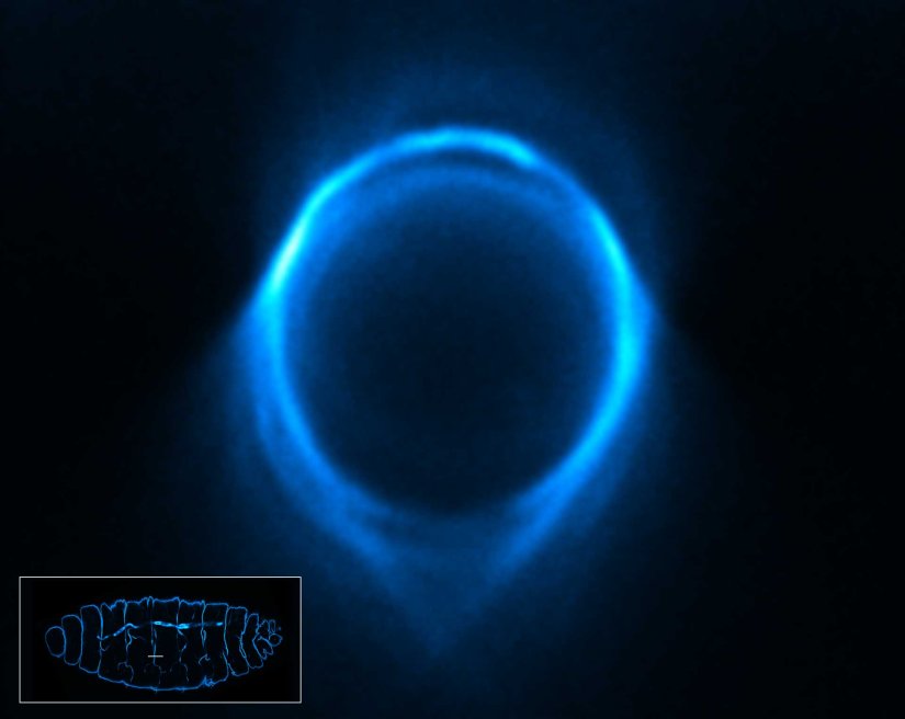
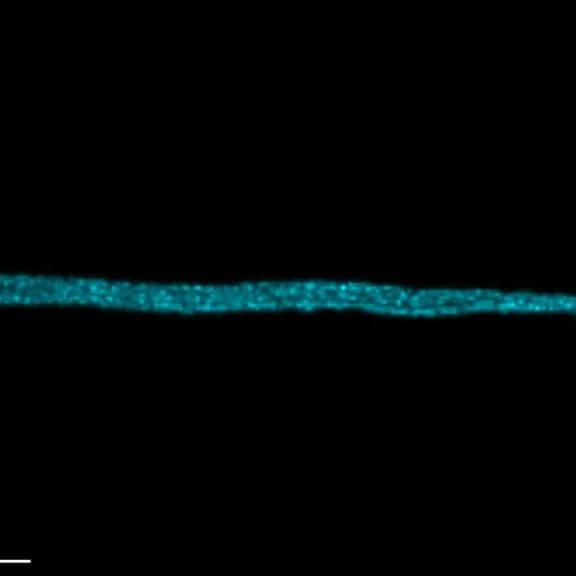
3D STED xz section of a Drosophila embryo trachea, imaged with RAYSHAPE and without abberation correction, depth 15 µm. Chitin was stained with abberior LIVE 610.
Modules:
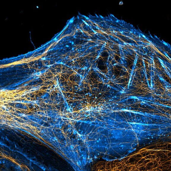
Description
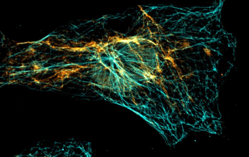
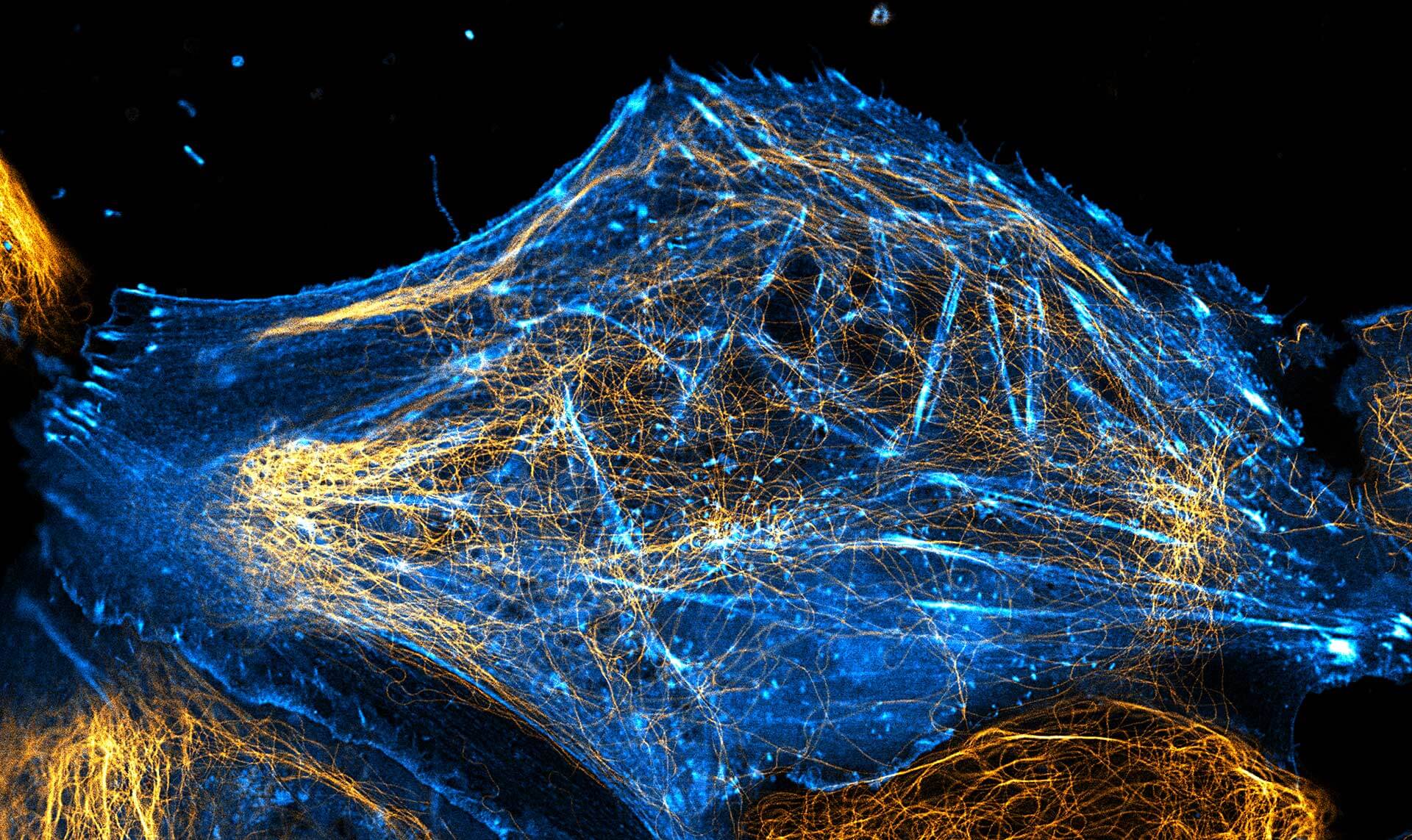
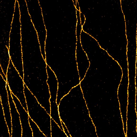
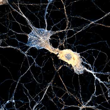
abberior STAR RED was coupled to polyclonal secondary nanobodies (alpaca VHH single domain antibodies) from Jackson ImmunoResearch and used to label tubulin in fixed mammalian cells via indirect immunofluorescence. The STED image was acquired with the STEDYCON system.

Description
Proteins of the nuclear pore complex and the golgi apparatus were stained by indirect immunofluorescence using abberior STAR RED (nuclear pore complex, magenta) and abberior STAR 580 (golgi, green) coupled to polyclonal secondary nanobodies (alpaca VHH single domain antibodies) from Jackson ImmunoResearch. Images of the fixed mammalian cells were acquired with the STEDYCON.
Description
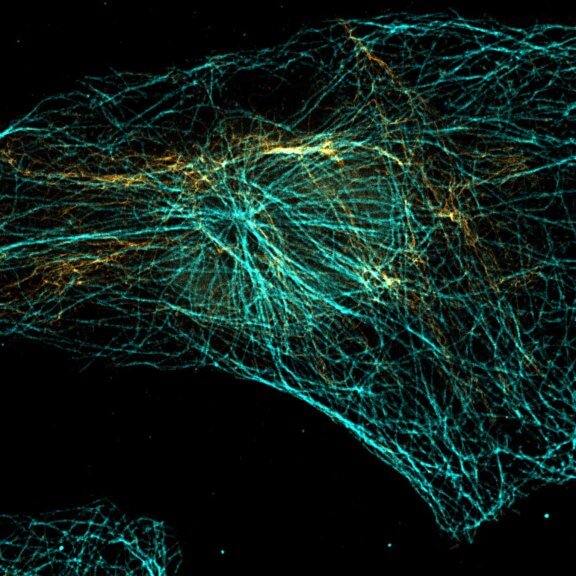
Vimentin and tubulin were stained by indirect immunofluorescence using abberior STAR RED (vimentin, orange) and abberior STAR 580 (tubulin, cyan) coupled to polyclonal secondary nanobodies (alpaca VHH single domain antibodies) from Jackson ImmunoResearch. Images of the fixed mammalian cells were acquired with the STEDYCON.
Description
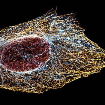
Indirect immunofluorescence staining with abberior STAR dyes coupled to polyclonal anti-mouse secondary nanobodies (alpaca VHH single domain antibodies) from Jackson ImmunoResearch shows excellent results in 3-color STED imaging. Vimentin (abberior STAR RED, green), tubulin (abberior STAR 580, magenta) and dsDNA (abberior STAR 460L, blue) were labeled in fixed mammalian cells and visualized with the STEDYCON.
Description
Description
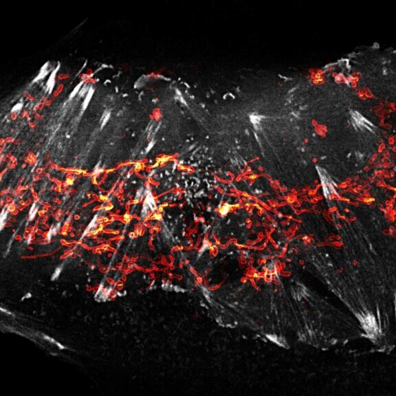
2-color STED image of a living cell expressing actin K118TAG incorporating TCO*A labeled with abberior LIVE 590 click (green). Plasma membrane is highlighted with abberior STAR RED membrane (magenta).
Description
2-color STED image of a living cell expressing actinK118TAG incorporating TCO*A labeled with abberior LIVE 550 click (cyan). Tubulin filaments were stained with our direct probe abberior LIVE 610 tubulin (yellow).


Discover every little detail –
the best microscopes for cell biology
3D MINFLUX visualizes the shape of peroxisomes in all dimensions. The peroxisomal membrane protein PMP70 is labeled with abberior FLUX 647 in fixed mammalian cells, the z scale is color-coded. abberior offers both the microscopes and the dyes to stain and resolve countless cellular structures and processes. And the optics we developed for the high demands of superresolution STED microscopy also bring confocal images to a new level.
Get a demo >MINFLUX – unrivaled resolution and speed
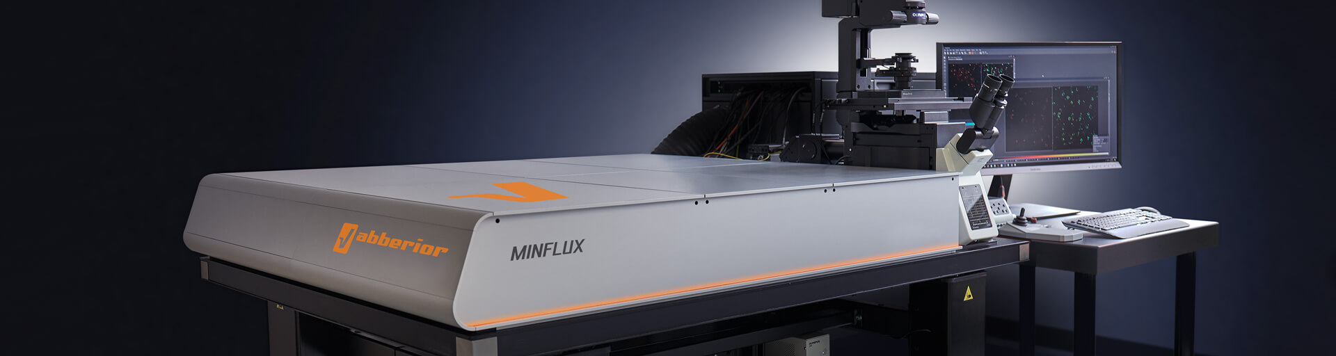
The MINFLUX platform offers an unprecedented array of imaging possibilities and allows you to resolve structures as small as a molecule, along all three dimensions. This unmatched resolution capability combined with unprecedented speed reveals sample details never seen before and helps to dissect fast and dynamic cellular processes in space and time. MINFLUX is the world’s most powerful fluorescence microscope. Details >
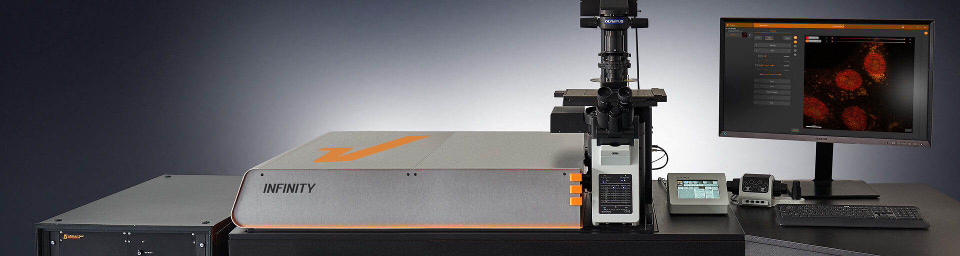
INFINITY – forever cutting edge
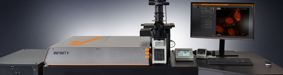
The INFINITY platform is the most customizable platform for all things microscopy and may be adapted to your particular demands depending on what aspect of cell biology you are working on. INFINITY is always up to the challenge. Just tell us what you need and we will build you a customized, continuously upgradable system specialized for your research. Details >
MIRAVA POLYSCOPE – one for all and all for one

MIRAVA® is the first true POLYSCOPE®. Every resolution – from millimeters down to 3 nm – combined in a singularly unique system. MIRAVA unites four microscopy technologies to cover an unprecedented resolution spectrum, extending over several orders of magnitude from diffraction-limited imaging all the way to true molecular resolution. Our LiGHTBOX software allows beginners to intuitively arrive at a top-notch image within three clicks, while also giving experts full control over the instrument. Analyze cellular architecture and function in detail with MIRAVA! Details >
STEDYCON 2 – confocal, STED, and lifetime… WOW!

The STEDYCON upgrades your existing widefield microscope to a confocal, STED, and lifetime machine with a resolution down to 30 nm. All that’s required is a free camera port and a good objective lens. With its super-intuitive user interface, the STEDYCON provides an intelligent microscope platform that enables everyone to acquire superb superresolution images after only minutes of training. Define your cell experiment and obtain premium confocal and STED images at the push of a button! Details >
Enjoy
winning combinations
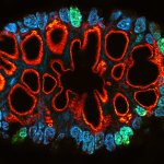
Every one of our modules adds another superpower to your microscope. When combined, they perform even better, like in this four-color staining of a fixed mammalian cell showing nuclear pores (NUP98, purple, abberior STAR RED), the golgi membrane (GM130, yellow, STAR 635), golgi medial rims (giantin, cyan, STAR ORANGE), and actin (gray, STAR 580 phalloidin). A single STED laser at 775 nm was used to achieve superresolution. STAR RED and STAR 635 as well as STAR ORANGE and STAR 580 were separated by their fluorescence lifetime with TIMEBOW, while MATRIX array detection physically removes background signal and increases resolution.
Ask for detailed information >Choose a superpower for your experiment – our modules
Every MINFLUX, INFINITY, and FACILITY microscope can be upgraded with modules to overcome the specific imaging challenges in your research. Do you need to reduce background signal? Do you want to separate dyes according to their fluorescence lifetime to expand your options in multicolor imaging? Or extent your choice of excitation and STED lasers? Our modules cover all these applications, and much more! And if you can’t find what you need on our website, get in touch with us, and we’ll develop it for you.
MATRIX Detector
Many eyes see more than one. The MATRIX detector drastically improves signal-to-background ratio, resolution, and dynamic range.
TIMEBOW Imaging
TIMEBOW lifetime imaging for stunning results at confocal and STED super-resolution.
FLEXPOSURE Illumination
Brings down the light dose on your sample and lables dramatically. Key ingredient for volume and live-cell superresolution.

RAYSHAPE Mirror
Dynamic aberration correction with a deformable mirror over about 200 µm z-range. 140 digital actuators adjust the mirror surface within milliseconds.


Custom Solutions
We offer solutions for even the most challenging applications. Everything that can be done, we will do.
Broaden your knowledge
of microscopes, dyes, and superresolution
What does it take to obtain a superresolution image like this one acquired with the STEDYCON and showing the golgi apparatus stained with abberior STAR 580 (cyan, GM130) and abberior STAR 635P (red, giantin)? What’s the difference between STED and other superresolution techniques like PALM and STORM? How does adaptive illumination protect your sample? Our knowledge base is your chance to close any gaps in fluorescence microscopy expertise you might have!
Tell me more >
PALM and STORM are often used as synonyms, and in fact they have a lot in common. But there are slight differences that can be important for your application. And then there are other superresolution techniques, too – like STED and MINFLUX. Details >

Every technique that allows to observe cells is more or less invasive and fluorescence microscopy is no exception. Many imaging situations profit from a reduction in light dose as provided by FLEXPOSURE adaptive illumination. Details >
Why do superresolution microscopists love alpacas?

It is a very simple yet very important fact: the localization precision of any superresolution microscope can only be as good as the size of the fluorescent staining allows. In other words, when your fluorescent dye is too big or too far away from the protein you want to label, you will never be able to reach a resolution that is higher than this offset. The good news is: there are ways to reduce the offset between target protein and fluorescent label. And one of these are nanobodies. Details >

For all the talk about criteria and definitions, measuring the resolution of a microscope is more nuanced than you’d think. The scales at which microscopes operate today are subject to noise and background that obscure and distort signals. What you use for the measurement can make a big difference. The second article in our “Resolution” series. Details >
STEDYCON: ease-of-use in a shoebox

A sleek, black-and-orange box transforms your widefield microscope into a confocal and a superresolution STED instrument and your exploration of subcellular structures into a seamless, discovery-rich experience. Carefully designed with masterly engineering, STEDYCON breaks the stereotype of the finicky, hard-to-use scope. It opens new possibilities at the press of a button for any user and almost any location. How does it do it? The secret’s in the box. Details >

Are you surprised that the very nature of light caps the resolution that we can achieve in microscope images? Luckily, there are workarounds to this limit. These workarounds push the amount of detail in an image by manipulating precisely where and when fluorophores are allowed to emit. As such, they provide us with a completely new set of tools to shrink the distance between two points while still being able to resolve them. Details >
Additional information and articles

TIMEBOW Lifetime Imaging – gives time color
Explore abberior’s TIMEBOW lifetime toolbox with our application experts Julia Menzel and Jan-Gero Schloetel.
MINFLUX webinar – Tracking with unprecedented spatio-temporal resolution!
Thanks MINFLUX it’s possible to track the movement of individual molecules in the cell, e.g. kinesin. See what EMBL scientists have achieved.

Isn’t it super difficult to operate a MINFLUX microscope?
No! You work with a normal microscope body and sample preparation is easy. Just rely on the active 3D sample stabilization and have fun.
RAYSHAPE special
RAYSHAPE dynamic aberration correction with a deformable mirror preserves resolution and increases brightness. 140 actuators adjust mirror surface within milliseconds for over 200 µm z-range.
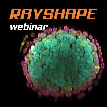
RAYSHAPE webinar – dynamic aberration correction for deep tissue imaging
Hello RAYSHAPE! Bye-bye, aberrations!

Lecture by Nobel laureate Stefan W. Hell on his MINFLUX concept.
MINFLUX allows an everyday 3D resolution of 2 – 3 nm and offers about 100 times faster tracking than with a camera-based system.
abberior FLUX special
Unlock the full potential of single-molecule localization microscopy (SMLM) with our abberior FLUX dyes.

Summer Symposia: MINFLUX superresolution post Nobel
Stefan Hell talks about his post Nobel development MINFLUX, reaching single digit molecular resolution and allows tracking with unprecedented spatio-temporal resolution.