


Measuring the physics of life – Biophysics
FAQ Video 02

“Why do we usually recommend APDs in our microscopes and why aren’t we worried about the supposedly lower dynamic range?”

FAQ Video 37

“How does TIMEBOW lifetime imaging work and why is MATRIX array detection a perfect match?”
FAQ Video 38

MINFLUX allows an everyday 3D resolution of 2 – 3 nm and offers about 100 times faster tracking than with a camera-based system.
NEWS
MINFLUX unravels the structural dynamics of PIEZO1 ion channels
Article and interview with Stefan Hell about kinesin tracking with MINFLUX’ latest technical developments
Article in Physics Today about MINFLUX and tracking kinesin.
English version of the Spektrum interview with Nobel Laureate Stefan W. Hell about STED, MINFLUX and PALM/STORM.
Observe motor proteins stepping along in living cells with MINFLUX
Technology feature in Nature about MINFLUX and tracking kinesin
TIMEBOW, MATRIX, STED and confocal imaging can be freely combined
Watch Recording: TIMEBOW lifetime imaging for stunning results with confocal and STED superresolution!
Biophysics applies approaches and methods traditionally used in physics to biological phenomena. It intersects with other disciplines such as biochemistry, molecular biology, and neuroscience. Biophysical research focuses on phenomena like electric current, for example in muscle tissue or neurons, temperature, molecular diffusion, mechanical stress, or structural dynamics to understand life’s underlying physical principles.
Another field of research is the development of biomaterials. Thanks to the advances in resolution, light microscopy plays an increasingly important role in biophysics. MINFLUX is nowadays applied for the study of structural dynamics in proteins and has been used to track the walk of kinesin-1 along axons and to dissect the movement of the PIEZO1 membrane channel in response to mechanical stimuli, to name just two examples.
Talk to a scientist >Following the forces at work
Flexible intruments for interdisciplinary resaearch
The image shows the tracks of a lipid-coupled Atto 647N molecule embedded in a supported lipid bilayer recorded with MINFLUX. The movement of the labeled lipid was tracked with a frequency of up to 10 kHz. More than most other disciplines, biophysics relies on highly precise instruments and sensitive detectors to be able to record subtle changes of the parameters under study. Microscopes need to be suited for live cell imaging and fast enough to capture dynamic processes within cells and tissues. abberior offers microscopes of superior quality in all components and their interplay. Supreme optics meet pulsed lasers and highly sensitive detectors.
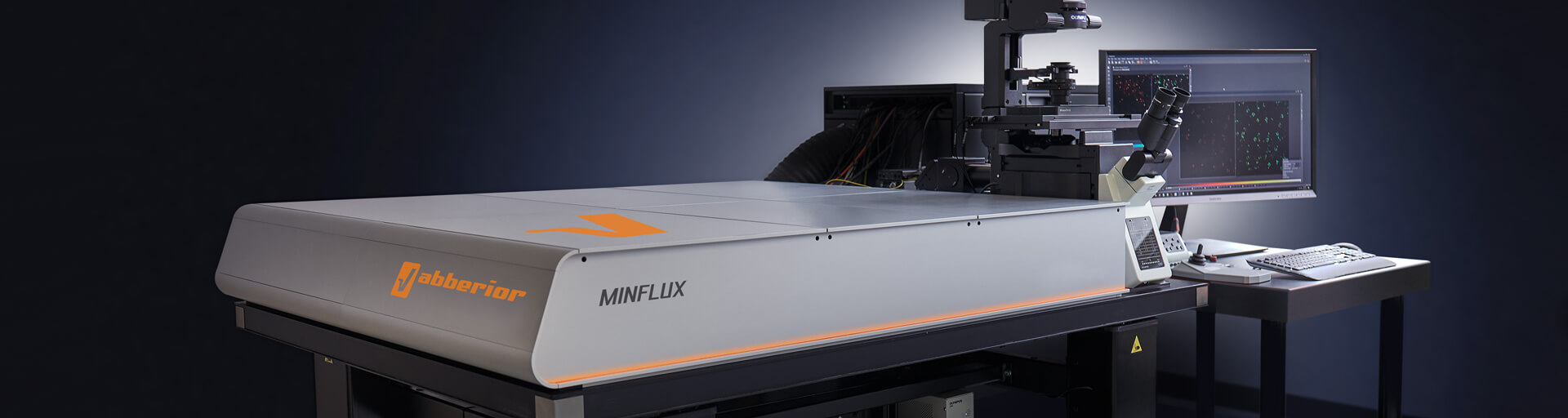
Get a demo >MINFLUX – unrivaled resolution and speed
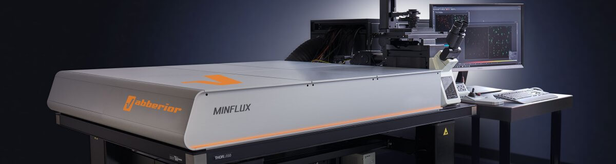
The MINFLUX platform offers an unprecedented array of imaging possibilities and allows you to resolve viruses and even single molecules like capsid proteins along all three dimensions.
This unmatched resolution capability combined with unprecedented speed reveals sample details never seen before and helps to dissect fast and dynamic cellular processes in space and time. Details >
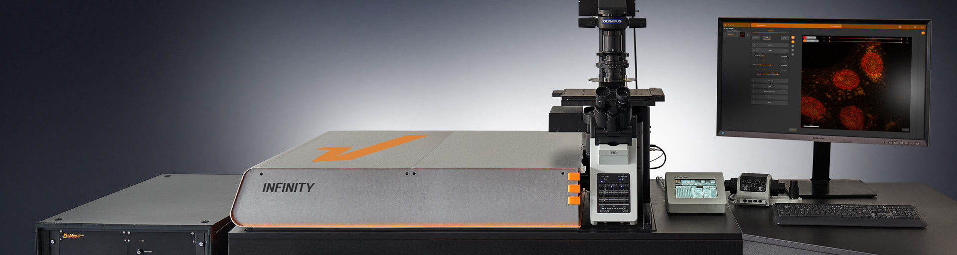
INFINITY – forever cutting edge
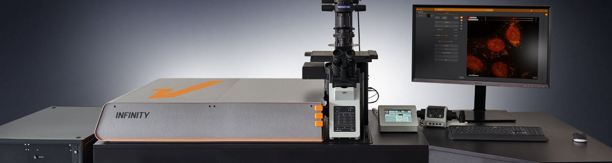
The INFINITY platform is the most customizable platform for all things microscopy and may be adapted to your particular demands in virological imaging. Optimize your microscope for the imaging of anything from subcellular structures to complex tissue. Even the most sophisticated biophysical experiment will become possible with INFINITY. Just tell us what you need and we will build you a customized, continuously upgradable system specialized for your research. Details >
MIRAVA POLYSCOPE– one for all and all for one

The MIRAVA® POLYSCOPE® combines every resolution – from millimeters down to 3 nm – in a singularly unique system. MIRAVA unites four microscopy technologies to cover an unprecedented resolution spectrum, extending over several orders of magnitude from diffraction-limited imaging all the way to true molecular resolution. Our LiGHTBOX software allows beginners to intuitively arrive at a top-notch image within three clicks, while also giving experts full control over the instrument. Details >
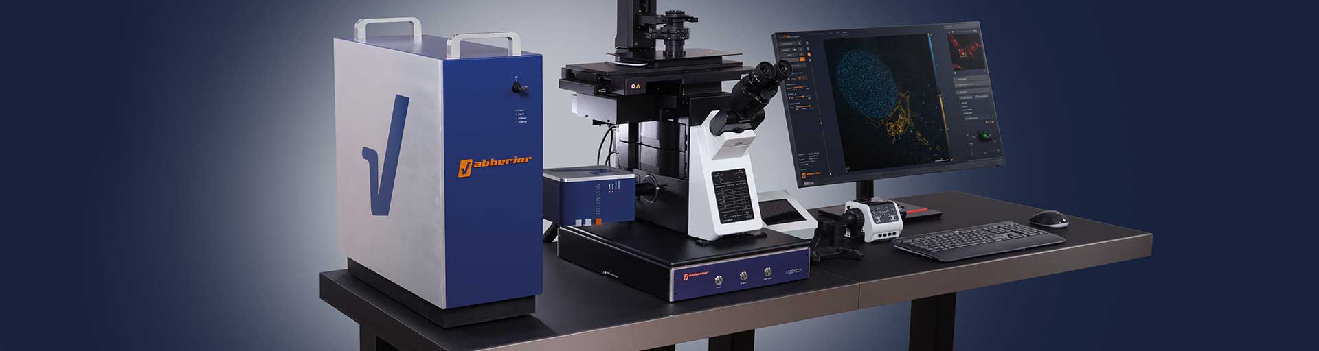
STEDYCON 2 – confocal, STED, lifetime… WOW!

The STEDYCON upgrades your existing widefield microscope to a confocal, STED, and lifetime machine with a resolution down to 30 nm. All that’s required is a free camera port and a good objective lens. With its super-intuitive user interface, the STEDYCON provides an intelligent microscope platform that enables everyone to acquire superb superresolution images after only minutes of training. Details >
Cutting-edge tools
to change the game
MINFLUX tracking of kinesin in living cells. Shown is the sum of all localized events (gray dots) and the trajectory of a kinesin molecule (orange line plot). Truncated kinesin-1 (HaloTag-K560) was labeled with JF646- HaloTag ligand in living U2OS cells treated with Taxol.
Ask for detailed information >Choose a superpower for your experiment – our modules
Configuring your microscope according to your specific needs is essential. Every MINFLUX, INFINITY, and FACILITY microscope can be upgraded with modules to overcome the specific imaging challenges in your research. You may, for instance, use TIMEBOW Imaging to monitor changes in a fluorophore’s nano-environment. FLEXPOSURE adaptive illumination may reduce the light burden on your sample by orders of magnitude, facilitating long live cell imaging sessions with little photobleaching or phototoxic effects. Autofocus and Autoalignment for all beams including STED guarantee best-possible imaging conditions at all times. And if you can’t find what you need on our website, get in touch with us, and we’ll develop it for you.
MATRIX Detector
Many eyes see more than one. The MATRIX detector drastically improves signal-to-background ratio, resolution, and dynamic range.
TIMEBOW Imaging
TIMEBOW lifetime imaging for stunning results at confocal and STED super-resolution.

FLEXPOSURE Illumination
Brings down the light dose on your sample and lables dramatically. Key ingredient for volume and live-cell superresolution.
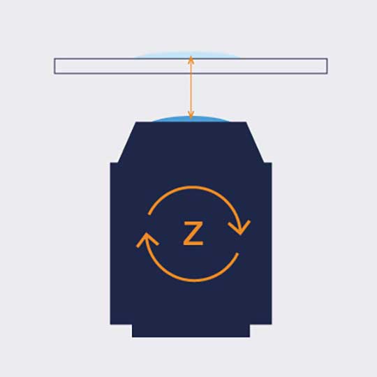
RAYSHAPE Mirror
Dynamic aberration correction with a deformable mirror over about 200 µm z-range. 140 digital actuators adjust the mirror surface within milliseconds.


Custom Solutions
We offer solutions for even the most challenging applications. Everything that can be done, we will do.
Broaden your knowledge
of microscopes, dyes, and superresolution
If you want to make your theoretical expertise on light microscopy grow, check out our knowledge base! It covers a vast range of topics from the very basics like “what is resolution?” or “how to measure resolution?” to sophisticated solutions for common imaging problems such as bleaching or challenges in staining. We invite you to read and learn!
Movement of fluorophore-labeled 30S ribosomes (colored) in two E. coli bacteria (grayscale) tracked with MINFLUX.
Tell me more >How the donut changed the world

For over a century, we stood at the edge of microscope resolution and cursed the inexorable blur of diffracted light. Instruments improved, but the fog never lifted. Then, one man stopped trying to control how light behaves. Armed with a donut-shaped laser beam, he instead commanded where it shines and untethered resolution forever. Details >
Which microscope has the best resolution?

The elctron microscope achieves the highest magnification and resolution. But does “highest” always equal “best”? Well, that depends on what you want to do with the resolution. Details >

Today’s high-end fluorescence microscopy is unthinkable without lasers. Reason enough to take a closer look at these sophisticated light sources. Details >

Every technique that allows to observe cells is more or less invasive and fluorescence microscopy is no exception. Many imaging situations profit from a reduction in light dose as provided by FLEXPOSURE adaptive illumination. Details >

MINFLUX reaches unprecedented spatio-temporal resolution in light microscopy and provides 2D and 3D localization precisions in the single-digit nanometer range. Details >
Superresolution for biology: when size, time, and context matter

The spatial resolution achievable with today’s light microscopes has unveiled life at the scale of individual molecules. Size is no longer a barrier to seeing biology at the most fundamental level. But life is not static. It emerges from movement and change. How do superresolution technologies hold up to the challenges of documenting dynamic biological mechanisms? Details >
Additional information and articles

TIMEBOW Lifetime Imaging – gives time color
Explore abberior’s TIMEBOW lifetime toolbox with our application experts Julia Menzel and Jan-Gero Schloetel.

abberior FLUX special
Unlock the full potential of single-molecule localization microscopy (SMLM) with our abberior FLUX dyes.
MINFLUX webinar – Tracking with unprecedented spatio-temporal resolution!
Thanks MINFLUX it’s possible to track the movement of individual molecules in the cell, e.g. kinesin. See what EMBL scientists have achieved.
Live-cell imaging – nanometer-sized fluorescent probes & tags
Ivana Nikić-Spiegel & Gražvydas Lukinavičius are giving a deeper inside of the design & use of nanometer-sized molecular probes for labeling living probes.