


Unveiling life in the leaf – Plant Science
FAQ Video 38

MINFLUX allows an everyday 3D resolution of 2 – 3 nm and offers about 100 times faster tracking than with a camera-based system.

FAQ Video 37

“How does TIMEBOW lifetime imaging work and why is MATRIX array detection a perfect match?”
NEWS
TIMEBOW, MATRIX, STED and confocal imaging can be freely combined
Watch Recording: TIMEBOW lifetime imaging for stunning results with confocal and STED superresolution!
abberior rocked @ EMBL – Imaging down to single-molecule resolution: STED and MINFLUX nanoscopy
EMBL Imaging Centre & abberior Instruments foster intense collaboration
Light microscopy is an indispensable tool in plant science, offering detailed insights into plant anatomy, physiology, metabolism, genetics, development, and more. It also facilitates the study of interactions between plants and other organisms such as symbiotic bacteria and fungi or parasites.
Imaging in botany spans several length scales, from whole organisms over tissue sections and single cells to sub-cellular structures and protein complexes. Thanks to superresolution microscopy, it is nowadays possible to image structures in unprecedented detail, even in living specimen, revealing so-far unavailable information on, for example, photosynthesis and stem cell differentiation in the meristem.
abberior offers microscopes for both confocal and STED imaging with superior performance as well as add-on modules addressing the particular challenges of plant microscopy such as autofluorescence. Furthermore, we facilitate unprecedented spatial and temporal resolution with MINFLUX in all three dimensions.
Our product portfolio is completed by a comprehensive selection of fluorescent dyes and labels suitable for various applications.
Talk to a scientist >From leaf to rhizome
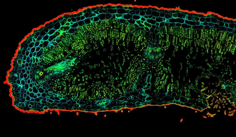
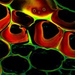
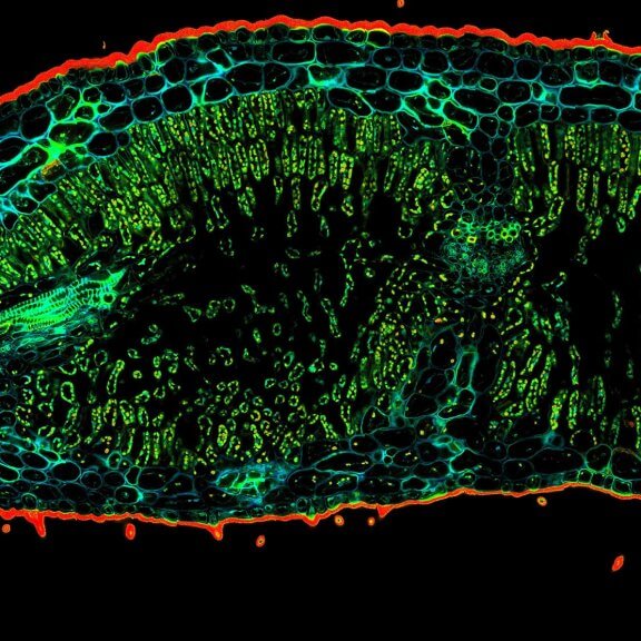
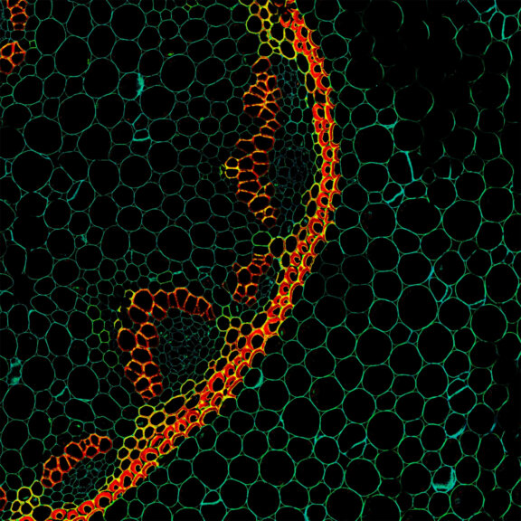
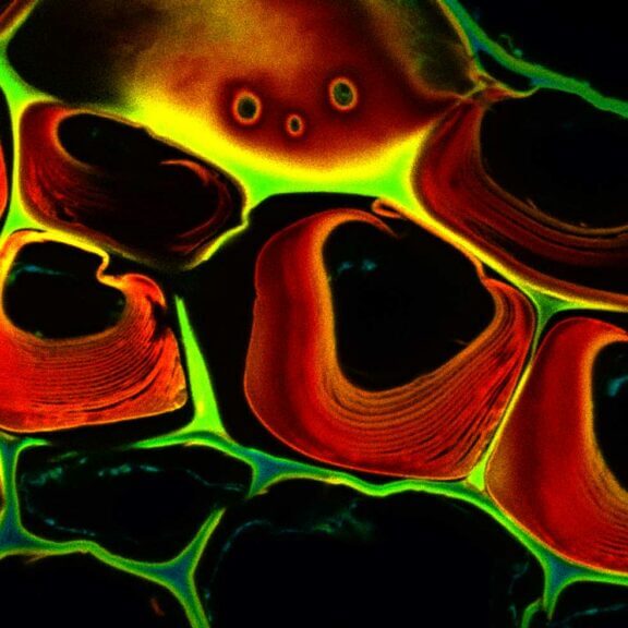
Regular shapes and patterns are typical for plant tissue and often lend microscopic images a distinctive aesthetic appeal, like in this stem cross section of Convallaria recorded with FACILITY and stitched from 10 by 10 tiles, each 125 µm by 125 µm. Autofluorescence was separated and colored according to the signal’s lifetime – which depends on the type of autofluorescent molecule and its nanoenvironment – with TIMEBOW lifetime imaging.
Test your sample >The green up close
Fluorescence microscopy reveals what is beyond the cell wall. Organelles like mitochondria, the vacuole, chloroplasts, golgi, nucleus, or the ER, and structures such as the cytoskeleton, chromatin, membranes, vesicles – countless components may today be visualized under the microscope. And imaging beyond the diffraction limit reveals details never seen before.
Description
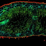
Confocal TIMEBOW acquisition of an Oleander leaf tip showing a range of autofluorescence lifetimes originating from different tissue components.
Description
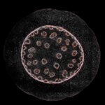
Confocal and STED image of a meiotic cell of Chinese spring wheat stained for two synaptonemal complex components.
Sample courtesy: Sepsi Adél, HUN-REN, Centre for Agricultural Research, Martonvásár, Hungary.
Modules:

Description
Autofluorescence in a plant stem cross section of Convallaria recorded with FACILITY. A large area, 10 by 10 tiles, each 125 µm by 125 µm, was imaged and stitched with image tiling in Fiji for ImageJ. This is combined with TIMEBOW lifetime imaging to show the differences in fluorescence lifetime due to the type of autofluorescent molecule and their nano-environment.
Modules:
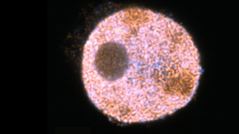
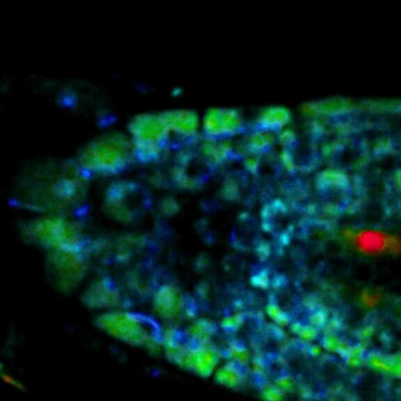
Description
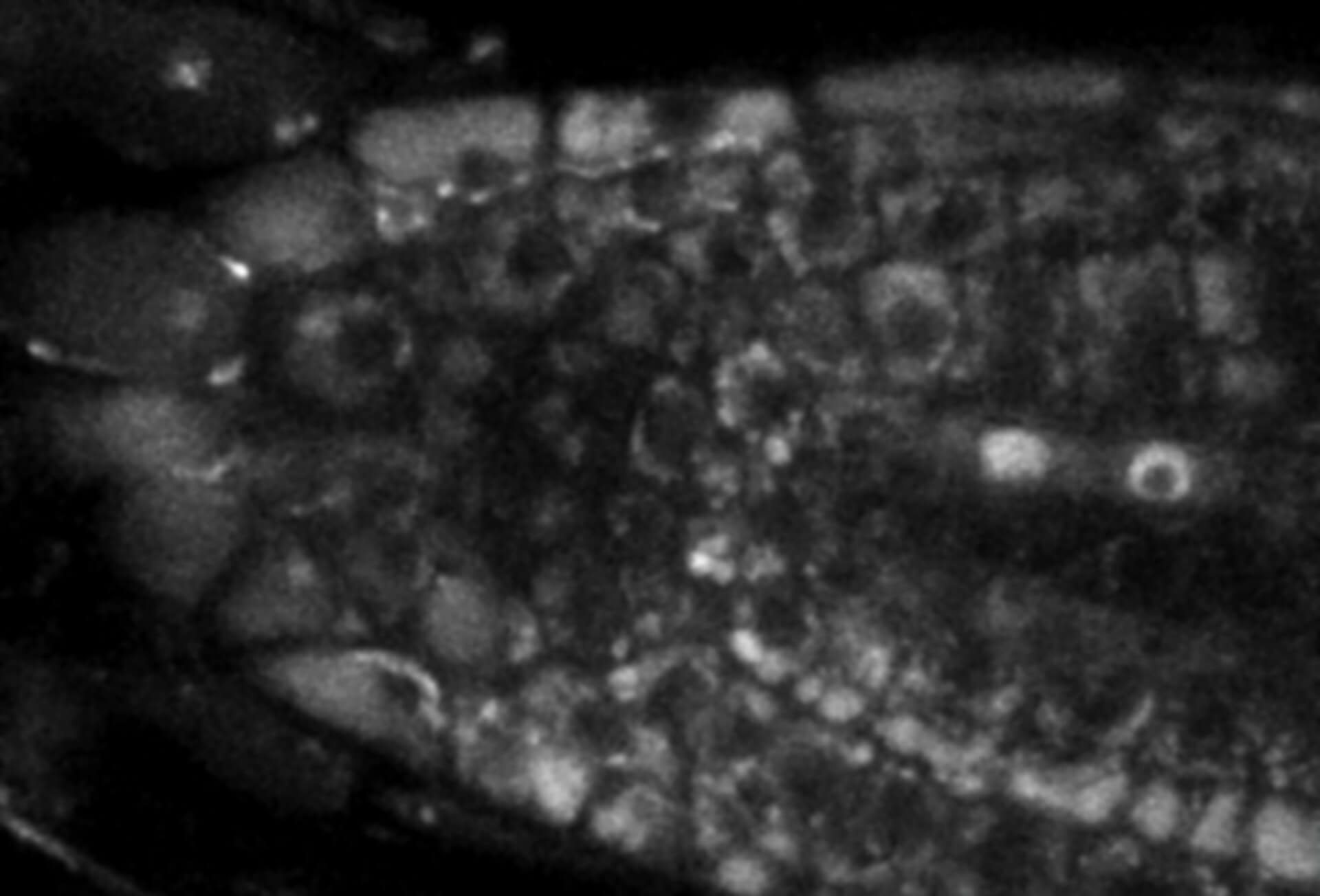
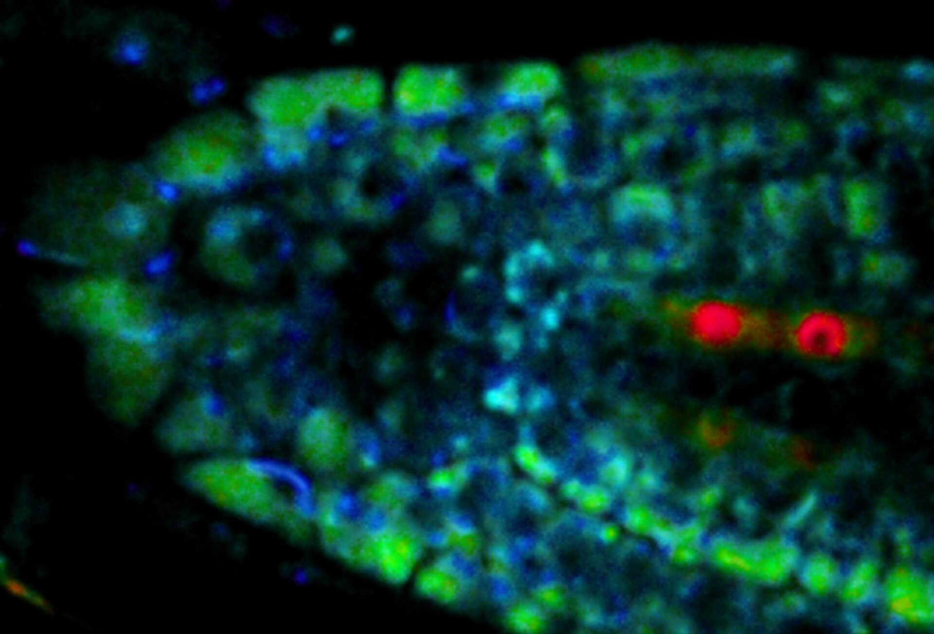
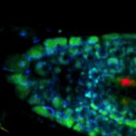
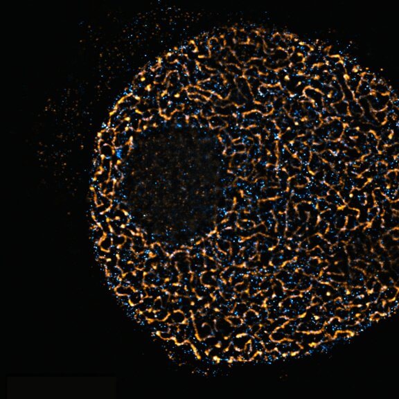
Live-cell sample of an Arabidopsis root tip suspended in water recorded with FACILITY. A subset of cells expresses a YFP construct. Lifetime imaging with TIMEBOW shows shifts in YFP fluorescence lifetime caused by the proteins nano-environment.
We thank Dr. Fábián Attila and Soós Vilmos (ATK, Brunszvik, HU) for providing this sample. Contact: soos.vilmos@atk.hu.
Modules:
Description
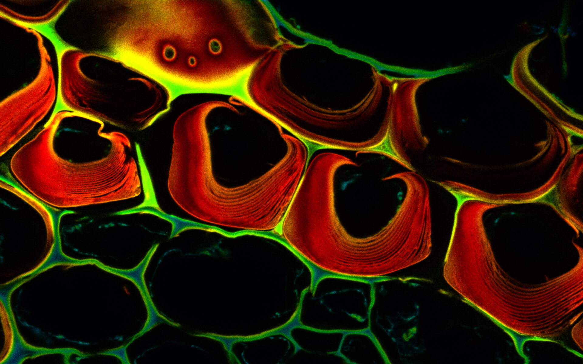
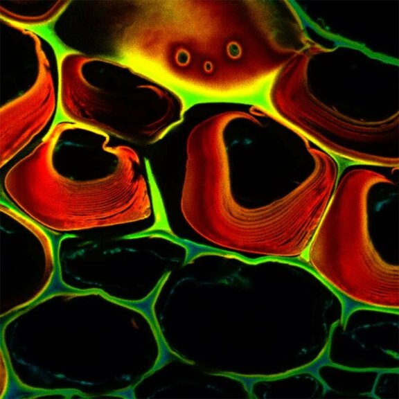
Autofluorescence in a plant stem cross section of Convallaria recorded with FACILITY. Image quality is improved by removing the background through MATRIX detection. This is combined with TIMEBOW lifetime imaging to show the differences in fluorescence lifetime due to the type of autofluorescent molecule and their nano-environment.
Modules:
Description
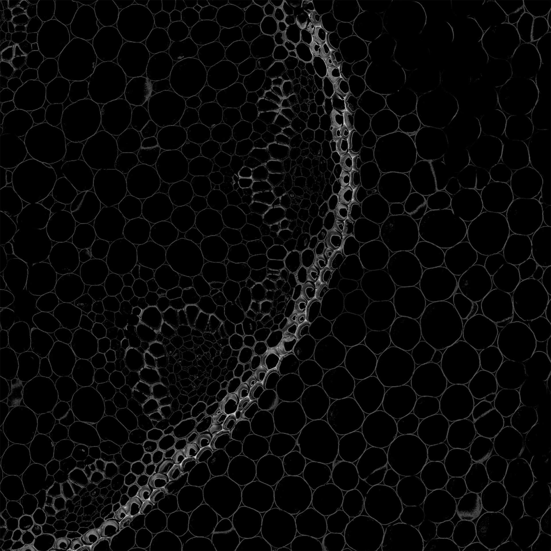
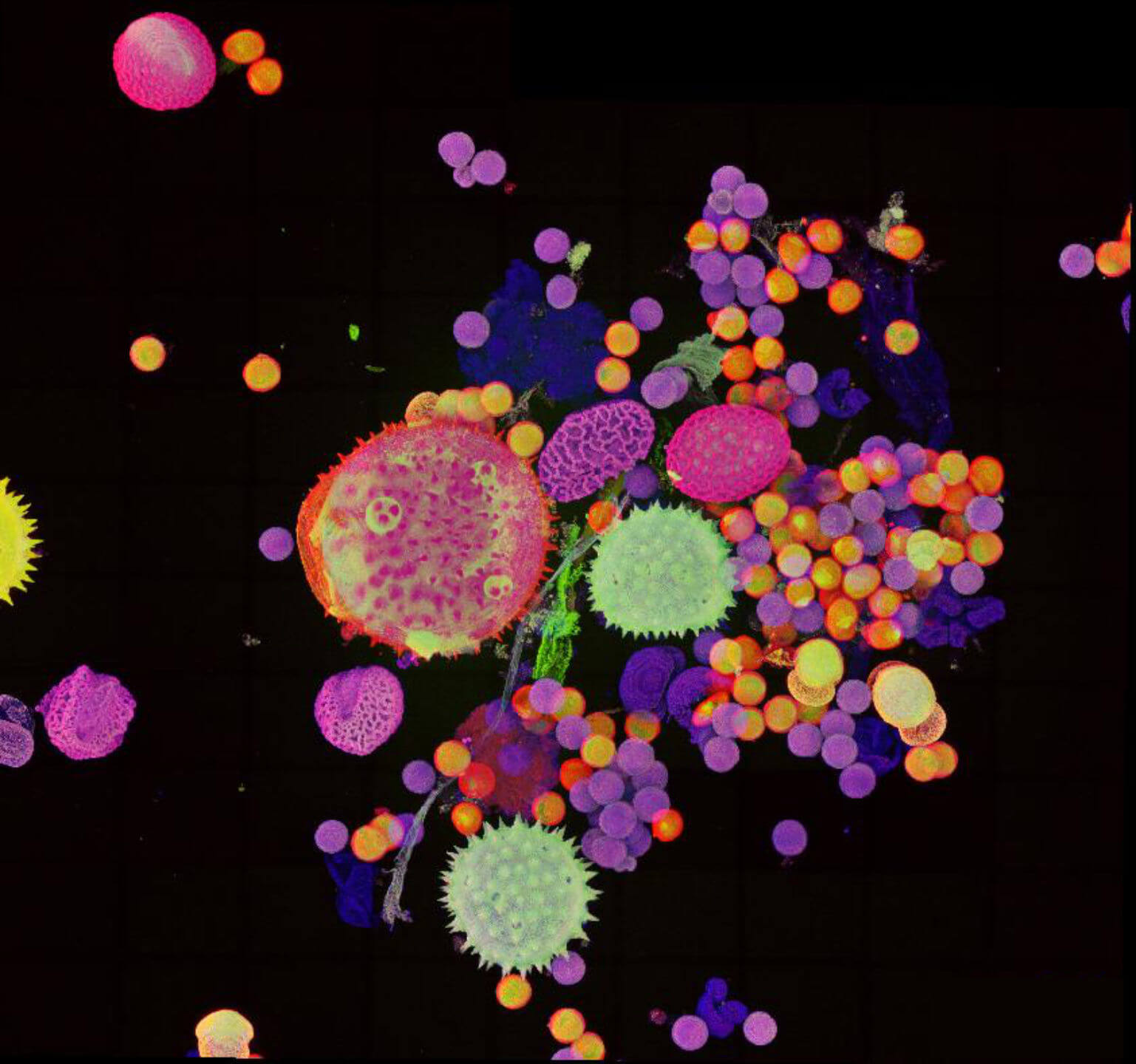
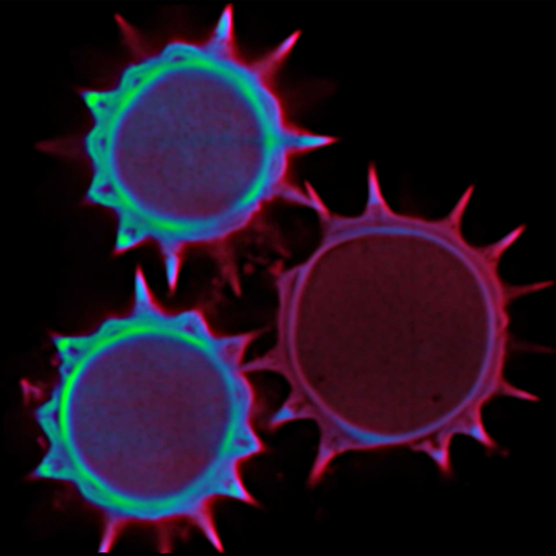
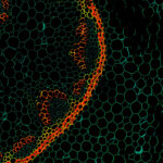
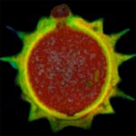
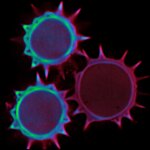
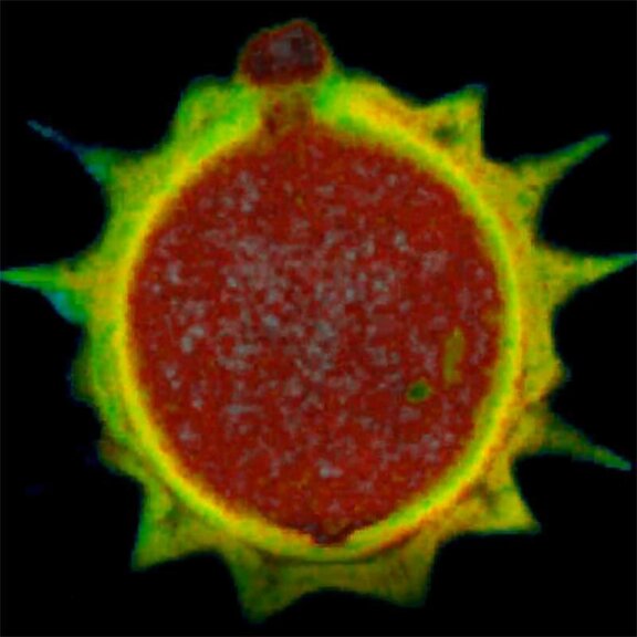
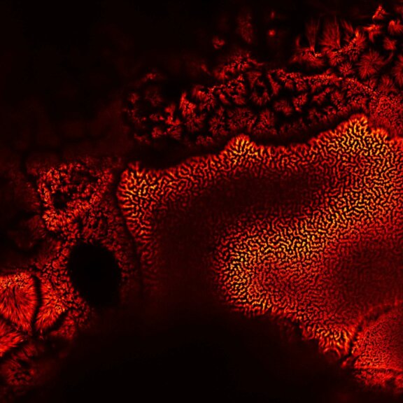
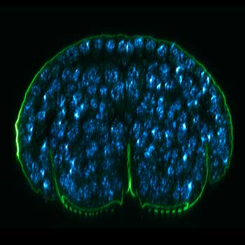
Pollen autofluorescence imaged with a STEDYCON on an IX83 microscope frame. z-stacks on 95 tiles were acquired and stitched with SVI Huygens.
Description
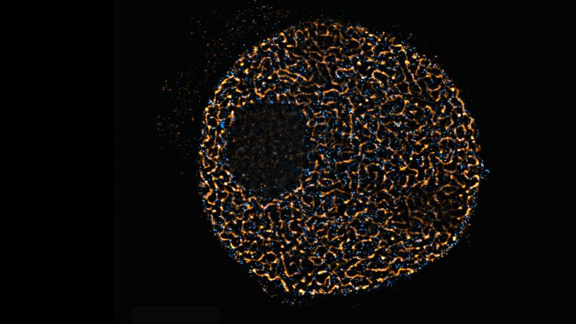
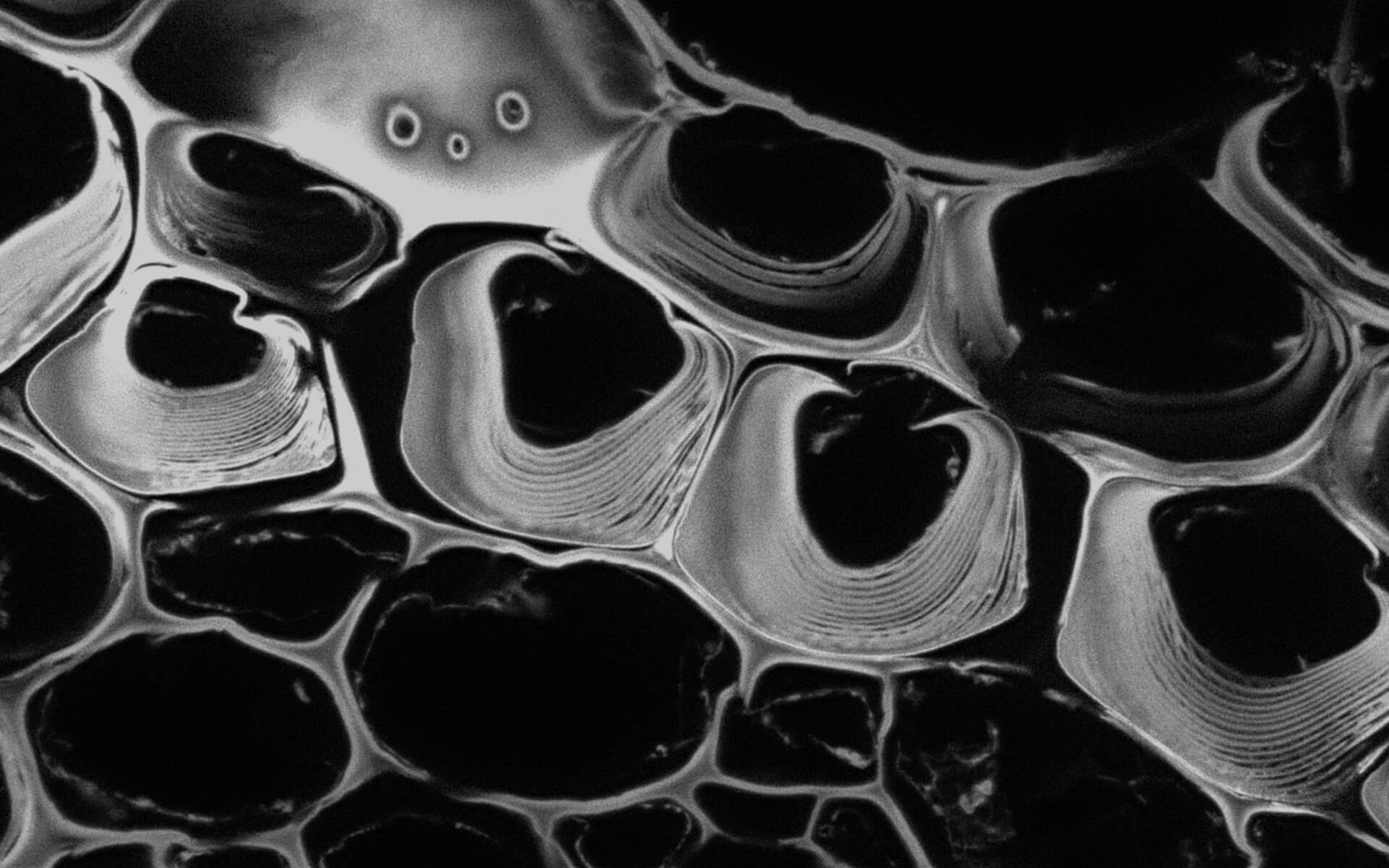
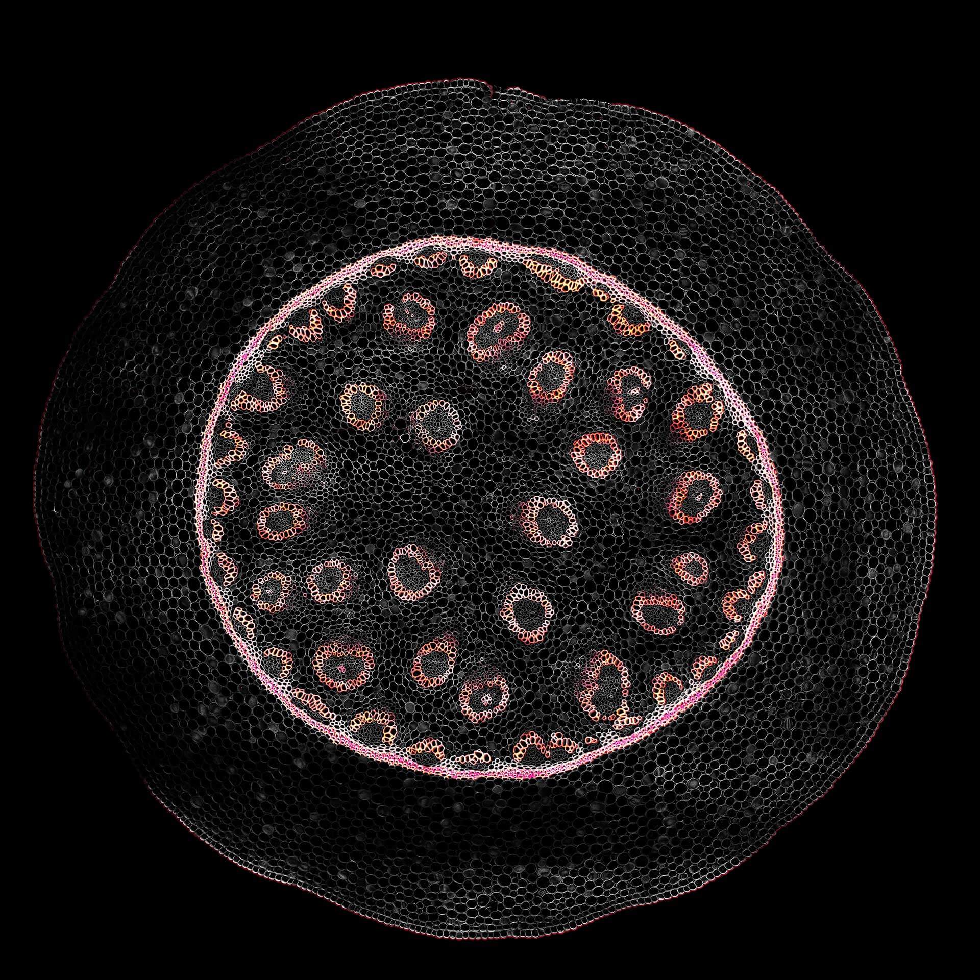
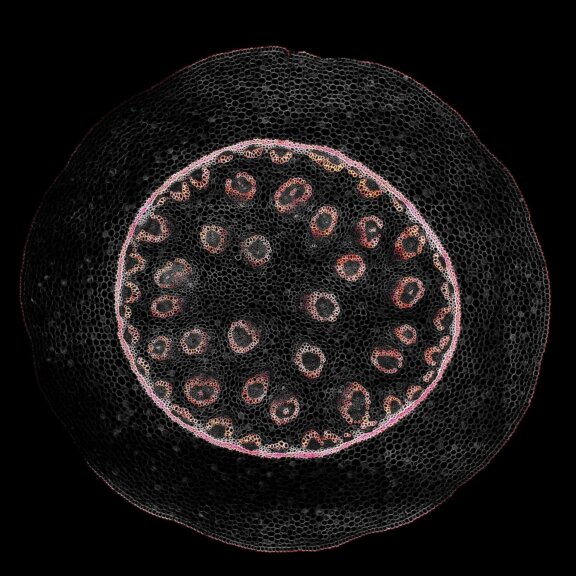
Confocal acquisition of Convallaria rhizome (cross-section) with a 20x oil objective; 3 channels, maximum intensity projection. The image consists of 14 z-planes in 9 by 9 tile pattern comprising a total area of 3.2 mm x 3.2 mm, stitched using SVI Huygens. Shown is a maximum intensity projection.


The color of life is green and …
For vibrant, colorful images, abberior offers microscopes ideally suited for the imaging of complex samples such as plant tissue – and with MINFLUX, we provide the most powerful fluorescence microscope in the world.
This live-cell sample of an Arabidopsis root tip was suspended in water and recorded with FACILITY. A subset of cells expresses a YFP construct. Lifetime imaging with TIMEBOW shows shifts in YFP fluorescence lifetime caused by the proteins’ nano-environment. MATRIX array detection improves optical sectioning by physically separating background from foreground.
Sample provided by Fábián Attila and Soós Vilmos, ATK, Brunszvik, Hungary.
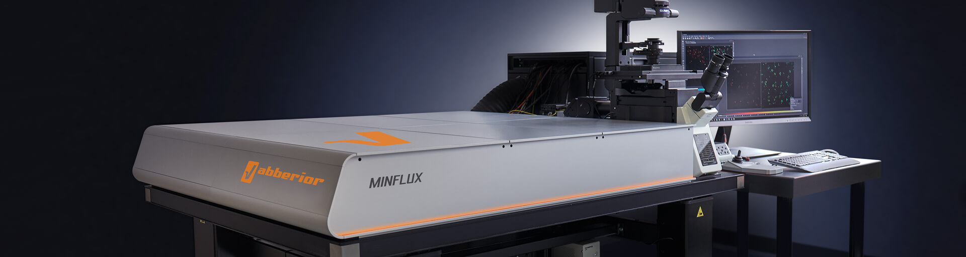
Get a demo >MINFLUX – unrivaled resolution and speed
The MINFLUX platform offers an unprecedented array of imaging possibilities and allows you to resolve molecular details along all three dimensions. This unmatched resolution capability combined with unprecedented speed reveals sample details never seen before and helps to dissect fast and dynamic cellular processes in space and time. With MINFLUX, it is now possible to follow single proteins over time with unprecedented precision and speed. Details >
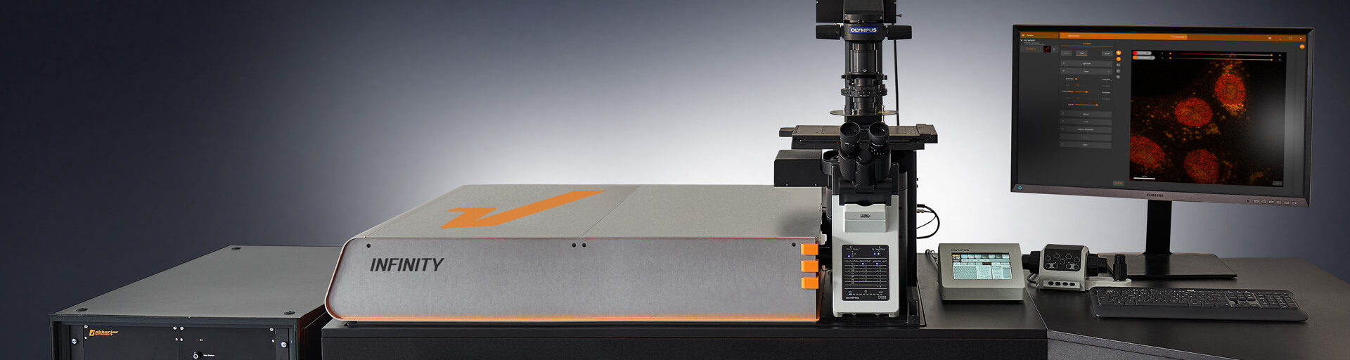
INFINITY – forever cutting edge
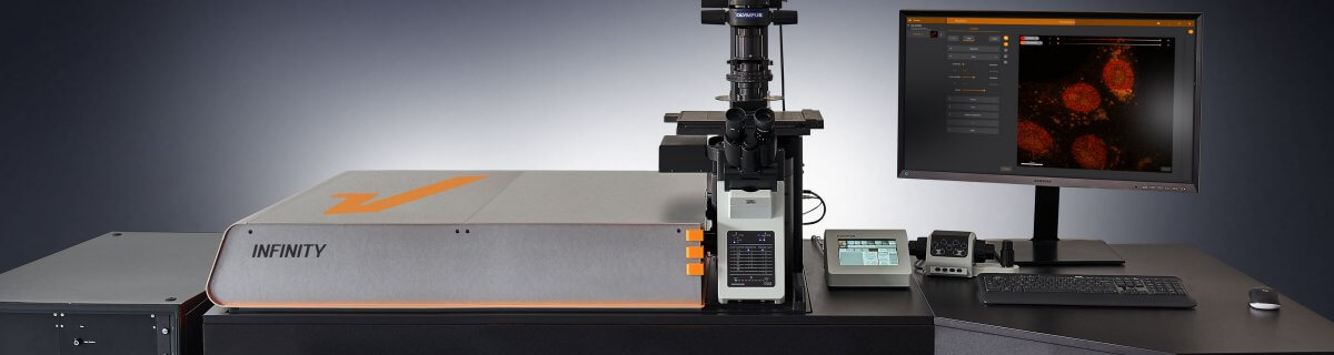
The INFINITY platform is the most customizable platform for all things microscopy and may be adapted to your particular demands in plant imaging. Optimize your microscope for the imaging of anything from subcellular structures to complex tissue. INFINITY is always up to the challenge. Just tell us what you need and we will build you a customized, continuously upgradable system specialized for your research. Details >
MIRAVA POLYSCOPE – one for all and all for one

The MIRAVA® POLYSCOPE® combines every resolution – from millimeters down to 3 nm – in a singularly unique system. MIRAVA unites four microscopy technologies to cover an unprecedented resolution spectrum, extending over several orders of magnitude from diffraction-limited imaging all the way to true molecular resolution. Our LiGHTBOX software allows beginners to intuitively arrive at a top-notch image within three clicks, while also giving experts full control over the instrument. Details >
STEDYCON 2 – confocal, STED, lifetime… WOW!

The STEDYCON upgrades your existing widefield microscope to a confocal, STED, and lifetime machine with a resolution down to 30 nm. All that’s required is a free camera port and a good objective lens. With its super-intuitive user interface, the STEDYCON provides an intelligent microscope platform that enables everyone to acquire superb superresolution images after only minutes of training. Details >
Plants light up by themselves –
handling autofluorecence made easy
Dealing with autofluorescence is a major challenge when imaging plant samples. TIMEBOW imaging allows to separate fluorescence according to the signals’ lifetime, as seen in this Convallaria cross section recorded with FACILITY. The differences in lifetime arise due to the fluorophores’ distinct molecular properties and their nanoenvironment. Image quality is improved by removing the background with MATRIX array detection.
Ask for detailed information >Choose a superpower for your experiment – our modules
Every MINFLUX, INFINITY, and FACILITY microscope can be upgraded with modules to overcome the specific imaging challenges in your research. You may, for instance, use TIMEBOW Imaging to separate signals in the same spectral channel by their fluorescent lifetime. This allows you to remove unwanted autofluorescence or extend your options for multicolor imaging. RAYSHAPE facilitates imaging deep inside the sample by dynamically correcting for aberrations. Autofocus and Autoalignment for all beams including STED guarantee best-possible imaging conditions at all times. And if you can’t find what you need on our website, get in touch with us, and we’ll develop it for you.
MATRIX Detector
Many eyes see more than one. The MATRIX detector drastically improves signal-to-background ratio, resolution, and dynamic range.
TIMEBOW Imaging
TIMEBOW lifetime imaging for stunning results at confocal and STED super-resolution.
FLEXPOSURE Illumination
Brings down the light dose on your sample and lables dramatically. Key ingredient for volume and live-cell superresolution.
RAYSHAPE Mirror
Dynamic aberration correction with a deformable mirror over about 200 µm z-range. 140 digital actuators adjust the mirror surface within milliseconds.


Custom Solutions
We offer solutions for even the most challenging applications. Everything that can be done, we will do.
Broaden your knowledge
of microscopes, dyes, and superresolution
This STEDYCON image shows pollen autofluorescence. z-stacks on 95 tiles were acquired and stitched with SVI Huygens. But do you know how fluorescence arises? How a donut-shaped laser beam controls this process to overcome the diffraction limit and to achieve superresolution in light microscopy? Or how a deformable mirror can be applied to compensate for aberrations and facilitate deep tissue imaging? Our knowledge base has all the answers!
Tell me more >How the donut changed the world

For over a century, we stood at the edge of microscope resolution and cursed the inexorable blur of diffracted light. Instruments improved, but the fog never lifted. Then, one man stopped trying to control how light behaves. Armed with a donut-shaped laser beam, he instead commanded where it shines and untethered resolution forever. Details >
How to correct for aberrations in light microscopy

Aberrations can give microscopists a hard time. They belong to microscopy like pathogens belong to life. There are ways to bring diverted rays back on track, but some are better than others. The question is: deformable mirror or correction collar? Details >

MINFLUX reaches unprecedented spatio-temporal resolution in light microscopy and provides 2D and 3D localization precisions in the single-digit nanometer range. Details >
Superresolution for biology: when size, time, and context matter

The spatial resolution achievable with today’s light microscopes has unveiled life at the scale of individual molecules. Size is no longer a barrier to seeing biology at the most fundamental level. But life is not static. It emerges from movement and change. How do superresolution technologies hold up to the challenges of documenting dynamic biological mechanisms? Details >

For all the talk about criteria and definitions, measuring the resolution of a microscope is more nuanced than you’d think. The scales at which microscopes operate today are subject to noise and background that obscure and distort signals. What you use for the measurement can make a big difference. The second article in our “Resolution” series. Details >

Are you surprised that the very nature of light caps the resolution that we can achieve in microscope images? Luckily, there are workarounds to this limit. These workarounds push the amount of detail in an image by manipulating precisely where and when fluorophores are allowed to emit. As such, they provide us with a completely new set of tools to shrink the distance between two points while still being able to resolve them. Details >
Additional information and articles

TIMEBOW Lifetime Imaging – gives time color
Explore abberior’s TIMEBOW lifetime toolbox with our application experts Julia Menzel and Jan-Gero Schloetel.
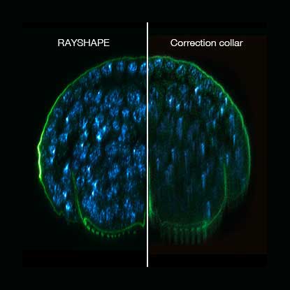
RAYSHAPE special
RAYSHAPE dynamic aberration correction with a deformable mirror preserves resolution and increases brightness. 140 actuators adjust mirror surface within milliseconds for over 200 µm z-range.
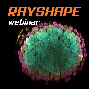
RAYSHAPE webinar – dynamic aberration correction for deep tissue imaging
Hello RAYSHAPE! Bye-bye, aberrations!

Online Symposium – Super-Res & Plant Science
Kathryn Wright presents tips and tricks from 30 years of plant cell imaging. Jan-Gero Schloetel images live tobacco leaves on our FACILITY microscope
MINFLUX
The world’s most powerful fluorescence microscope MINFLUX, based on Stefan Hell’s invention, allows to resolve structures as small as a molecule in 3D.

MATRIX STED reloaded
MATRIX array detection is particularly well suited for imaging densely labelled samples and providing excellent resolution in areas normally hidden in haze.
MINFLUX webinar – Tracking with unprecedented spatio-temporal resolution!
Thanks MINFLUX it’s possible to track the movement of individual molecules in the cell, e.g. kinesin. See what EMBL scientists have achieved.