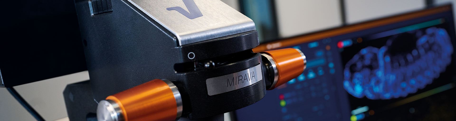
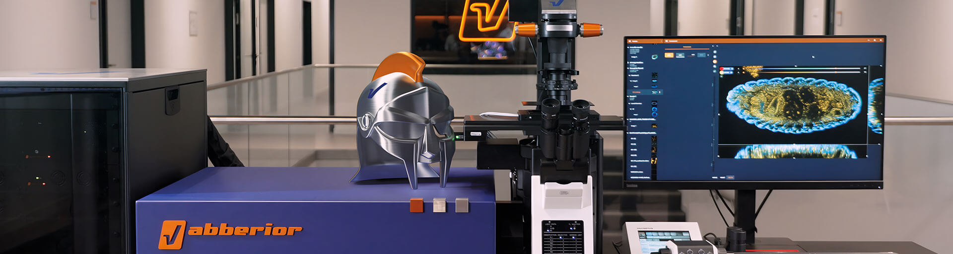
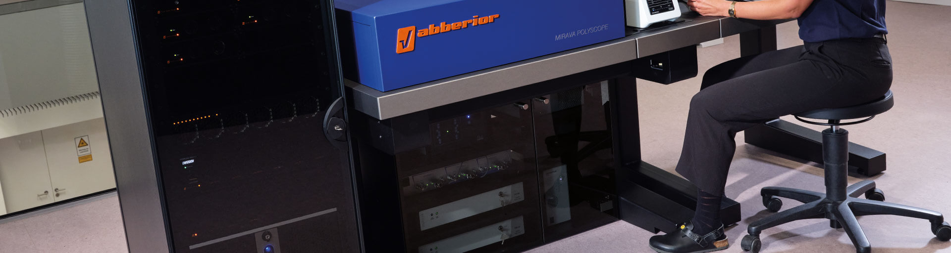


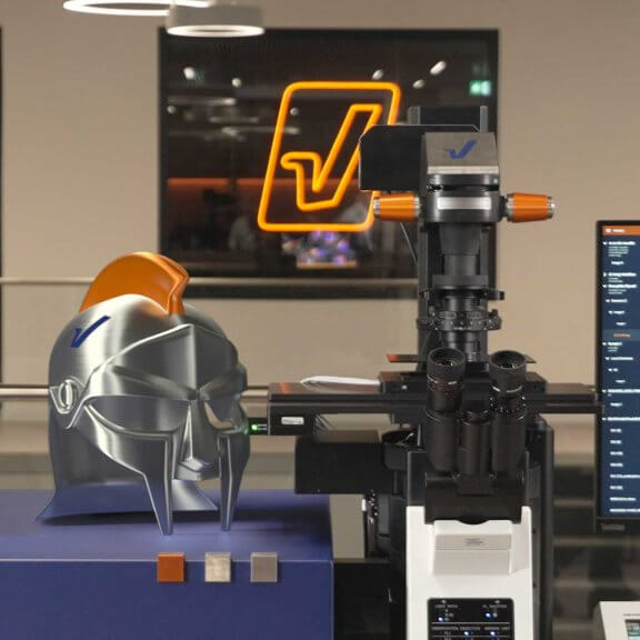
MIRAVA POLYSCOPE
Every resolution – from millimeters down to 3 nm – combined in a singularly unique system. 1)
abberior’s MIRAVA® POLYSCOPE® unites four microscopy technologies to cover an unprecedented resolution spectrum, extending over several orders of magnitude from diffraction-limited imaging all the way to true molecular resolution.
one for all and all for one: the perfect image
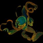
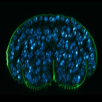
Description
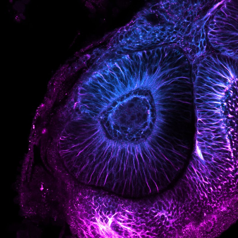
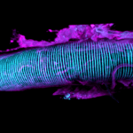
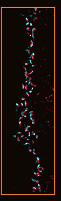
Confocal image of a developing zebrafish eye showing tubulin stained with abberior STAR RED and abberior STAR ORANGE.
Image courtesy of Graziamaria Paradisi, Marco Tartaglia, Antonella Lauri, Ospedale Pediatrico Bambino Gesù, Rome, Italy
Modules:
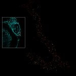
Description
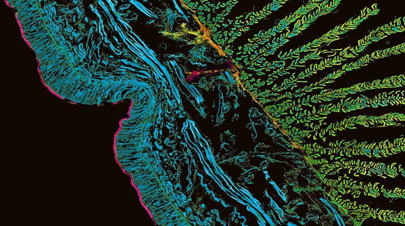
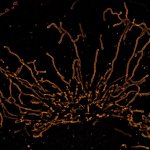
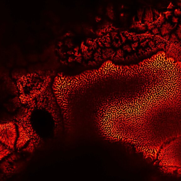
Confocal image of autofluorescence in a cross-section of the earthworm Lumbricus terrestris. TIMEBOW lifetime imaging detects differences in fluorescence lifetime, which depend on the type of autofluorescent molecule and its nano-environment, and visualizes them in distinct colors.
Modules:

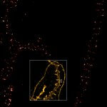
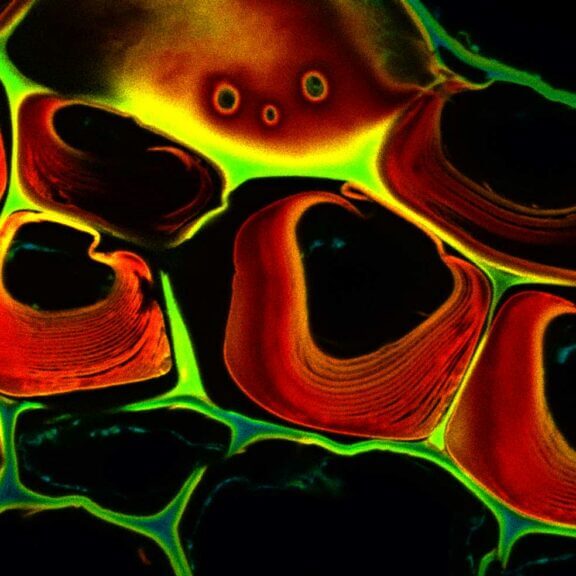
Description
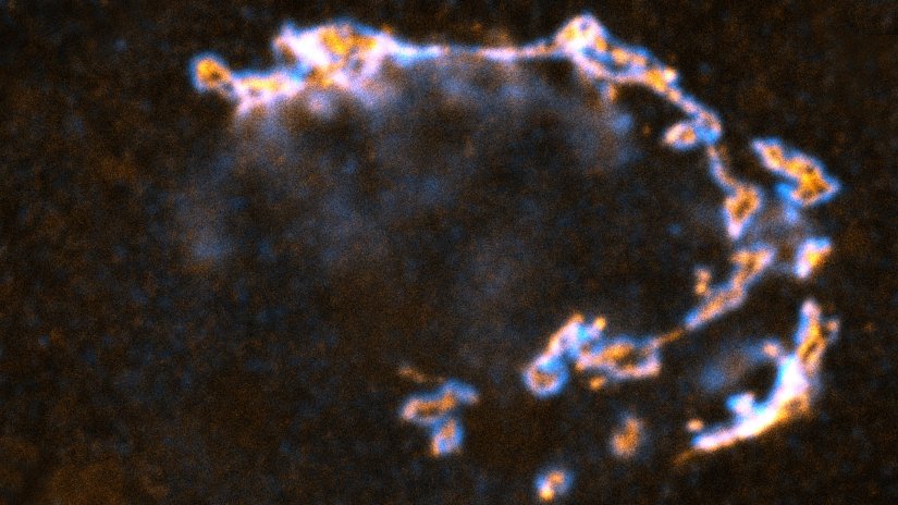
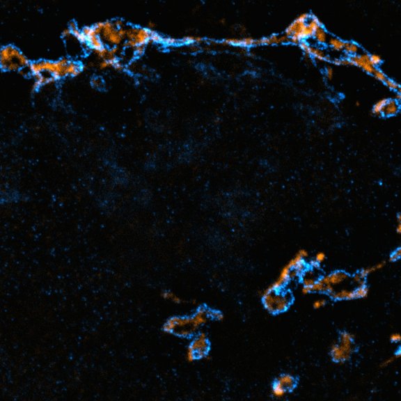
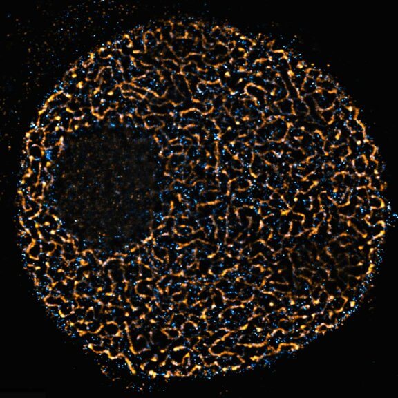
STED image of the golgi apparatus in a fixed mammalian cell, recorded with MATRIX array detection and deconvolved with TRUESHARP image boosting. Stained are the golgi proteins GM130 (cyan, abberior STAR RED) and giantin (orange, abberior STAR ORANGE).
Modules:

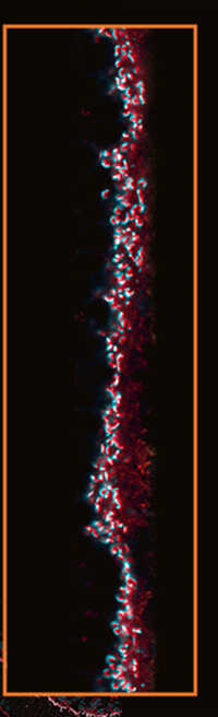
Description
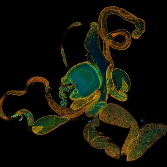
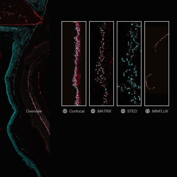
Imaging across scales from diffraction-limited to molecular resolution: ribbon synapses in fixed mouse retina tissue stained for VAMP1 (abberior STAR RED) and CtBP2 (abberior STAR ORANGE) (confocal, MATRIX, STED) or for Bassoon (MINFLUX).
Sample courtesy: Arlene Hirano, PhD, University of California Los Angeles, Los Angeles, CA, USA (confocal, MATRIX, STED), and by Dr. Chad Grabner and Prof. Dr. Tobias Moser, Max Planck Institute for Multidisciplinary Sciences, Göttingen, Germany (MINFLUX).
Modules:
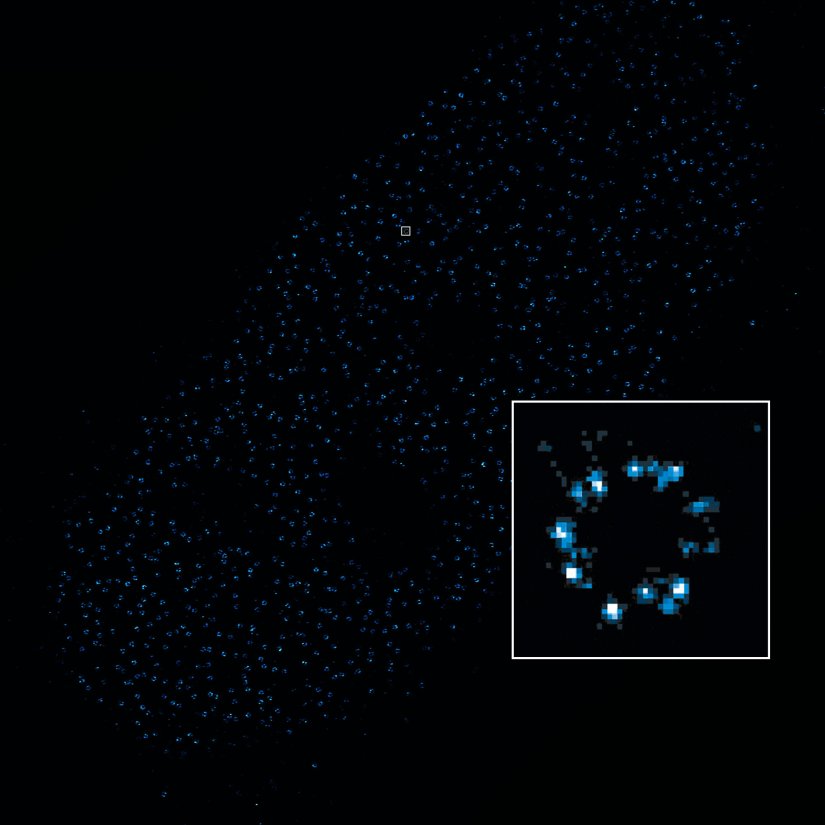
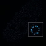
Description
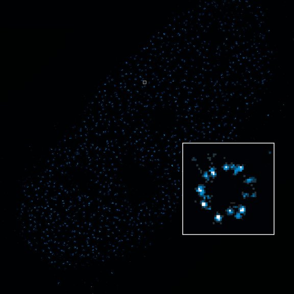
2D MINFLUX image of nuclear pore complex subunits, imaged in fixed mammalian cells expressing GFP-tagged NUP96 stained with abberior DNA-PAINT 660. The molecular resolution of the MINFLUX module of the MIRAVA POLYSCOPE allows visualizing the shape and arrangement of individual subunits of the nuclear pore complex.
Modules:
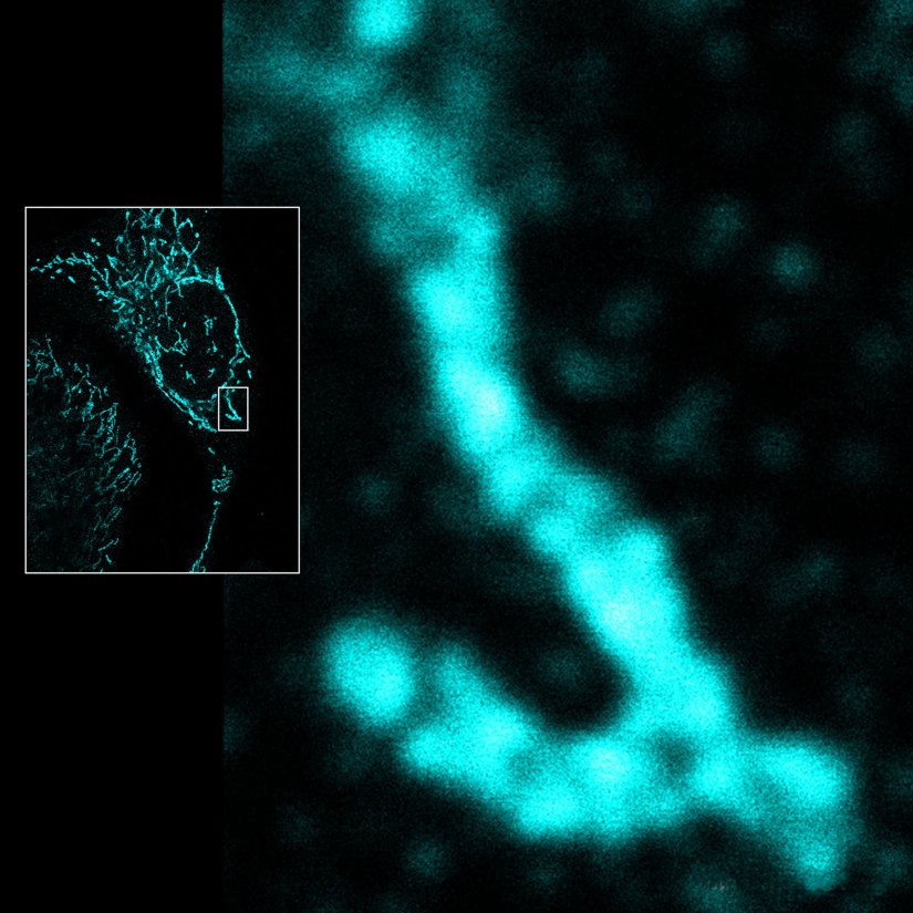

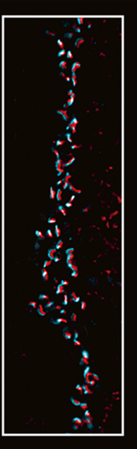
Description
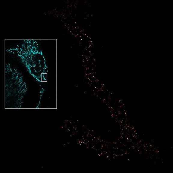
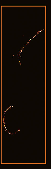
Mitochondria labeled for the outer membrane protein TOMM20, imaged in confocal mode and with the MINFLUX module of the MIRAVA POLYSCOPE.
Modules:
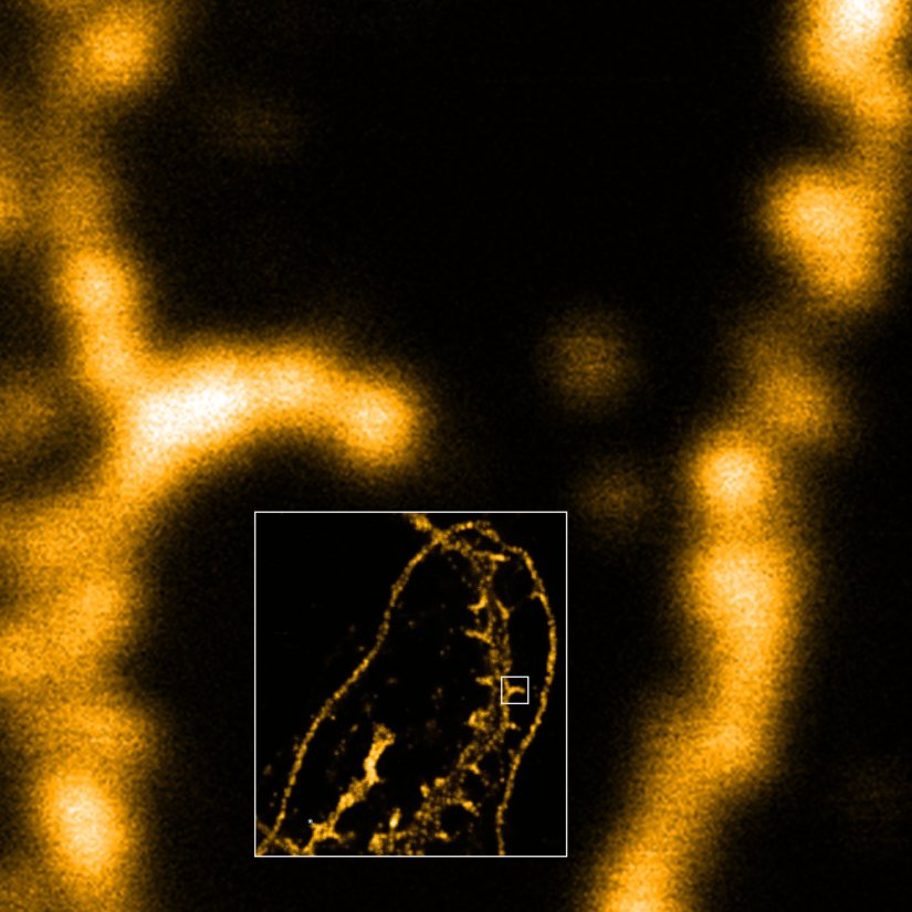
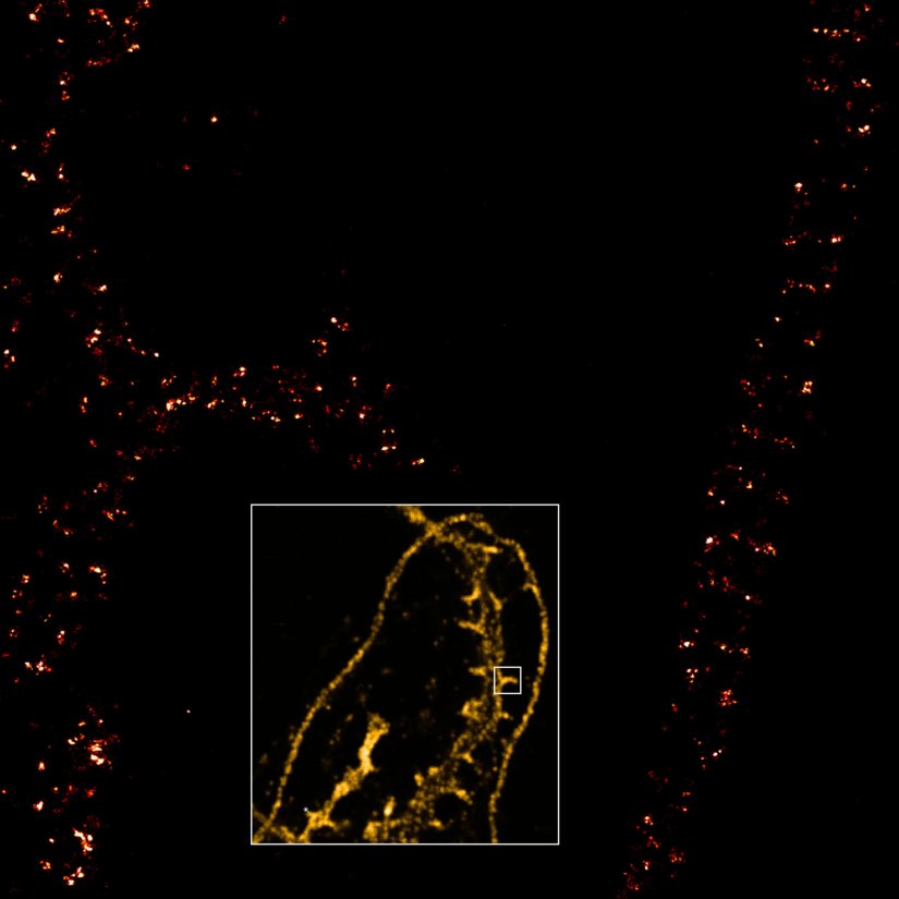
Description
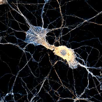
Dendrite with spines of a neuron grown on a coverslip, stained for βII spectrin and imaged in confocal mode and with the MINFLUX module of the MIRAVA POLYSCOPE.
Modules:


MIRAVA POLYSCOPE @ ELMI, Heidelberg
Many thanks for visiting us and attending our workshops! We had a lot of fun talking to you.The next opportunity to see our MIRAVA POLYSCOPE live in action will be at MMC 2025, July 1. to 3. in Manchester, UK. We look forward to seeing you there!


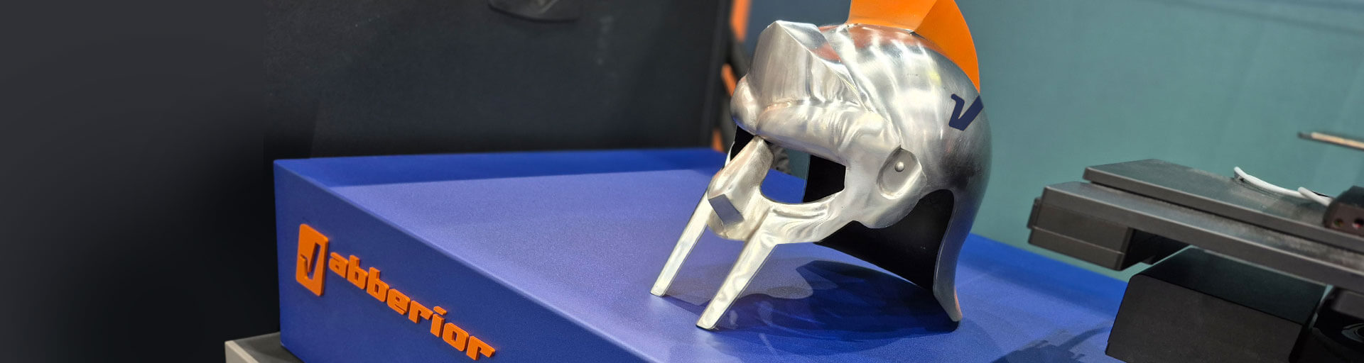
Safety first at ELMI 2025!
Our latest tool for dealing with razor-sharp and crystal-clear images taken with our MIRAVA POLYSCOPE!


Focus on your research, we take care of the rest
Confocal imaging
Resolution down to 200 nm
MIRAVA’s superpowers
FLEXPOSURE adaptive illumination
RAYSHAPE aberration correction
Get a demo >The MIRAVA POLYSCOPE takes confocal imaging to the next level.
With an extensive range of state-of-the-art features, you will enjoy unparalleled performance that meets the highest of imaging demands. No matter what your project is, be it live cell, deep tissue, or multicolor imaging, MIRAVA empowers users to achieve their goals with unmatched comfort and flexibility.
Equipped with seven excitation laser lines, MIRAVA guarantees straightforward and versatile multicolor imaging. It’s exceptionally gentle to the sample: our avalanche photodiode (APD) detectors require only half the excitation power to collect the same signal as hybrid detectors.2) With abberior’s unique module for adaptive illumination we take it to the next level of sample care. With FLEXPOSURE you will bring down the light dose by up to orders of magnitude, without compromising on contrast and resolution.
Apply one or more of MIRAVA’s many modules to create the perfect image for your needs: get rid of the haze while enhancing resolution with our MATRIX array detector. Dive deeper into complex samples without worrying about aberrations eating up your signal, thanks to RAYSHAPE dynamic aberration correction. Use TIMEBOW to add the dimension of fluorescence lifetime to your experiment, giving you more information about your sample on many levels.
And if you require more detail, you can simply switch to STED or MINFLUX technology without needing to switch to another microscope. That’s imaging across all scales with MIRAVA.

Many eyes see more than one
MATRIX imaging
Resolution down to 70 nm
MIRAVA’s superpowers
FLEXPOSURE adaptive illumination
RAYSHAPE aberration correction
Get a demo >Imaging with MATRIX array detection means resolution beyond the diffraction limit and unsurpassed signal-to-background ratio.
Thick or dense samples with a lot of background become clear and sharp as the MATRIX isolates relevant signal from the rest. This allows you to process and analyze data with much greater confidence and achieve the best results.
Our MATRIX detector consists of 23 individual detector elements. On its own, each one of them is a small pinhole, able to record at the highest diffraction-limited resolution. At the same time, all elements taken together, give you the full signal of a large-area detector. The best out of both worlds, high signal and high resolution in one single shot!
And it gets even better. A single detector, such as a photomultiplier or a hybrid detector, collects signal from the focal plane, but also from other planes above and below. It cannot tell whether a detected photon is wanted in-focus light or irritating background.
In contrast, each of the many MATRIX detector elements looks at the sample from a different angle, allowing for precise differentiation between in-focus and out-of-focus signals. The result is crystal-clear images with an unparalleled signal-to-background ratio, even in challenging imaging conditions.
MATRIX becomes even more powerful when paired with TRUESHARP image boosting, abberior’s answer to the challenge of reliable deconvolution.
In contrast to common deconvolution algorithms, TRUESHARP works without a-priori assumptions to remove noise and background and to enhance resolution. Instead, it uses measured information from the image itself, such as data recorded with MATRIX or TIMEBOW. That way, TRUESHARP gets rid of disturbances and pushes resolution without falsifying the image.
Enjoy 100% measured superresolution and stunning signal-to-background ratio with MATRIX.

Nobel prize technology from its inventors
STED imaging
Resolution down to 20 nm
MIRAVA’s superpowers
FLEXPOSURE adaptive illumination
RAYSHAPE aberration correction
Get a demo >MIRAVA STED – 30 years of experience meets attention to detail.
Performing STED microscopy on the MIRAVA means applying top-notch technology for best results. Use gentle STED laser power together with TRUESHARP image boosting for a significant increase in resolution compared to confocal imaging, without stressing the sample. Increase power if you need an even more detailed image and use FLEXPOSURE adaptive illumination to keep the light dose down. Or push it to the max and combine all our STED technologies to reach 20 nm resolution. Whatever you need, with MIRAVA, it’s all just a matter of a few clicks.
Even 3D STED is no more difficult. abberior’s dynamic beam shaper modulates the STED donut and optimally adapts it to different objective lenses.
abberior’s superpowers to master any task.
RAYSHAPE dynamic aberration correction allows you to look deep into complex samples like tissue or whole organisms without compromising on image quality due to aberrations. Using adaptive optics with a deformable mirror, RAYSHAPE puts diverted rays back on track, guaranteeing bright and crisp images from top to bottom – regardless of embedding medium, immersion medium, or specimen.
We established pulsed STED lasers as a gold standard long ago to reduce bleaching and phototoxicity and further reduced these harmful effects with the development of FLEXPOSURE adaptive illumination. With FLEXPOSURE, you are still recording long-term experiments over volumes or many frames, while ordinary always-on-lasers have long bleached the sample to death. Instead of illuminating the specimen with full intensity everywhere, we are gentle to the sample and turn all lasers on and off, shining light only where and when it has an effect and nowhere else.
TIMEBOW lifetime imaging gives time a color. Every fluorescence signal contains lifetime information that travels with the photons from the sample to the detector. TIMEBOW allows you to reveal previously hidden structural details by separating fluorophores emitting in the same spectral channel. This gives you great additional possibilities when it comes to multicolor labeling and imaging. Additionally, TIMEBOW STED exploits the spatial information encoded in the lifetime to increase image resolution.
Add the MATRIX array detector and increase resolution and image quality even further, by using the information that is encoded in the simultaneous recording of 23 images from different angles.
Our wonder weapon is TRUESHARP image boosting. TRUESHARP offers next generation deconvolution you can trust. It removes noise and background signal from your recorded data and strongly enhances resolution, sharpness, and brilliance of your images by incorporating experimentally measured information.
Of course, you can combine the capabilities of all modules for getting the perfect picture.
100x sharper than a confocal
MINFLUX imaging
Resolution down to 3 nm
MIRAVA’s superpowers
FLEXPOSURE adaptive illumination
Highest resolution at the lowest light dose, accessible to every researcher.
The MINFLUX module of the MIRAVA POLYSCOPE delivers nanometer-scale resolution that was previously achievable only with electron microscopes, but with the gentler touch of light.
MINFLUX stands out in the single molecule localization microscopy field. Other techniques follow the principle “the more, the merrier” and require huge numbers of fluorescence photons to achieve maximum resolution. MINFLUX, in contrast, localizes fluorophores by finding out where they do
not emit when excited with a hollow beam – minima-seeking, not maxima-bleaching. Our MIRAVA POLYSCOPE achieves molecular resolutions of a few nanometers without pushing the photon budget.
From sample preparation to imaging and analysis, MINFLUX uses simple, well established workflows. Labeling works as with any other type of single molecule localization microscopy.
Since abberior always takes a holistic approach, we offer FLUX dyes that are designed for superior performance in MINFLUX imaging applications.
They combine exceptional photostability, very high brightness, and low background labeling with photoswitching kinetics optimized for MINFLUX imaging. Conjugates of our dyes show excellent solubility in aqueous buffers and allow background-free labeling in cells or tissue.
Straightforward, easy, and reliable: unmatched resolution out-of-the-box with MINFLUX.
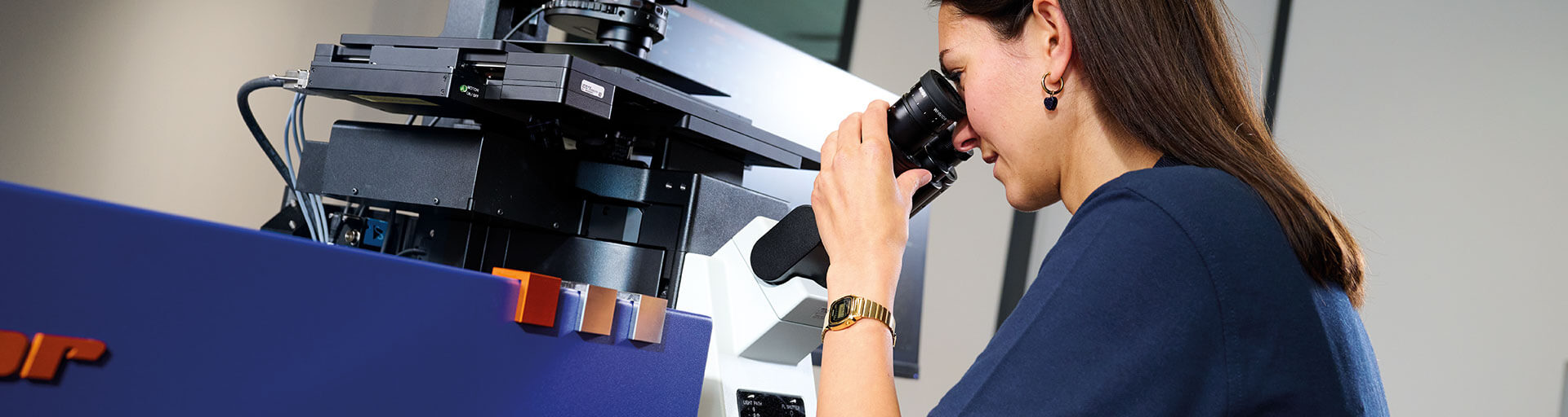
With the MINFLUX module of the MIRAVA POLYSCOPE, imaging at the molecular level is easier than ever. Set up and control your MINFLUX measurement with just a few clicks, thanks to seamless integration into our sleek LiGHTBOX software.
The MINFLUX hardware is fully integrated into the MIRAVA system, yielding unmatched robustness and optical quality. It constantly monitors and corrects even subtle, sub-nanometer drifts caused by environmental changes (e.g. ambient temperature), keeping your sample perfectly still for days.
MIRAVA sets the stage for effortless microscopy on the molecular level. Focus on your measurement and let MIRAVA do the MINFLUX.
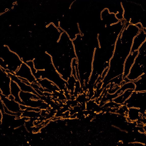
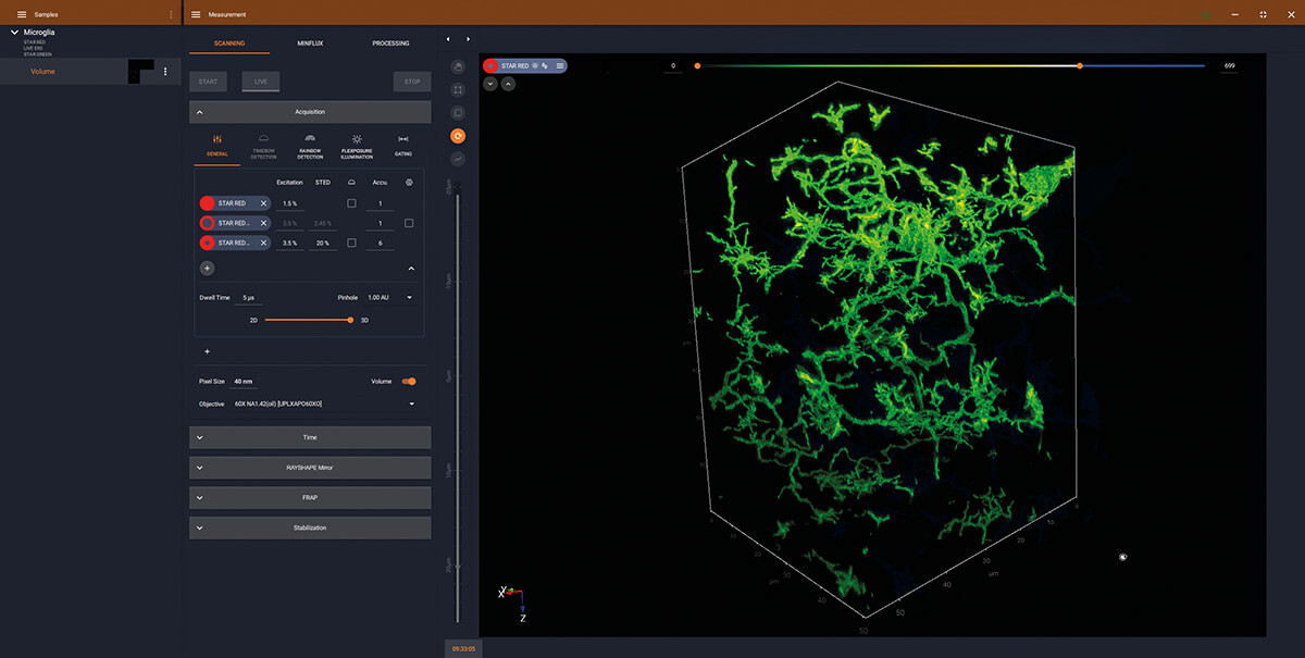
3D STED volume of microglia and processes labeled for a specific cell surface receptor in mouse neocortical tissue. Sample courtesy: Csaba Cserép and Ádám Dénes, Laboratory of Neuroimmunology, HUN-REN Institute of Experimental Medicine, Budapest, Hungary
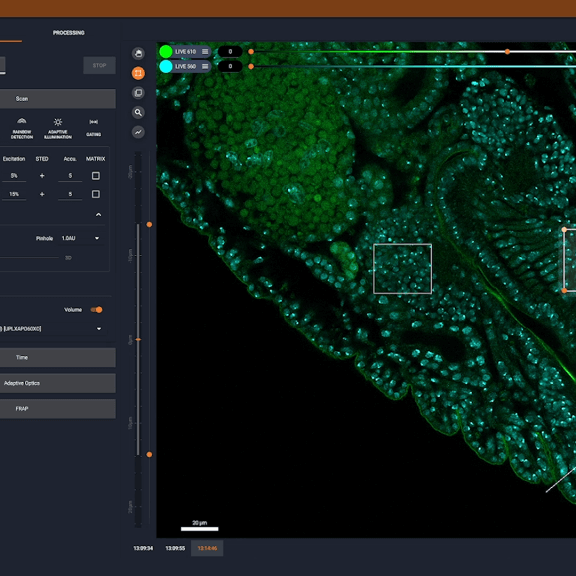
LiGHTBOX software
it’s all about ease of use
MIRAVA’s superpowers
FLEXPOSURE adaptive illumination
Designed for seamless operation and unparalleled in its clear structure, abberior’s LiGHTBOX ensures that even the most advanced imaging technologies are accessible to all users, regardless of their experience. All major settings are just one click away, and the intuitive software design facilitates quick learning, while it grants experts full control over all aspects of their measurement. You can even automate image acquisition and analysis by running Python scripts.
Thanks to LiGHTBOX you can focus on your research because the software handles the tedious tasks on its own, like detection bands, alignment, aberration correction while focusing, Nyquist sampling, focus, exposure settings…
Switching between confocal imaging and MATRIX, STED, or MINFLUX is a matter of seconds. And setting up the measurement doesn’t take much longer, no matter what imaging technique you choose.
LiGHTBOX is the most comprehensive and user-friendly solution for highest imaging demands.
1) Sample-dependent localization precision
2) 4x higher Photon Detection Efficiency (PDE) compared to conventional multi-alkali Photomultiplier Tubes (PMT) and 6x higher in the extended red range.
- Confocal imaging with resolution down to 200 nm
- MATRIX imaging with resolution down to 70 nm
- STED imaging with resolution down to 20 nm
- MINFLUX imaging with resolution down to 3 nm
MIRAVA POLYSCOPE – the world’s only all-in-one solution for confocal, MATRIX, STED, and MINFLUX microscopy with resolutions from millimeters down to 3 nm
MATRIX Detector
Many eyes see more than one. The MATRIX detector drastically improves signal-to-background ratio, resolution, and dynamic range.
LiGHTBOX
Quickly and intuitively obtain top-notch STED images with abberior’s LiGHTBOX software. Enjoy ease of use in every aspect – no ifs, ands, or buts!
RAYSHAPE Mirror
Dynamic aberration correction with a deformable mirror over about 200 µm z-range. 140 digital actuators adjust the mirror surface within milliseconds.
TIMEBOW Imaging
TIMEBOW lifetime imaging for stunning results at confocal and STED super-resolution.
FLEXPOSURE Illumination
Brings down the light dose on your sample and lables dramatically. Key ingredient for volume and live-cell superresolution.
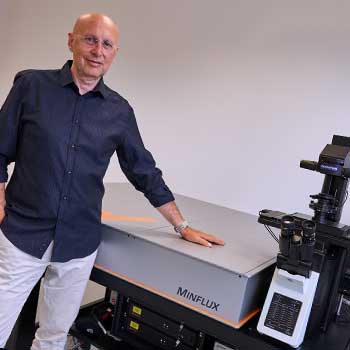
MINFLUX
The world’s most powerful fluorescence microscope MINFLUX, based on Stefan Hell’s invention, allows to resolve structures as small as a molecule in 3D.
MIRAVA POLYSCOPE
@ FOM, Taipei
Great people, fantastic conference, super feedback – thank you very much!


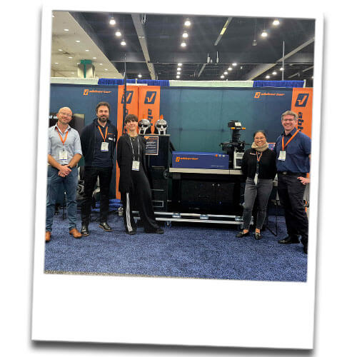
MIRAVA POLYSCOPE
@ BPS, Los Angeles
Thank you for visiting us at the BPS 2025. We enjoyed meeting you and talking to you.