



STEDYCON 2
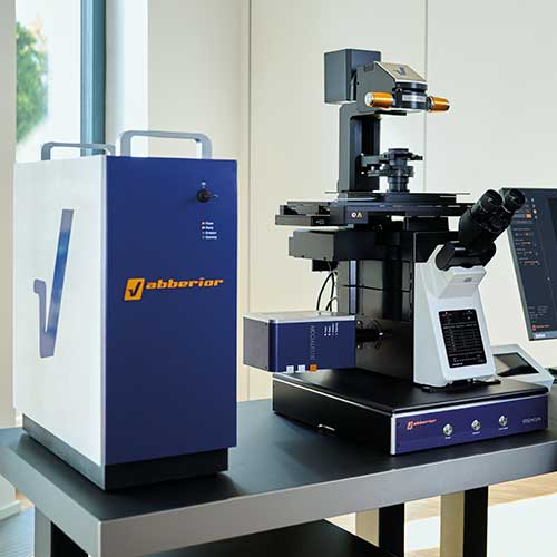
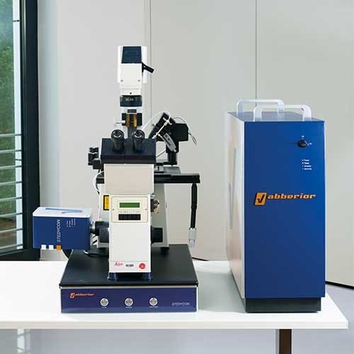
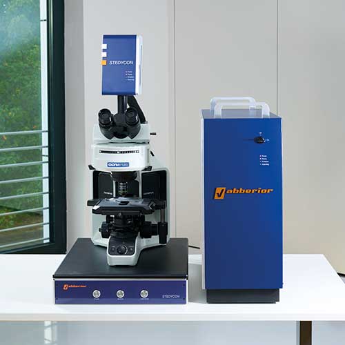
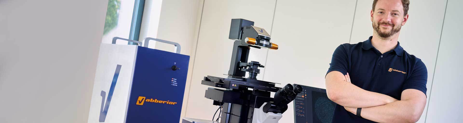
The new STEDYCON® 2 brings you uncompromised performance in a shoebox format. It transforms any widefield system into a powerful confocal, superresolution STED, and lifetime imaging machine. With its super-intuitive user interface, the STEDYCON provides an intelligent microscope platform that enables everyone to acquire superb superresolution and FLIM images after only minutes of training. Define your experiment and enjoy fine imaging at the push of a button!
Do you have an outdated microscope in your lab? Give it a new life as a STEDYCON with EVERGREEN, abberior’s sustainability initiative!
confocal, STED, lifetime … WOW!
Description
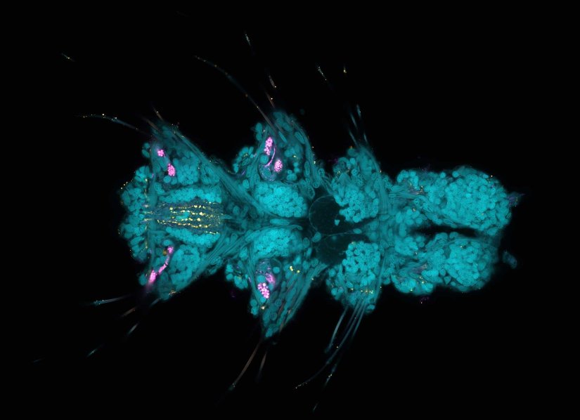
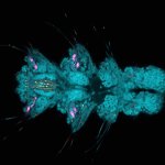
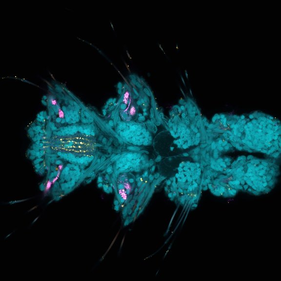
Larva of Platynereis dumerilii acquired in a confocal overview. Nuclei are shown in cyan (DAPI), tubulin in magenta, and serotonin-positive neurons in yellow.
Description
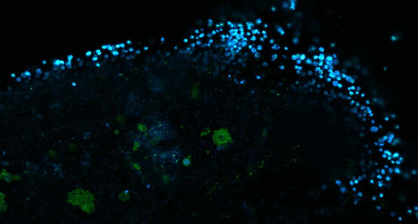
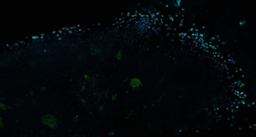
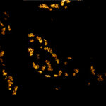
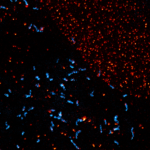
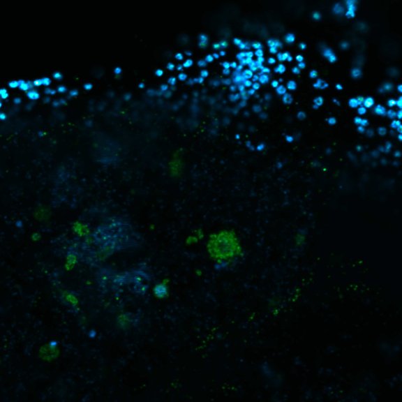
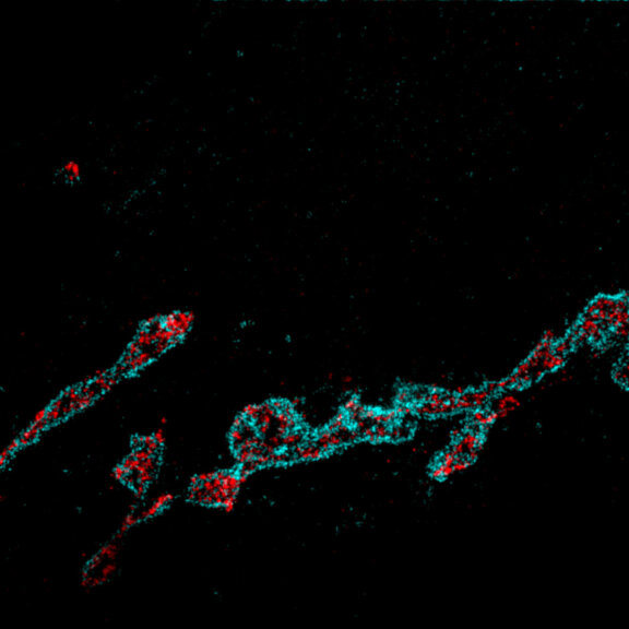
Human cytomegalovirus particles in infected human foreskin fibroblasts, labeled for a viral glycoprotein (abberior STAR RED, blue) and a tegument protein (abberior STAR 580, green). The sample was 4x gel-expanded and imaged with STED.
Description
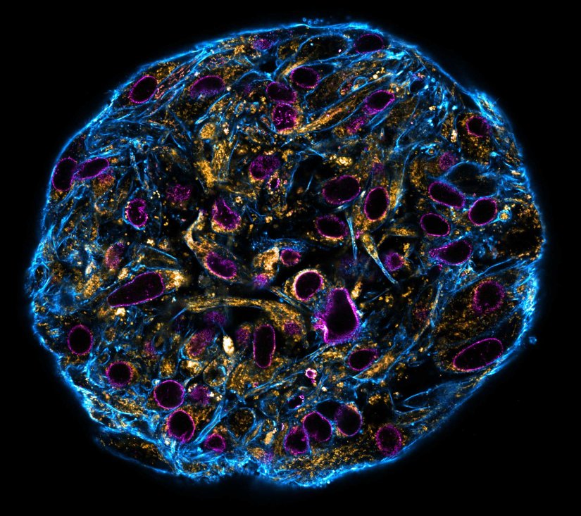
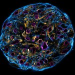
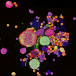
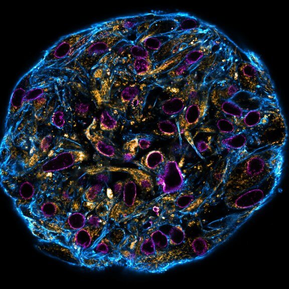
Confocal stitched image of a spheroid stained for nuclear pores (magenta), actin cytoskeleton (blue), and mitochondria (orange).
NIH-313 spheroid, fixed with 4% PFA, kindly provided by ibidi and prepared on µ-Slide VI 0.4 µ-Pattern ibiTreat.
Description
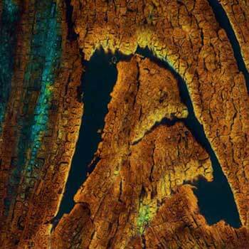
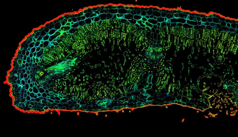
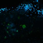
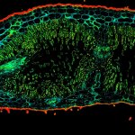
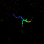
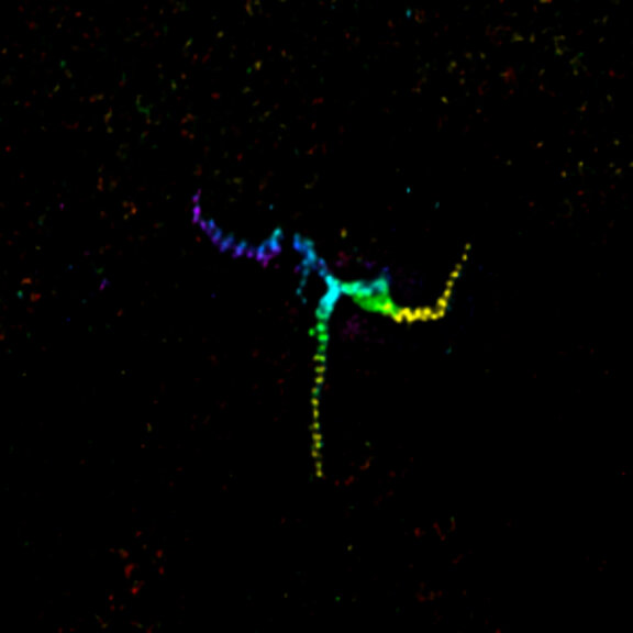
Confocal TIMEBOW acquisition of an Oleander leaf tip showing a range of autofluorescence lifetimes originating from different tissue components.
Description
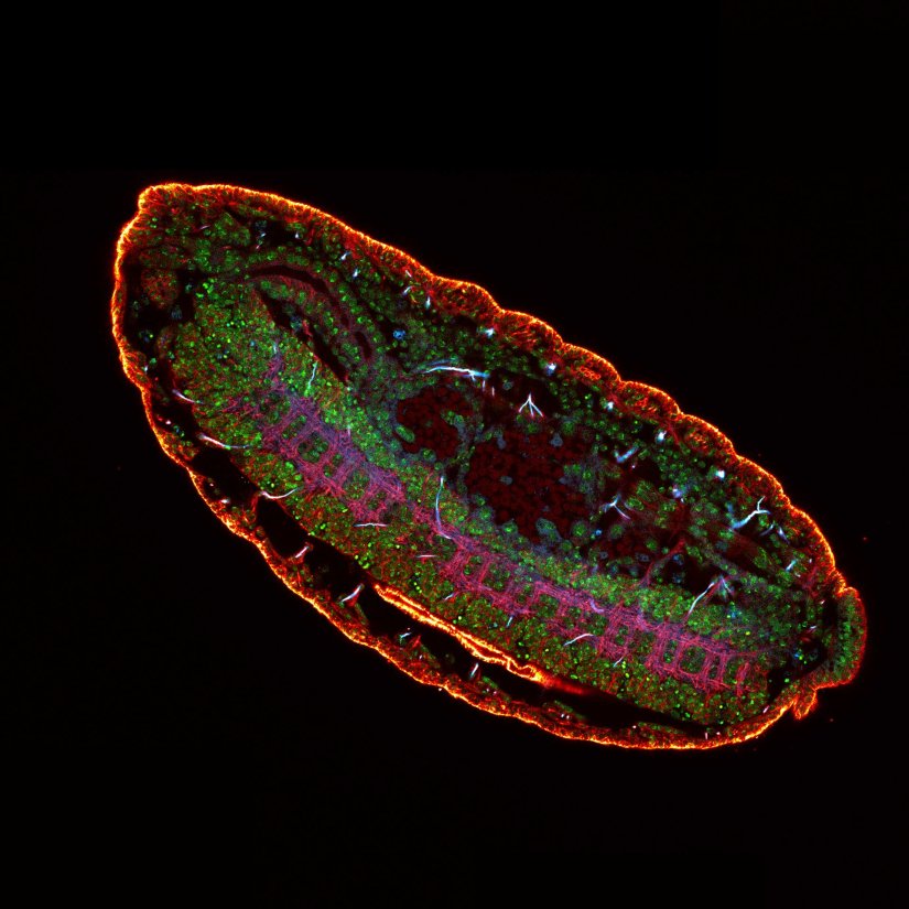
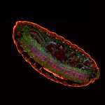
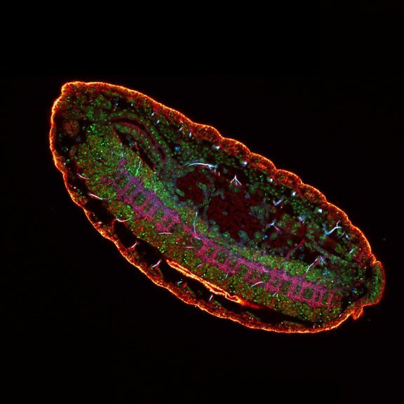
3 color confocal image of a Drosophila embryo stained for chitin (abberior STAR RED, cyan), tubulin (abberior STAR ORANGE, magenta), and DNA (PicoGreen, green).
Description
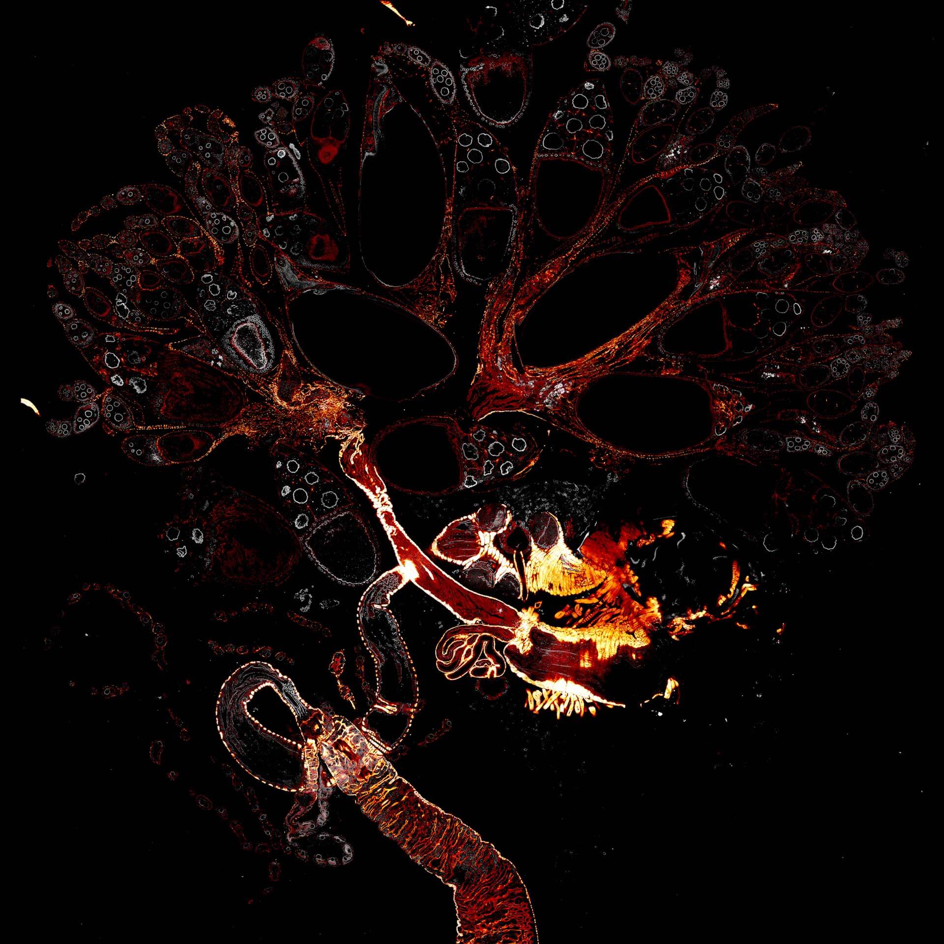
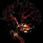
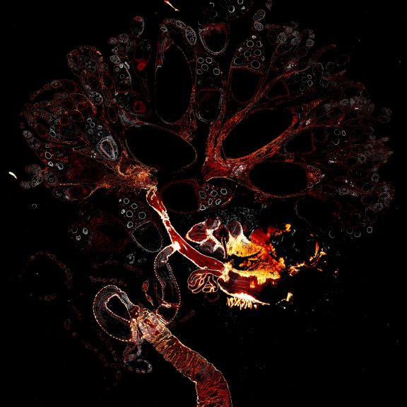
Drosophila female reproductive system stained for F-actin (red) with abberior STAR RED phalloidin. abberior STAR ORANGE is highlighting a nuclear pore protein (gray).
Image was acquired with the STEDYCON tiling feature and assembled with the SVI Huygens Stitcher.
Description
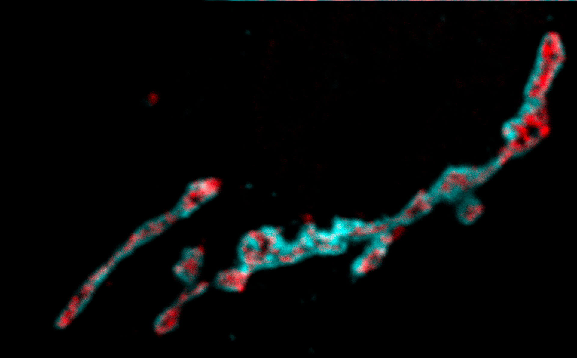
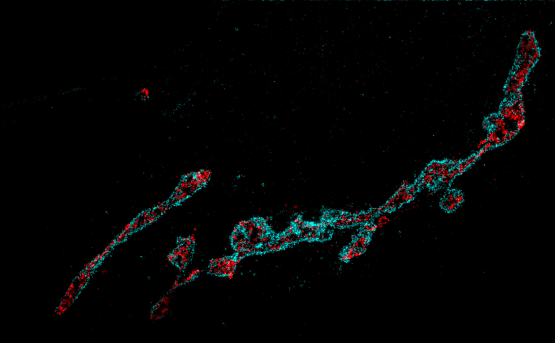
Two proteins in the Golgi apparatus were immunolabelled using primary antibodies specific for GM130 and Giantin and secondary antibodies coupled to abberior STAR 580 and abberior STAR 635P. Shown is RAW DATA. Images were acquired using a STEDYCON attached to a Zeiss Axio Imager Z2.
Description
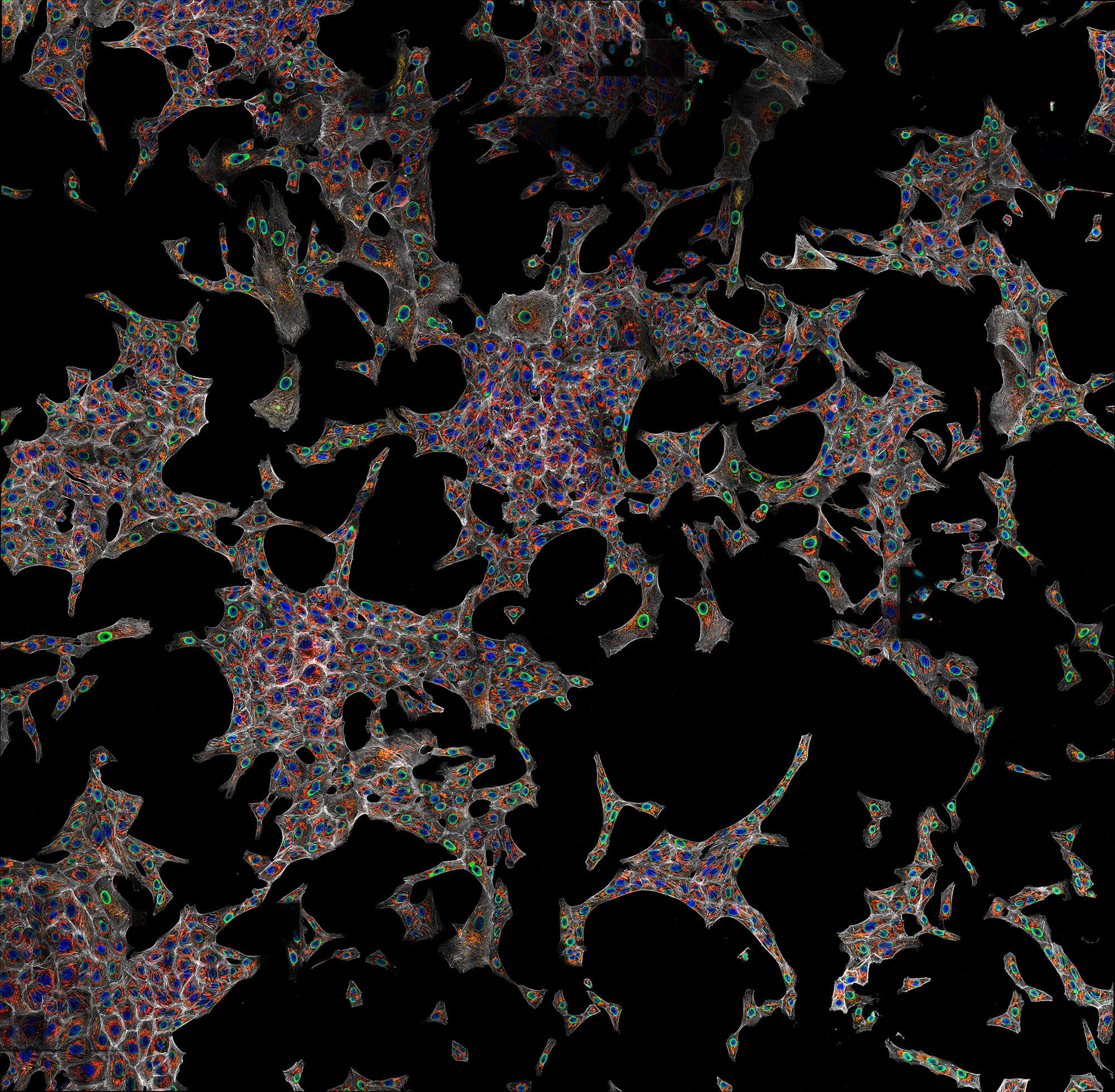
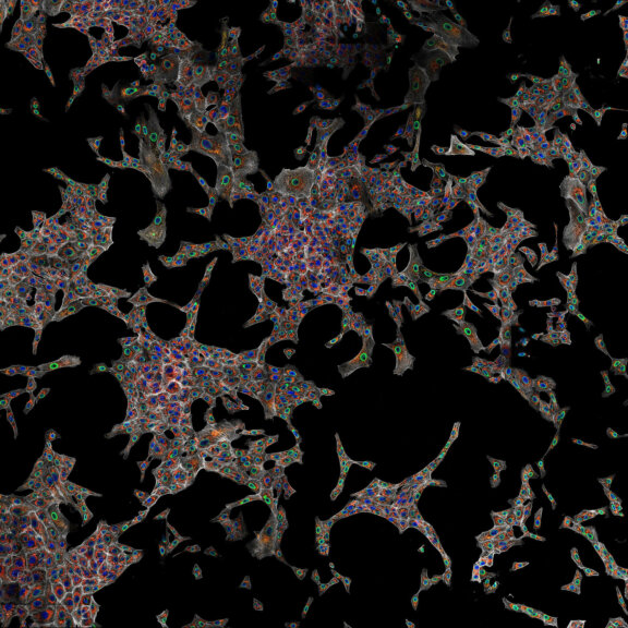
Confocal imaging of a large sample region. Shown is a 2.8 mm x 2.5 mm region, acquired using 35 by 29 tiles using the STEADYFOCUS and stitched with SVI Huygens. Sample: mammalian cells immunolabelled for the mitochondrial protein TOM20 (abberior STAR RED, red), double-stranded DNA to visualize mitochondrial DNA (abberior STAR ORANGE, green), phalloidin to label F-actin (abberior STAR GREEN, gray), and DAPI to label nuclei. Sample preparation by abberior GmbH.
Description
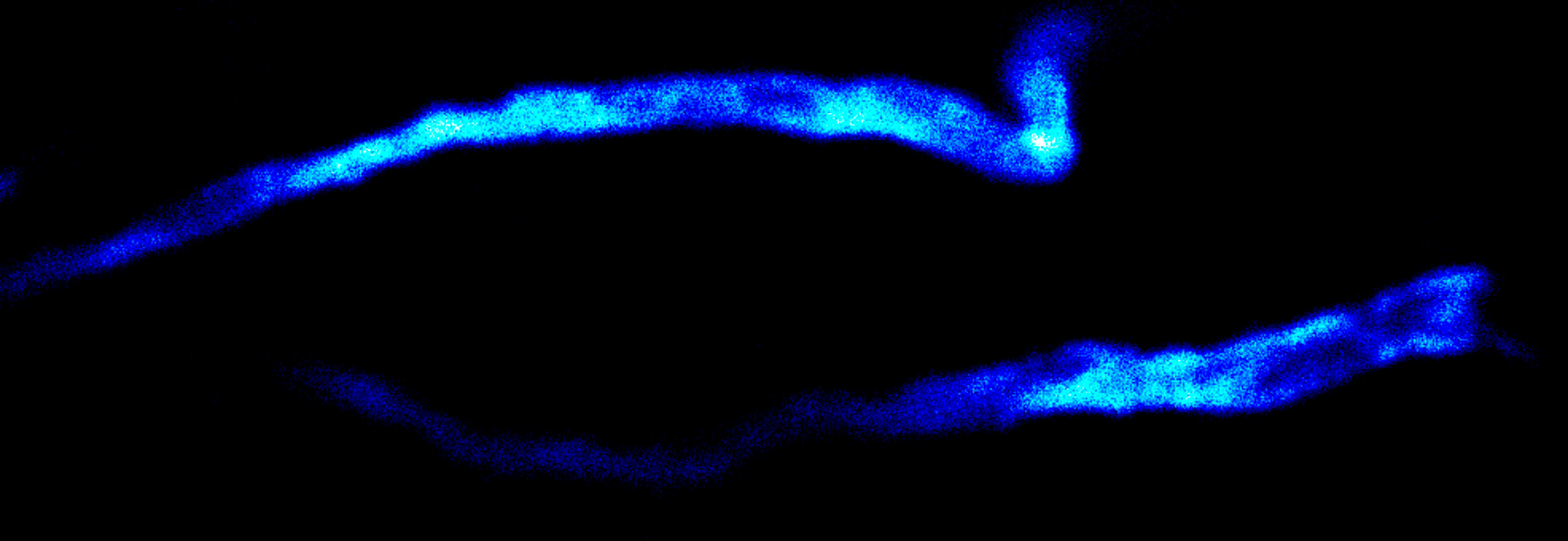
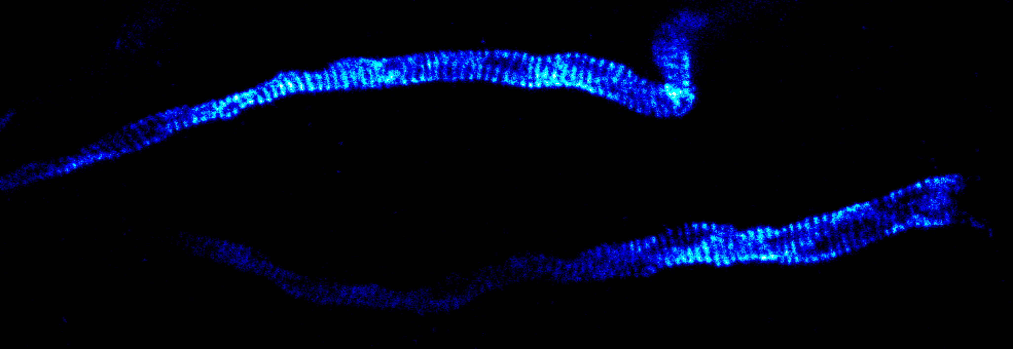
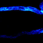
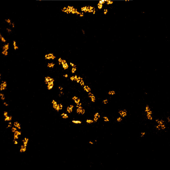
Immunolabelling of βIV-spectrin, a scaffolding protein of the axon initial segment, using a self-made primary antibody and Alexa 594 secondary antibody. Cryo-section of mouse neocortex cut at 20 µm. Images were acquired using a STEDYCON attached to a Zeiss AxioImager Z2. The sample was kindly provided by Dr. Maren Engelhardt, Institute of Neuroanatomy, Medical Faculty Mannheim, Heidelberg University.
Description
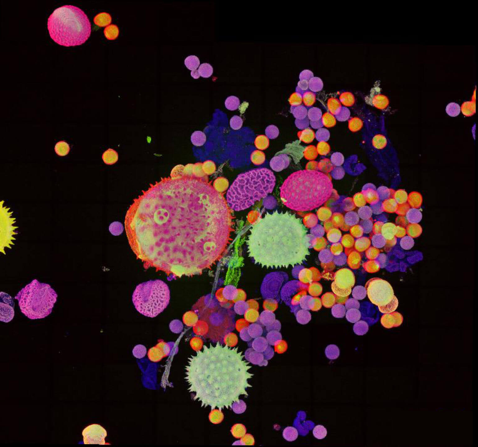
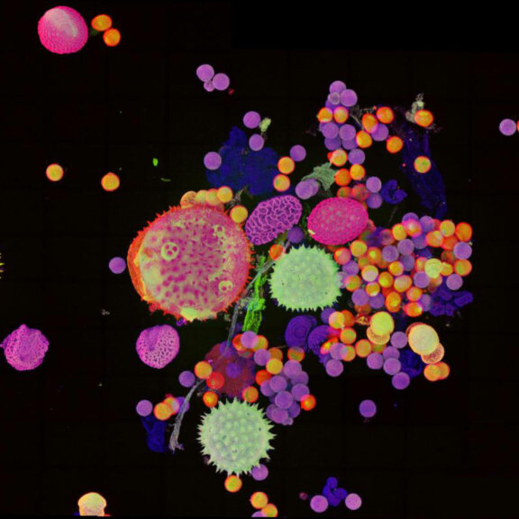
Pollen autofluorescence imaged with a STEDYCON on an IX83 microscope frame. z-stacks on 95 tiles were acquired and stitched with SVI Huygens.
Description
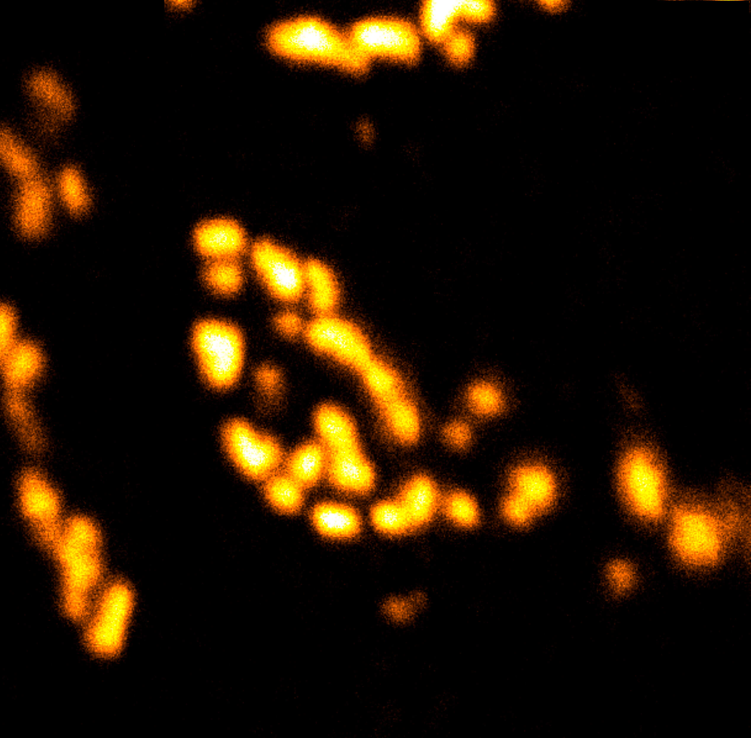
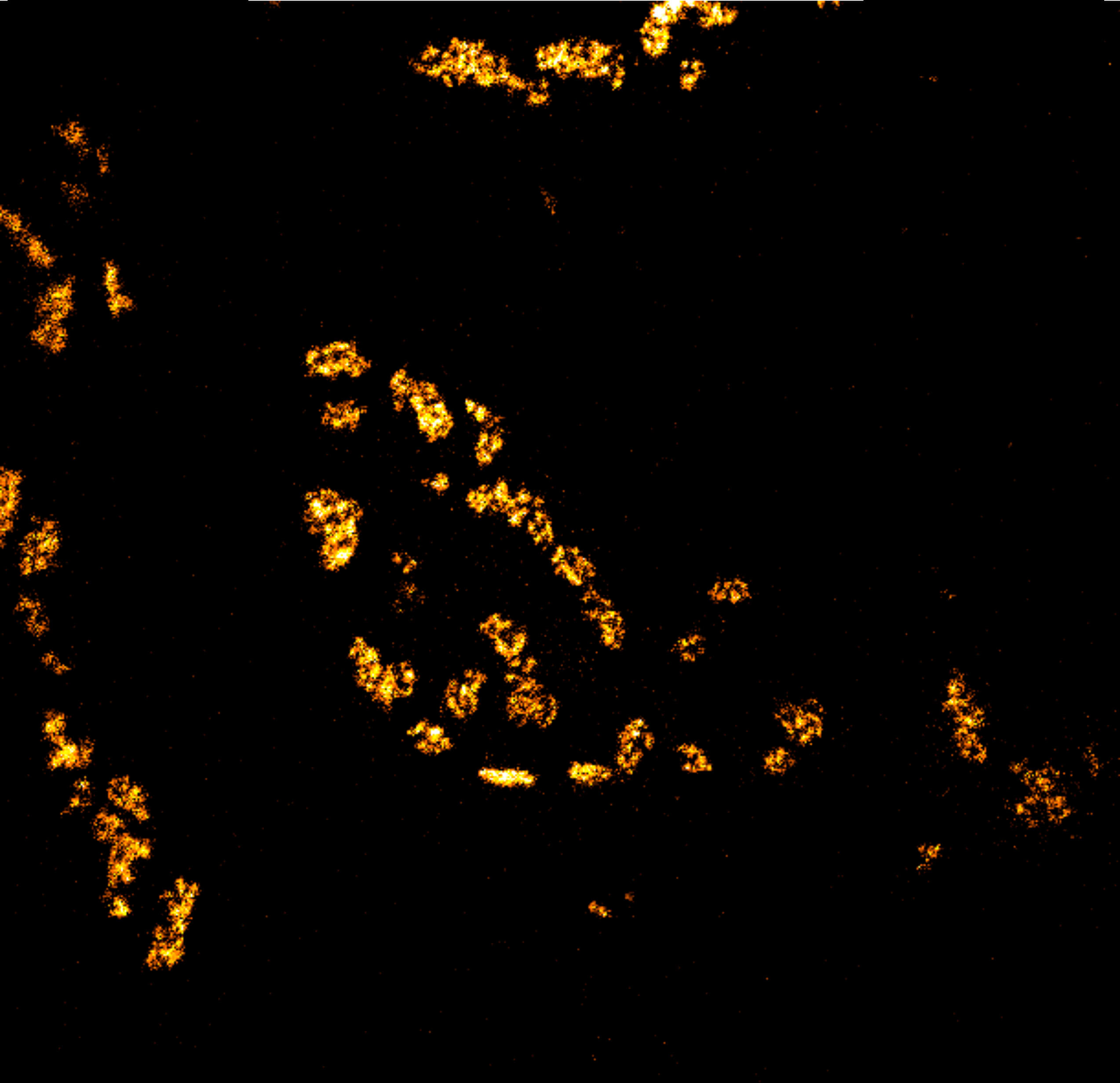
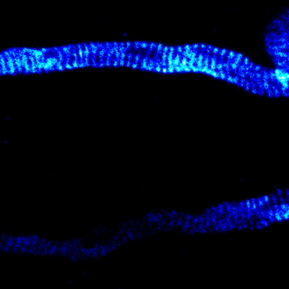
Body wall preparation of a 3rd instar Drosophila melanogaster larva. Synapses at the neuromuscular junction are visualized by immunostaining against Bruchpilot (Brp), an integral component of active zones in Drosophila. Images were acquired on a STEDYCON attached to an Olympus IX81. Images were kindly provided by Dr. Nadine Ehmann, Institute of Physiology, Neurophysiology, University of Würzburg.
Description
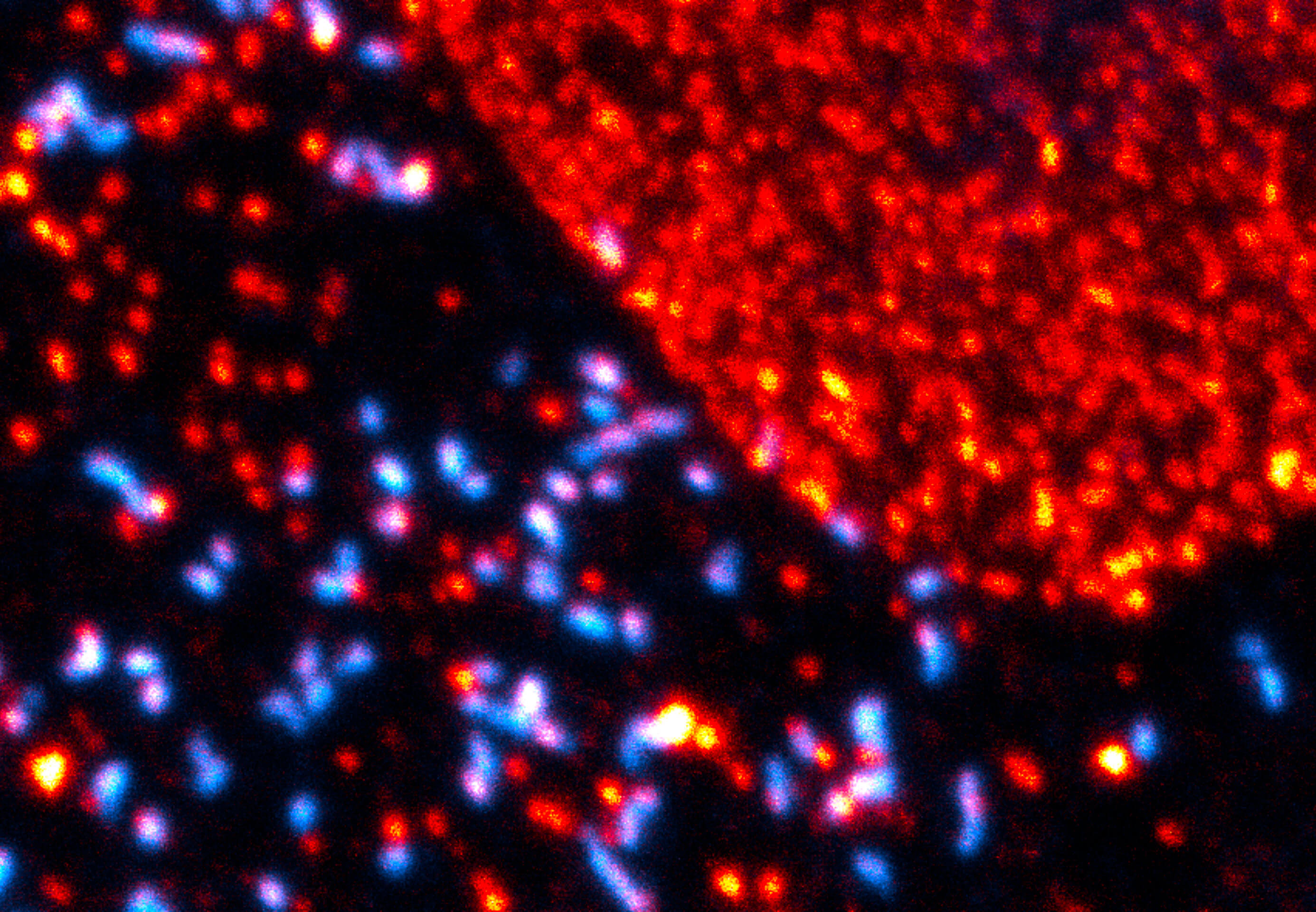
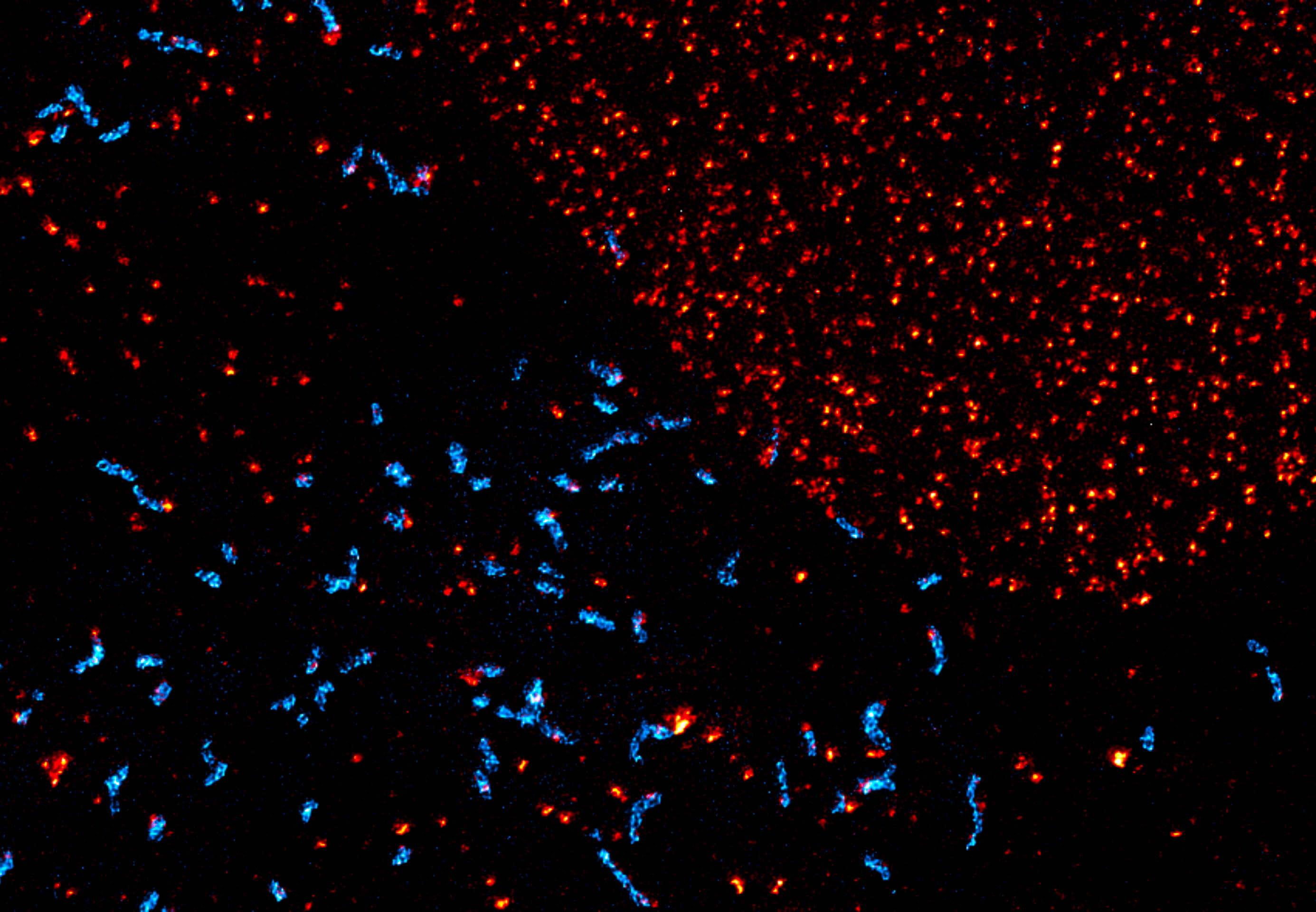
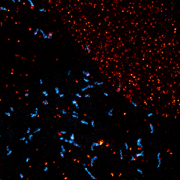
Maximum intensity projection of a z-stack showing nuclear pore complex protein (red, abberior STAR RED) and peroxisomes (cyan, Alexa 594) in mammalian cells. Imaged with abberior Instruments’ STEDYCON and deconvolved with SVI Huygens optimized for STEDYCON.
Description
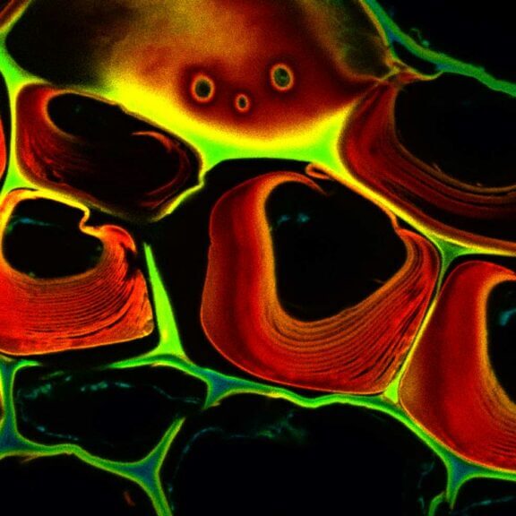
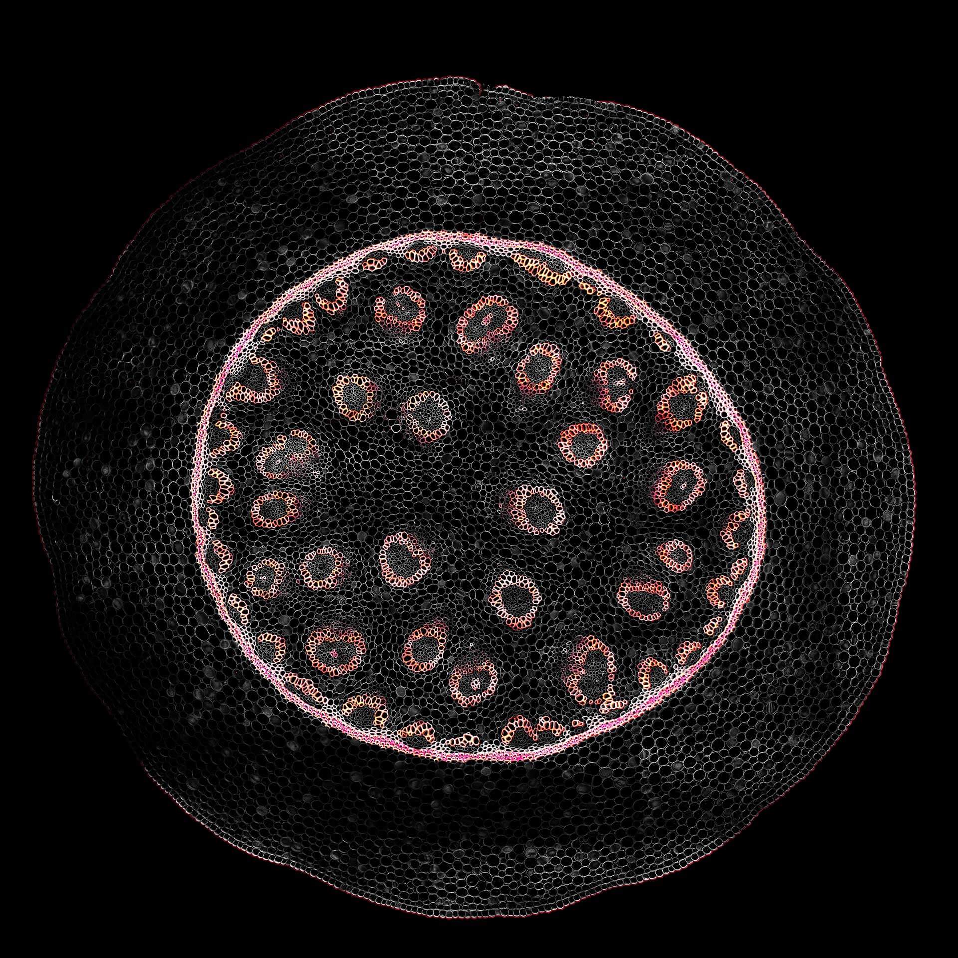
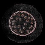
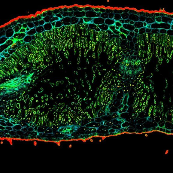
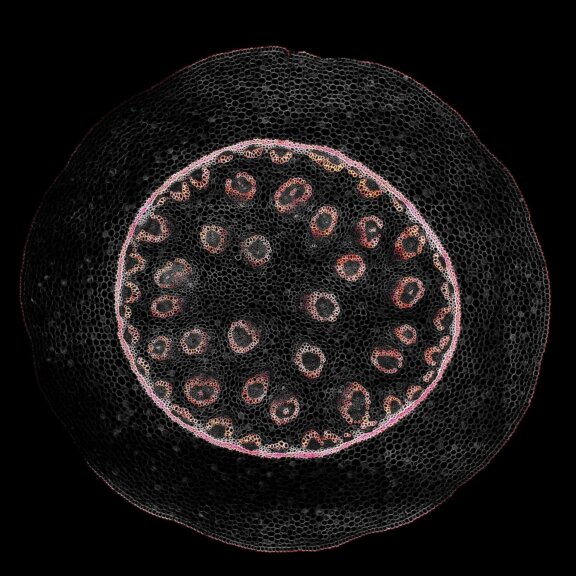
Confocal acquisition of Convallaria rhizome (cross-section) with a 20x oil objective; 3 channels, maximum intensity projection. The image consists of 14 z-planes in 9 by 9 tile pattern comprising a total area of 3.2 mm x 3.2 mm, stitched using SVI Huygens. Shown is a maximum intensity projection.
Description
Description
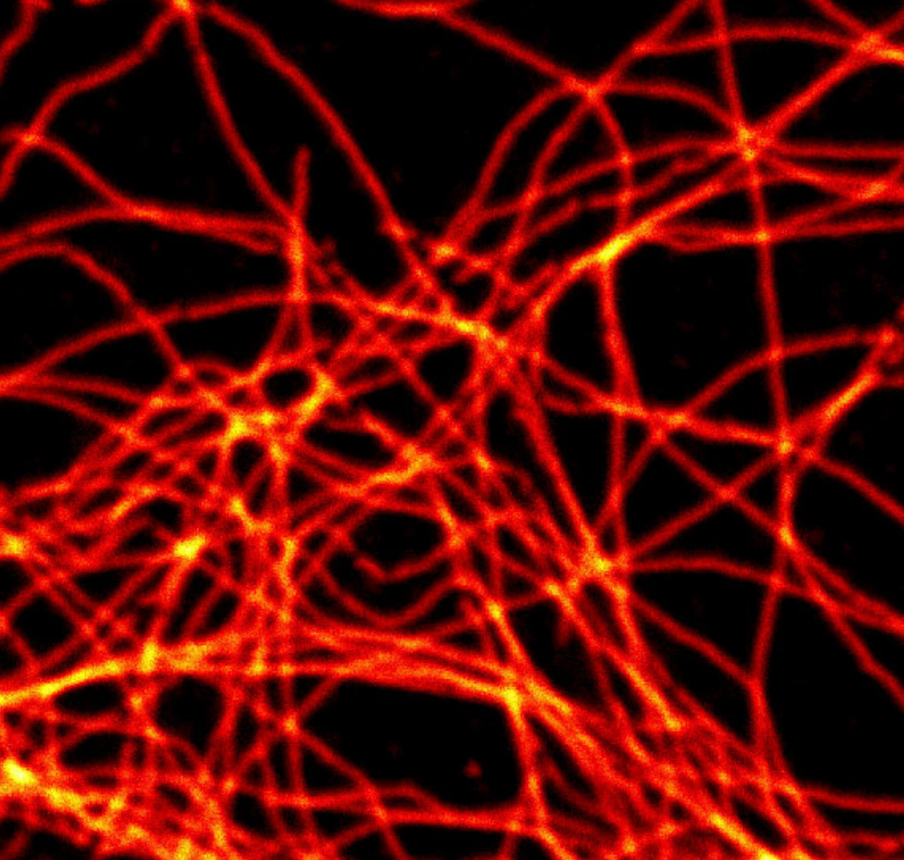
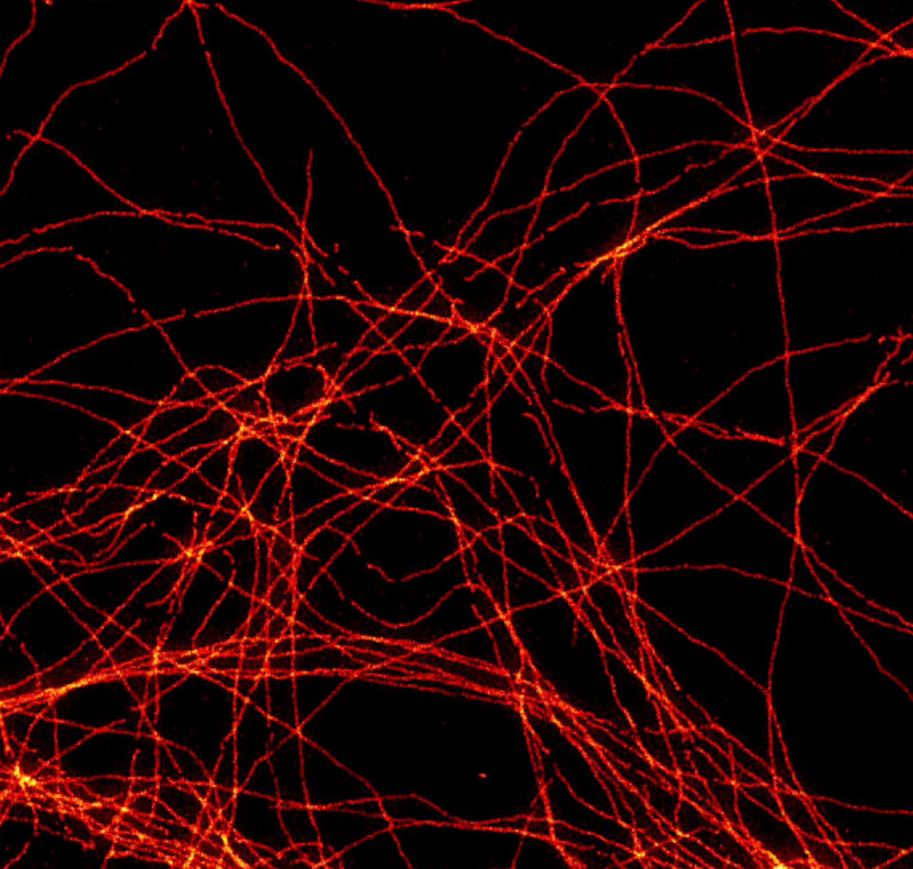
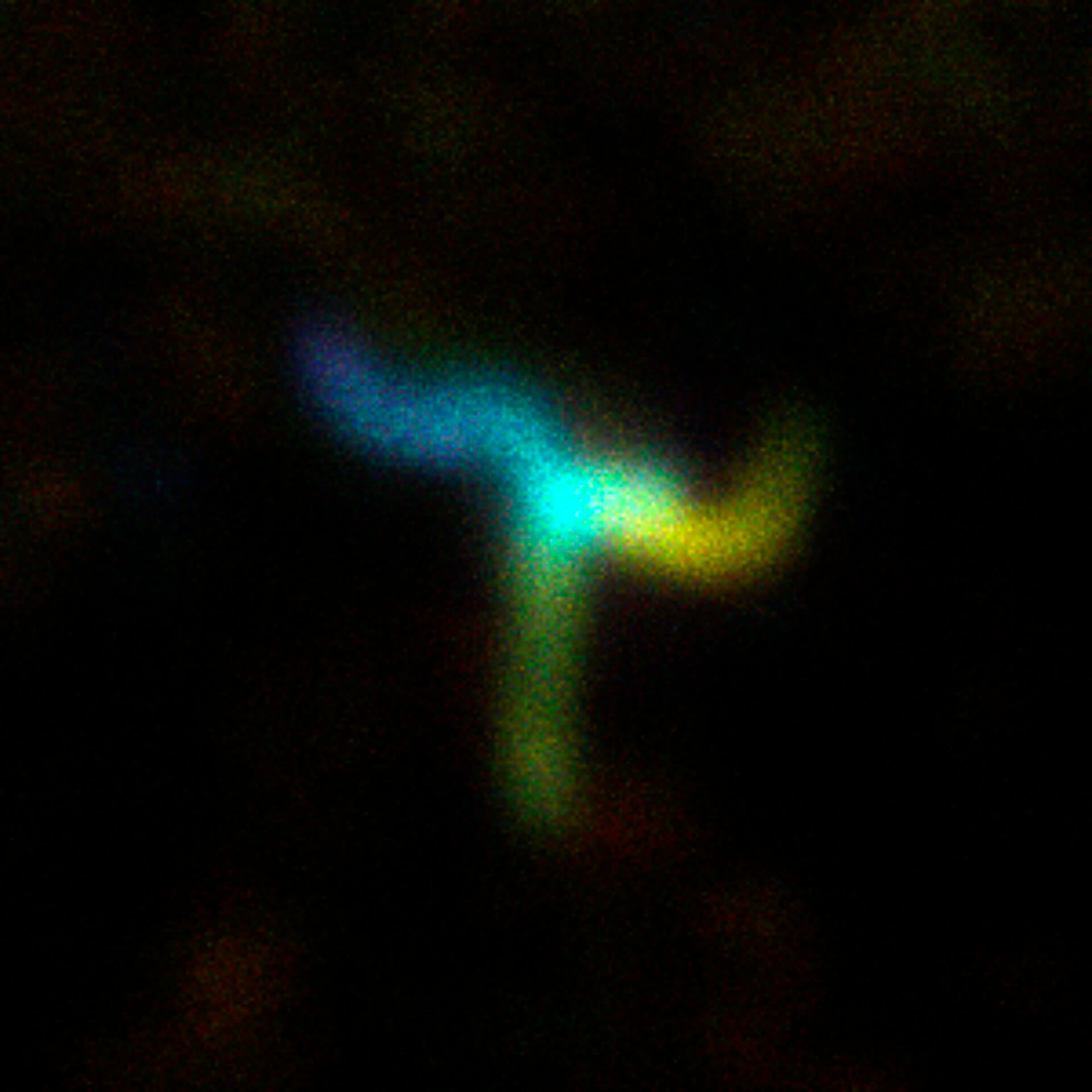
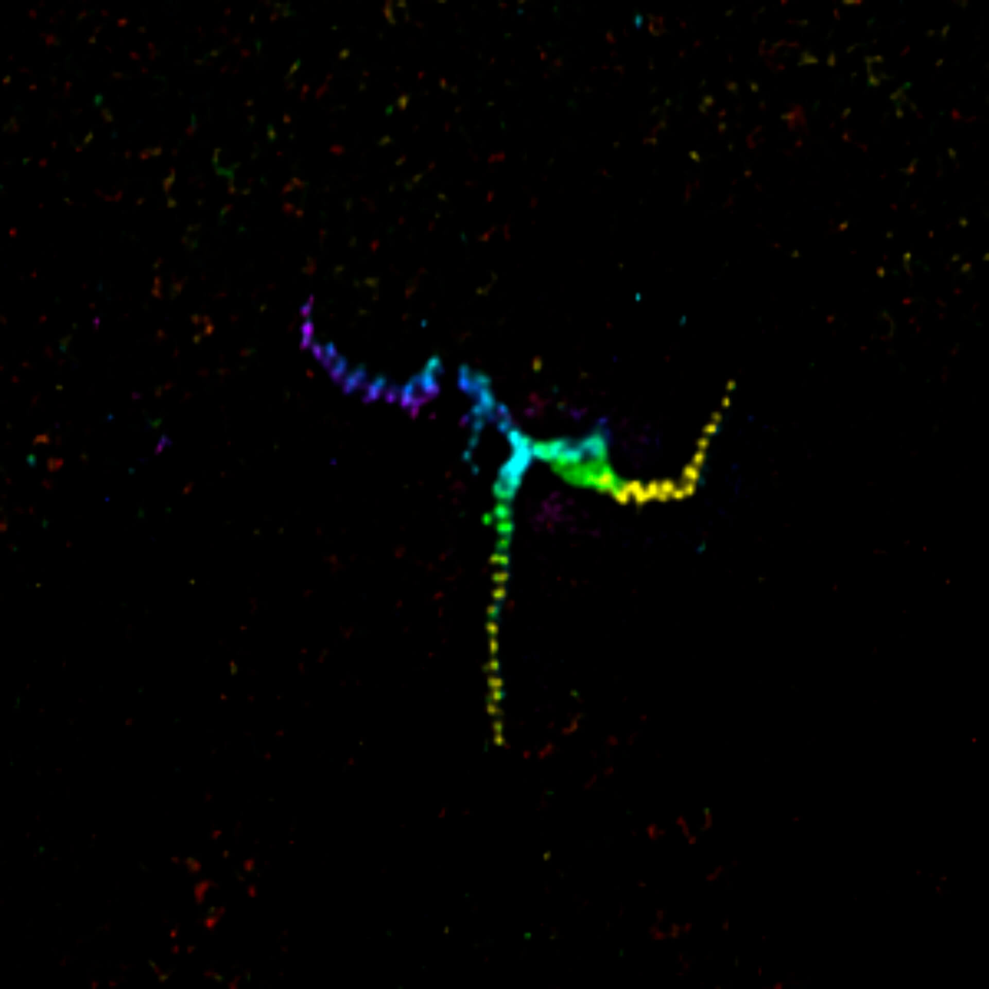
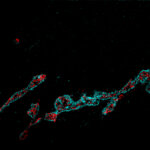
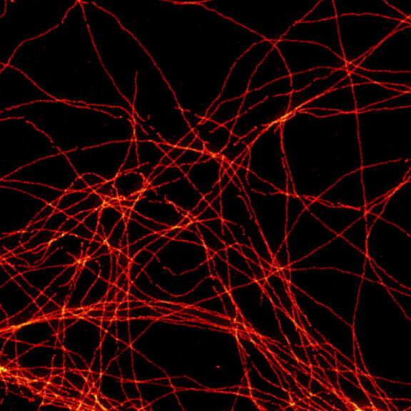
Centrosome linker. U2OS cells in which the centrosome-linker-protein rootletin was immunolabelled using secondary antibodies coupled to abberior STAR RED. The sample was prepared by R. Vlijm at MPI for Medical Research, Heidelberg, Germany. Imaged with abberior Instruments’ STEDYCON and deconvolved with SVI Huygens optimized for STEDYCON.
Description
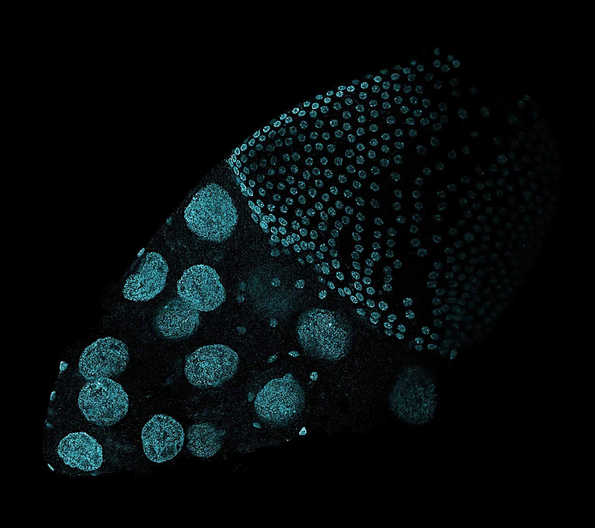
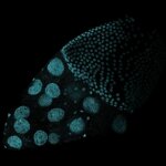
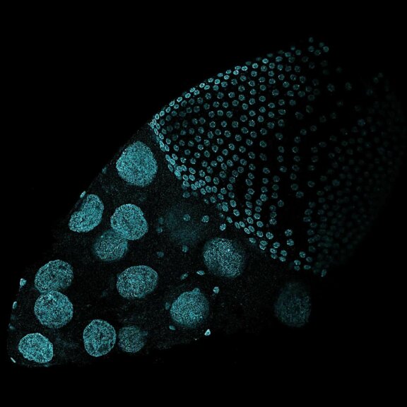
Drosophila ovariole stained with abberior LIVE 560 DNA, showing the different cell types of the egg chamber. The image was stitched using SVI Huygens.
Description
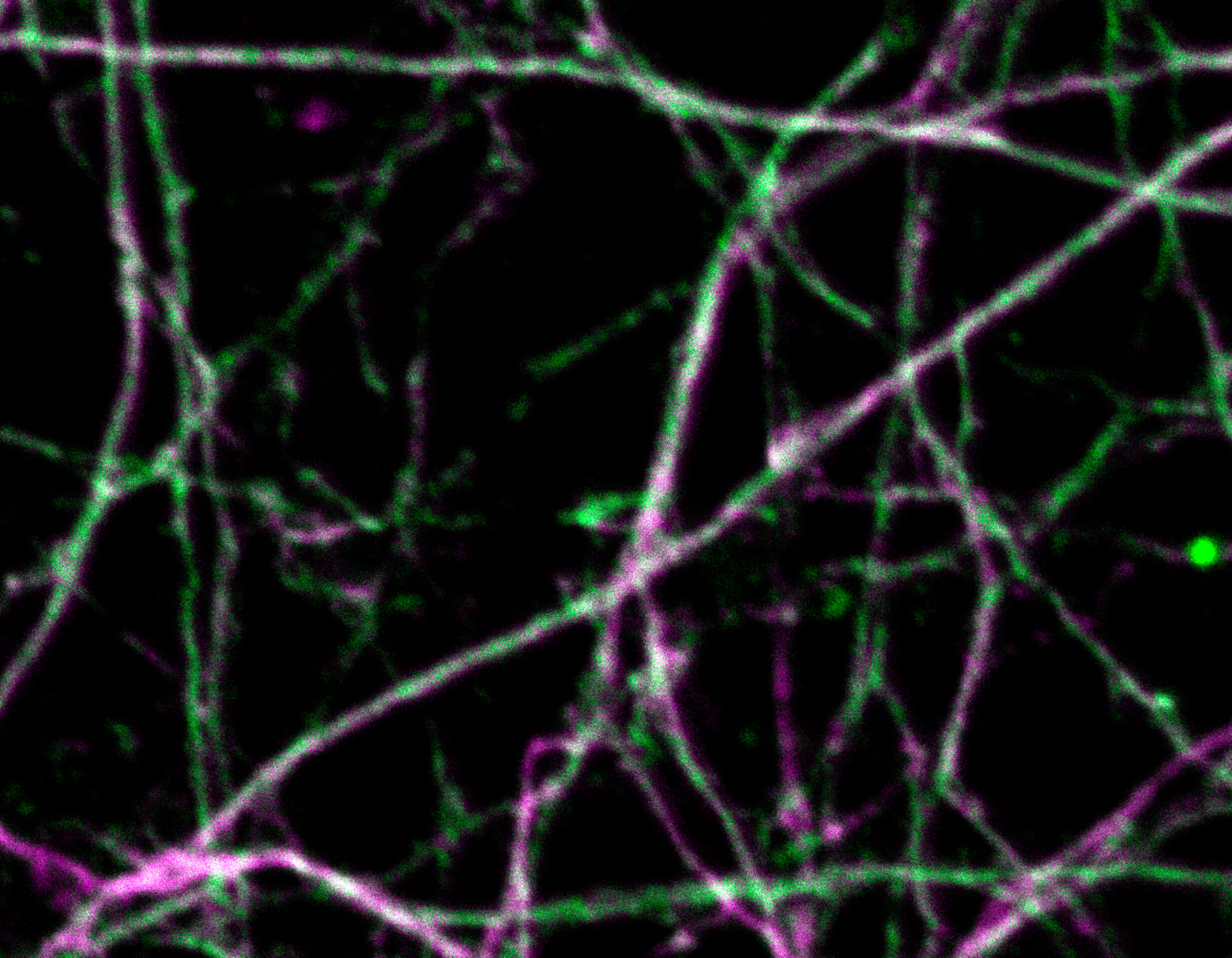
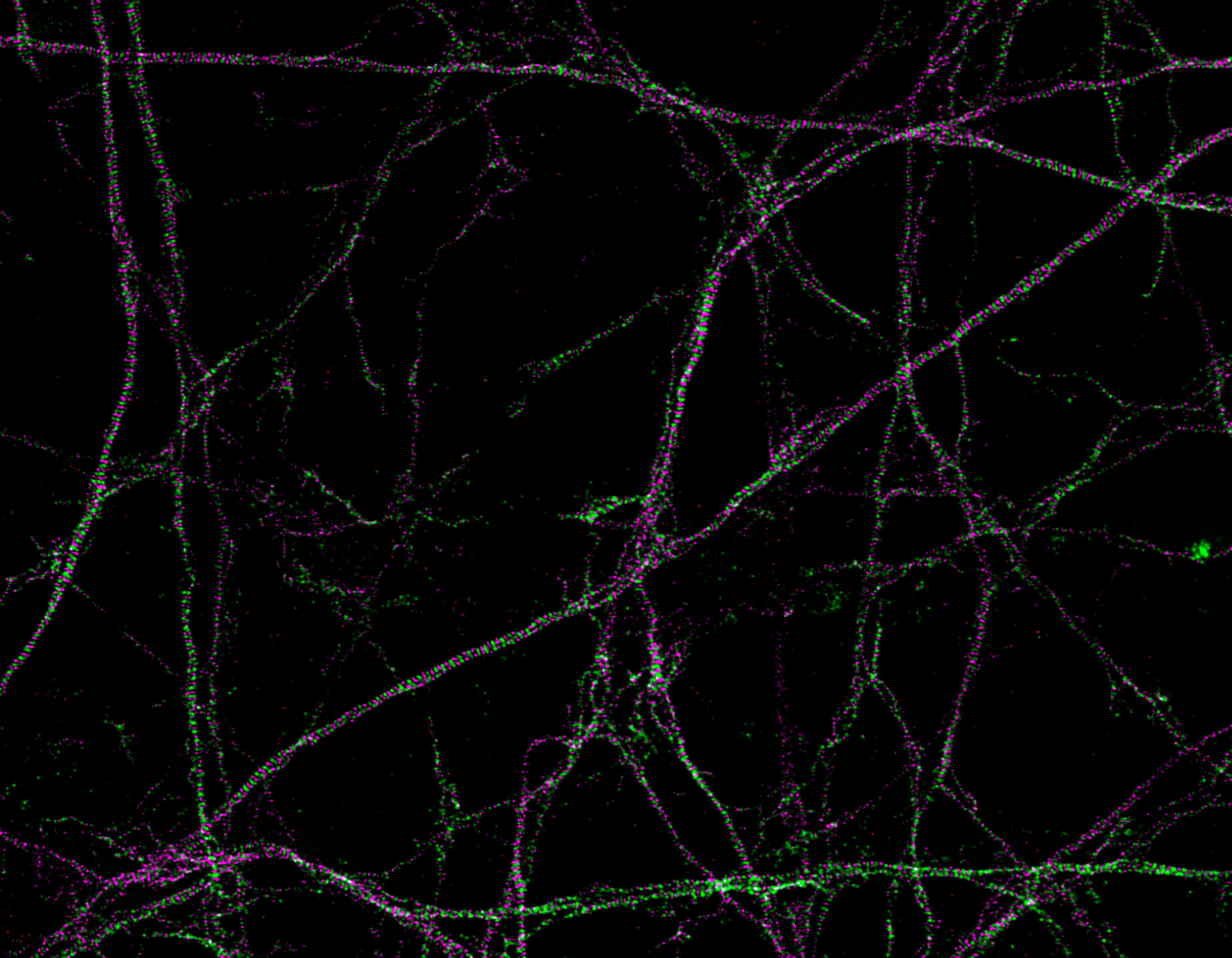
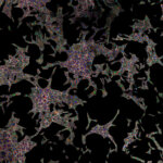
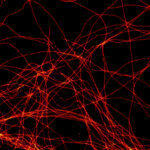
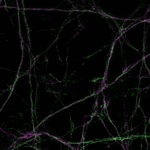
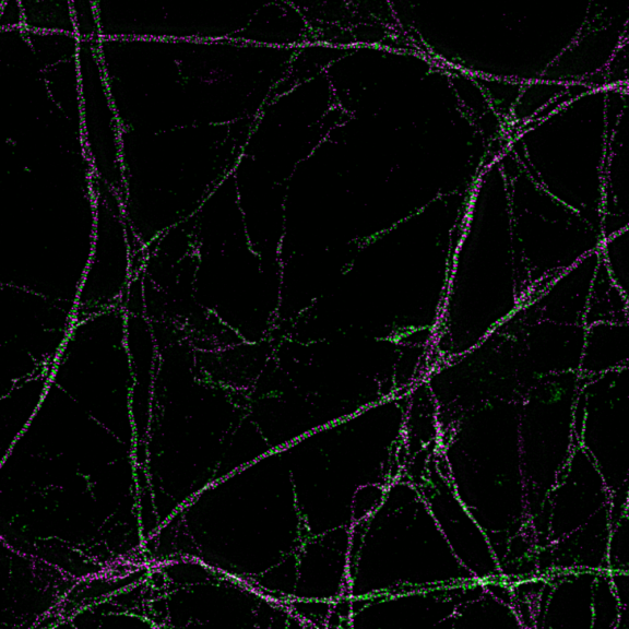
Primary hippocampal neurons with cytoskeleton proteins labeled (magenta, alpha-Adducin, abberior STAR 635P and green, ßII spectrin, Alexa 594). Imaged with abberior Instruments’ STEDYCON and deconvolved with SVI Huygens optimized for STEDYCON.
FAQ Video 19

“How did you manage to fit both, a STED and a confocal microscope, in a shoe box?”
STEDYCON‘s
highlights
- STED superresolution down to 30 nm
- TIMEBOW lifetime imaging, FLIM fully integrated
- Revolutionary design and flexibility, upgrades any microscope frame
- Reliability and stability as a principle
- Ease-of-use at its best with intuitive, browser-based software
- Low cost of ownership compared to other confocal and STED microscopes
From zero to hero
Plug-and-play for serious science
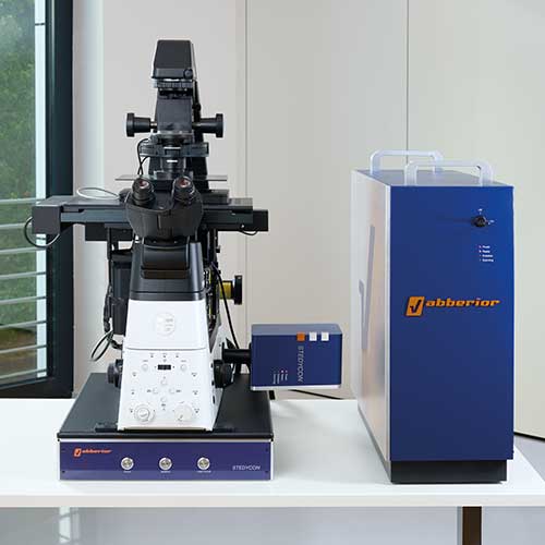
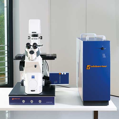
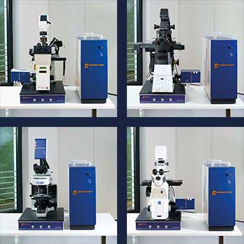
STEDYCON transforms almost any microscope frame into a full-fledged confocal and superresolution system, independent of the manufacturer, whether it is upright or inverted, old or new. All it needs is a free camera port and a good objective lens. Lasers, scanners, and detectors are already inside the STEDYCON box. That’s why connecting the STEDYCON takes just a few minutes.
Get in touch with us to upgrade your system!
STEDYCON will never let you down
Runs anywhere. Works every time.
STEDYCON, with its uniquely compact design, is engineered for ultimate robustness. Its lasers – including the STED laser – are coupled into a single optical fiber – meaning that they are aligned by design, eliminating beam drift and ensuring consistent, high-performance imaging without the need for recalibration, ever.
So no matter where you work – whether in climatized laboratories or standard office environments – STEDYCON will always deliver a rock-solid performance. It is virtually maintenance-free and all parts that require fine-tuning are motorized, allowing most adjustments to be handled remotely.
Beautifully simple and surprisingly powerful
STEDYCON’s smart control software
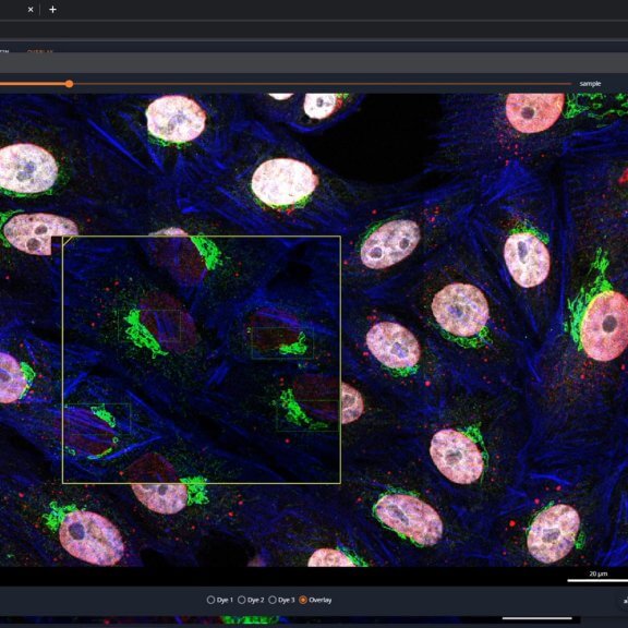
Discover the elegance of STEDYCON’s smart control software – devised to get you from system start to stunning superresolution in no time. Of course, with confocal imaging along the way. All that’s required is a few minutes of training, and off you go!
Switching between confocal mode and superresolution STED is just one click, and lifetime data is always within reach with the fully integrated TIMEBOW module.
And if you have a complex imaging task that takes a couple of hours, you won’t need to spend the time in front of the microscope. Monitor progress from the back of the seminar room, from home – anywhere with internet access. As the software is browser-based, you can control it from any device, anywhere, at any time.
Confocal, STED, and lifetime imaging
STEDYCON has it all
Experience first-class confocal imaging with state-of-the-art features tailored for a wide range of applications. Capture dynamic biological processes with live cell imaging, analyze the distribution of your protein of interest, perform multicolor experiments, and acquire z-stacks and volumetric data. Swiftly adapt the system to your specimen, from single cells to tissues and whole organisms. Of course, you can also mount delicate expanded specimens.
If your experiment demands more detailed images, switch to superresolution STED with a single click. Start with gentle, low STED power for an already significant increase in resolution. Add lifetime information using TIMEBOW STED for an additional resolution boost without harming your sample. Need even more detail? Push the resolution to 30 nm for best results.
Do you need lifetime information about your sample? Instantly access TIMEBOW lifetime imaging. Lifetime imaging on the STEDYCON is powered by PicoQuant’s established MultiHarp 150 hardware. Fully integrated into the STEDYCON smart control software, command is seamless and intuitive with no need for external tools or expertise.
STEADYFOCUS
in focus, forever
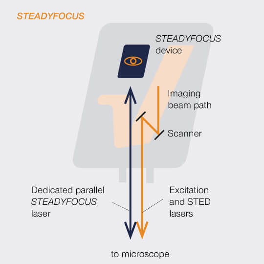
The STEADYFOCUS laser runs side by side with the imaging beam path. No dichroics are required and there is absolutely no interference with imaging.
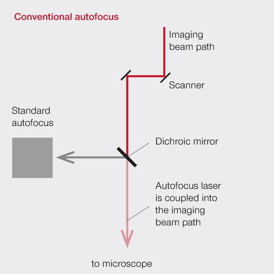
The autofocus laser is coupled into the beam path using a dichroic mirror, which interferes with imaging.
Our STEADYFOCUS keeps the focus position stable. Forever. During STED imaging, when browsing through the sample, or for hours of time-lapse imaging, there is no need for refocussing, the STEADYFOCUS does the work for you. And everything you need for it is integrated inside the STEDYCON head, so it works on upright microscopes, too!

.
.
STEADYFOCUS
tiling and stitching is a piece of cake
Discover your samples! Acquire a quick tiled overview as a basis for tiled scans with overlap or for multi-position imaging. Define your experiment, let the microscope image for you, with active drift correction thanks to our STEADYFOCUS!
After tiled image acquisition, send over the data to Huygens, for easy, fast and reliable stitching – all needed parameters are automatically transferred.
Get the video.
.
Multipositioning
let our STEDYCON do the work
Discover your whole sample, and then take your time to define your experiment – at which position would you like to acquire a z-stack, or a single image, or a tiled scan? Then let the microscope do the imaging for you. Increase your working efficiency, let it run over night, or already prepare your next experiment! It saves time and endless clicks …