Sample gallery
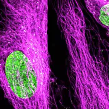
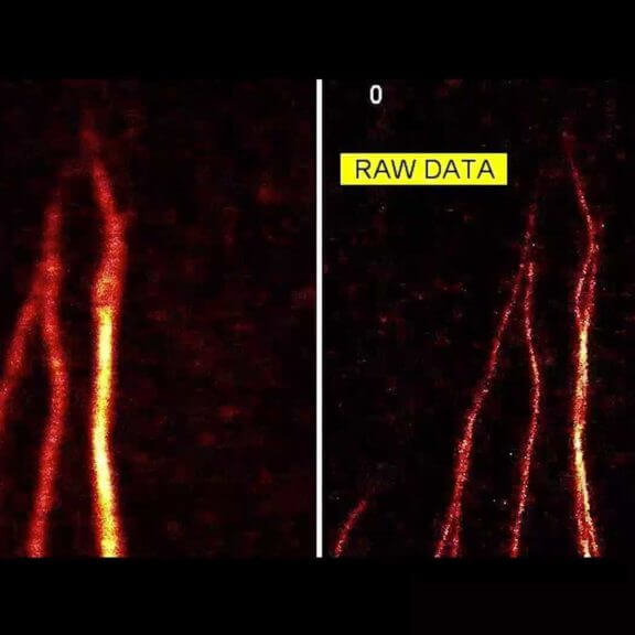
Fluorescence imaging, whether at confocal, STED or MINFLUX resolution, guarantees unique insights into the function and structure of life at the molecular level. Besides the scientific information content, some sample portraits provide simply beautiful images. Enjoy browsing our sample gallery.
the fine art of science
Description
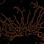
Video of mitochondria in living U2OS cells imaged for 1.5 hours with STED and deconvolved with TRUESHARP image boosting. Mitochondria were visualized with abberior LIVE SiR HaloX staining the outer mitochondrial membrane marker TOMM20. FLEXPOSURE adaptive illumination was used to reduce the light burden on the sample.
Description
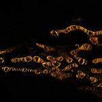
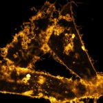
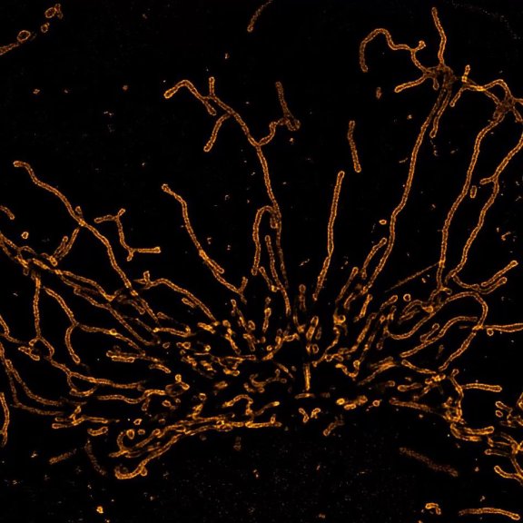
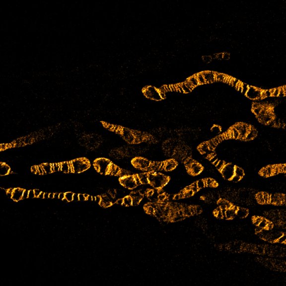
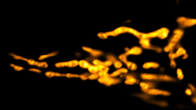
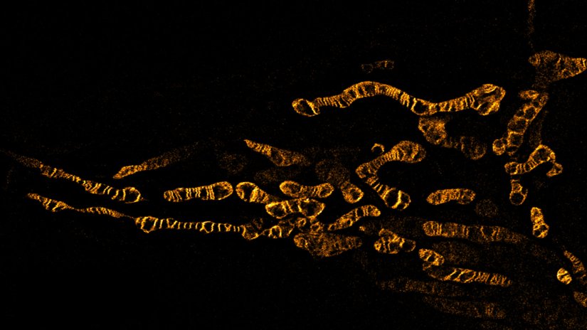
Living HeLa cells stained with the mitochondrial membrane marker abberior LIVE ORANGE mito, visualizing both outer and inner membranes. Confocal and STED images where deconvolved with TRUESHARP image boosting.
Modules:
Description
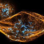
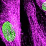
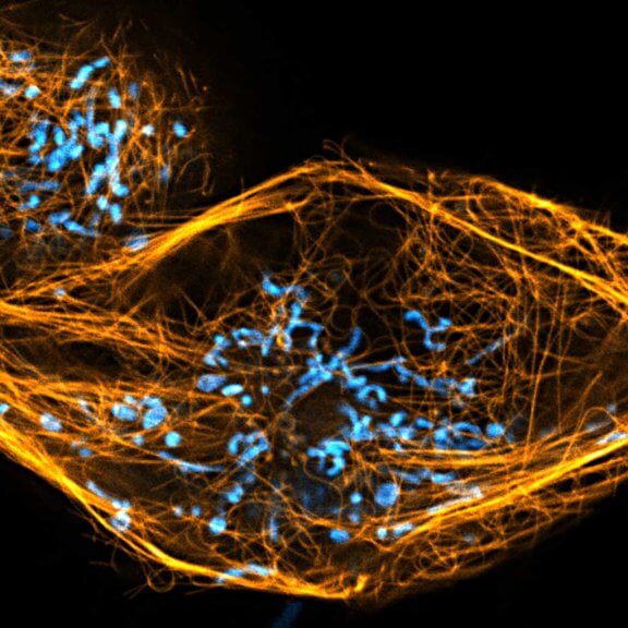
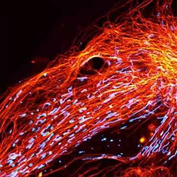
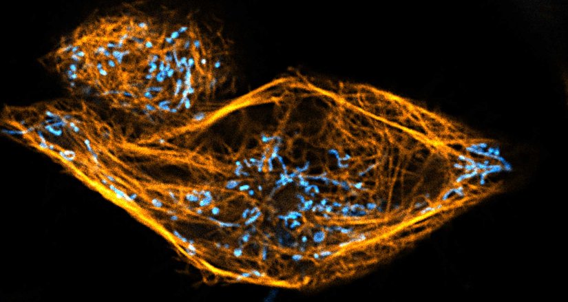
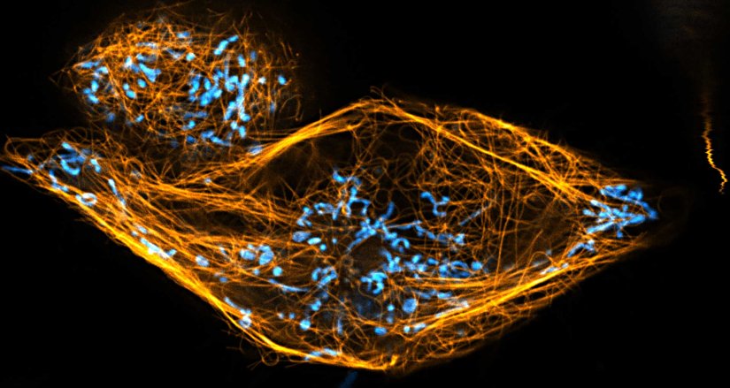
Two color live-cell confocal and STED image of a mammalian cell directly labeled with abberior LIVE 510 mito (cyan), and LIVE RED tubulin. This image was acquired with a STEDYCON microscope.
Description
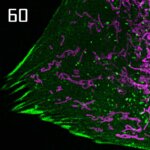
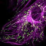
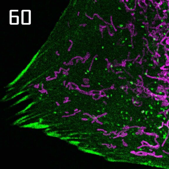
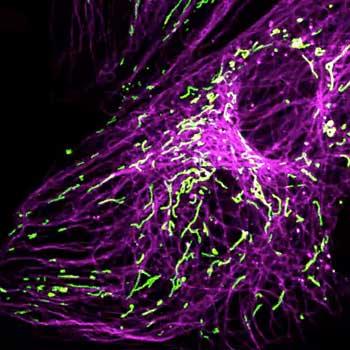
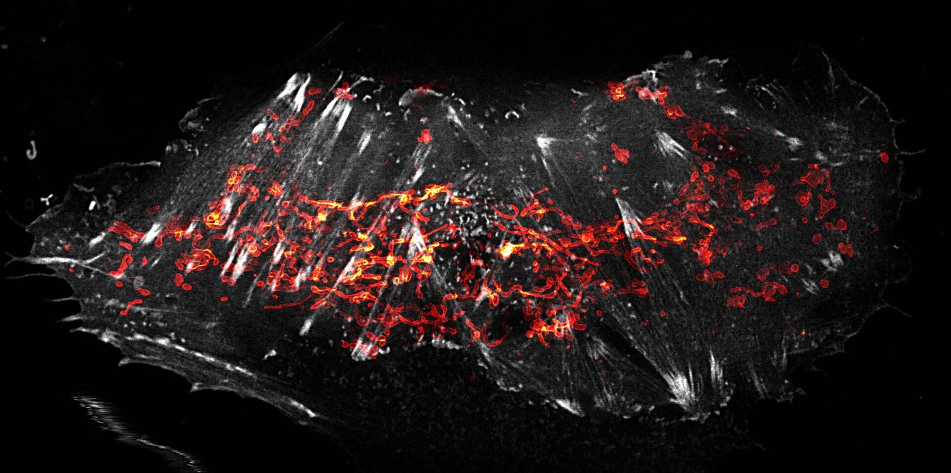
OMP25-SNAP protein in mitochondria labeled with abberior LIVE 610 SNAP (magenta) and Actin^K118TAG labeled with abberior LIVE 550 click (green). Movie with 60 frames.
Description
Description
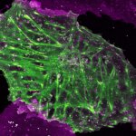
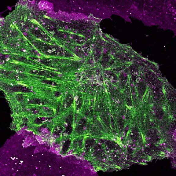
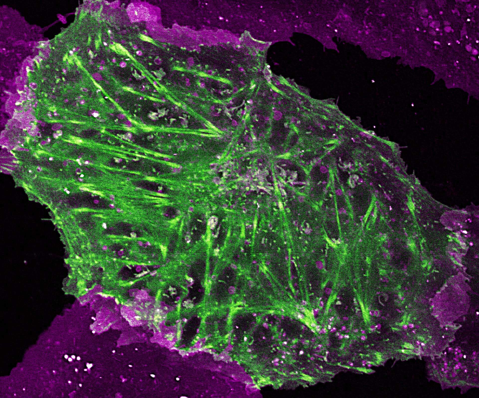
2-color STED image of a living cell expressing actin K118TAG incorporating TCO*A labeled with abberior LIVE 590 click (green). Plasma membrane is highlighted with abberior STAR RED membrane (magenta).
Description
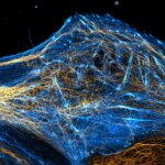
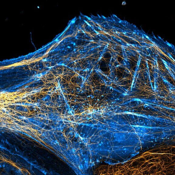
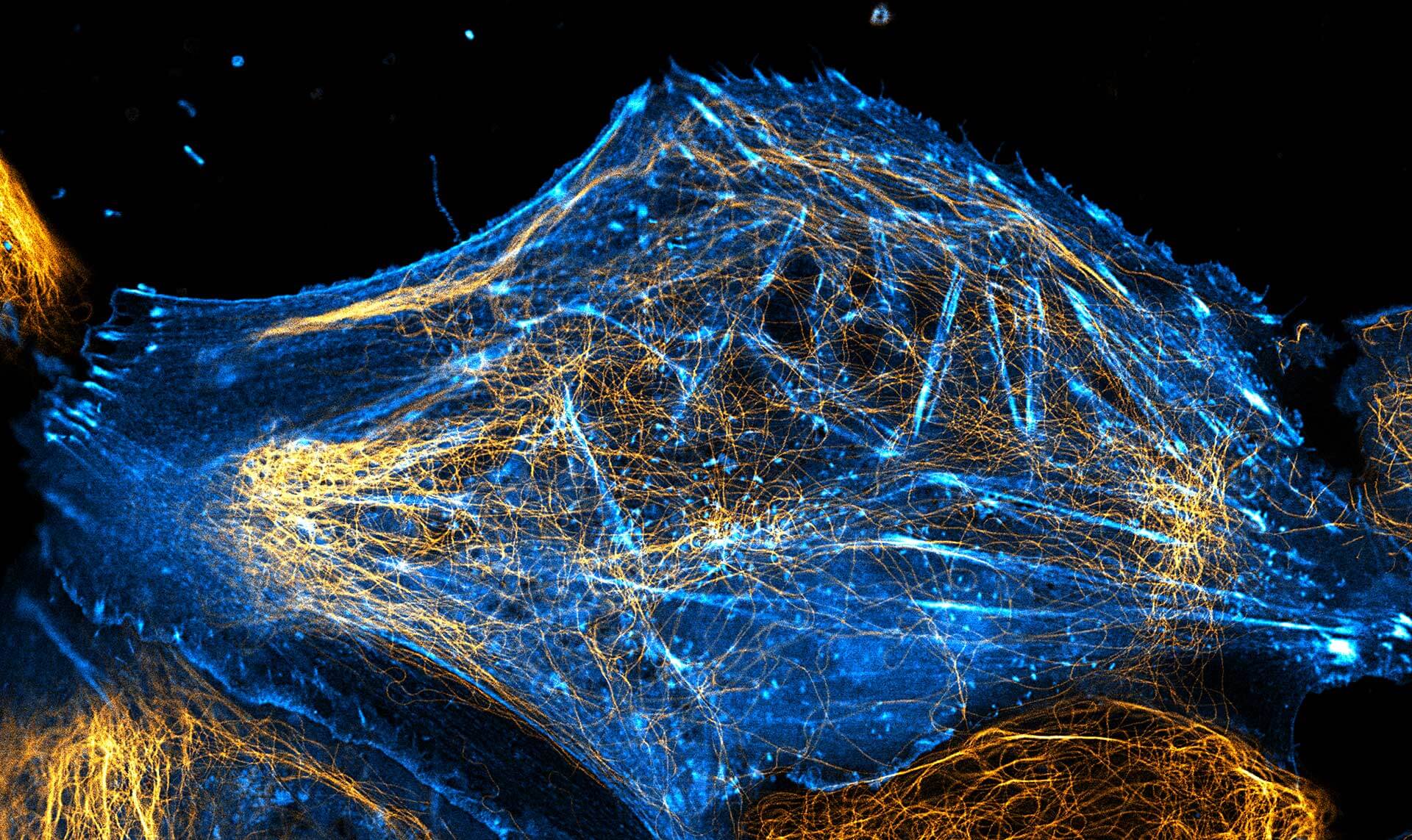
2-color STED image of a living cell expressing actinK118TAG incorporating TCO*A labeled with abberior LIVE 550 click (cyan). Tubulin filaments were stained with our direct probe abberior LIVE 610 tubulin (yellow).
Description
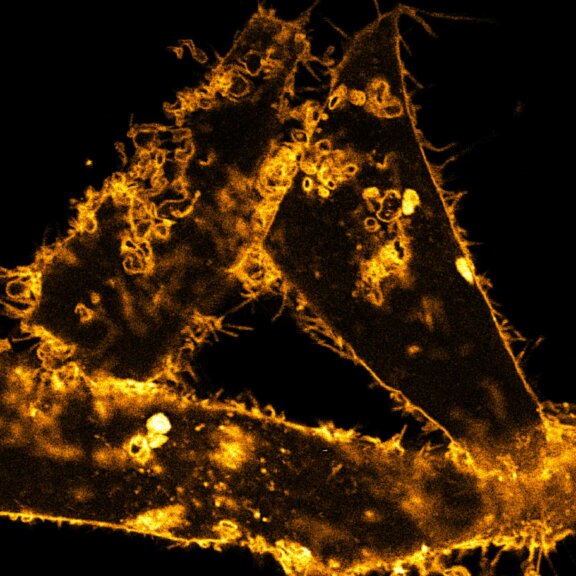
STED time-lapse of living mammalian cell stained with abberior STAR ORANGE membrane.
Description
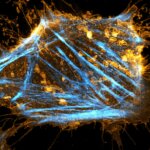
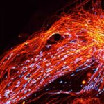
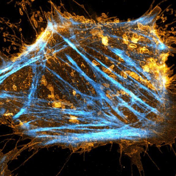
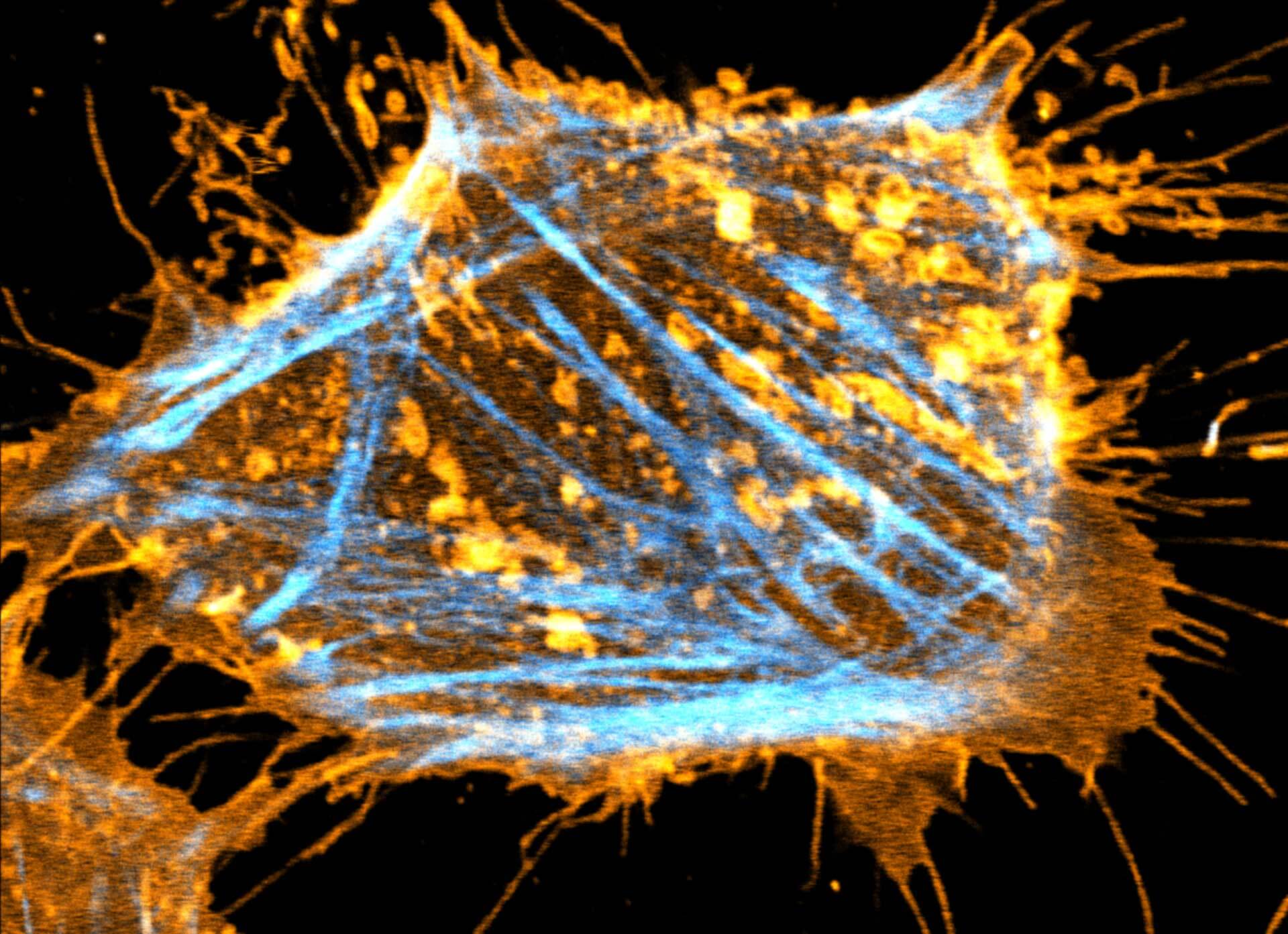
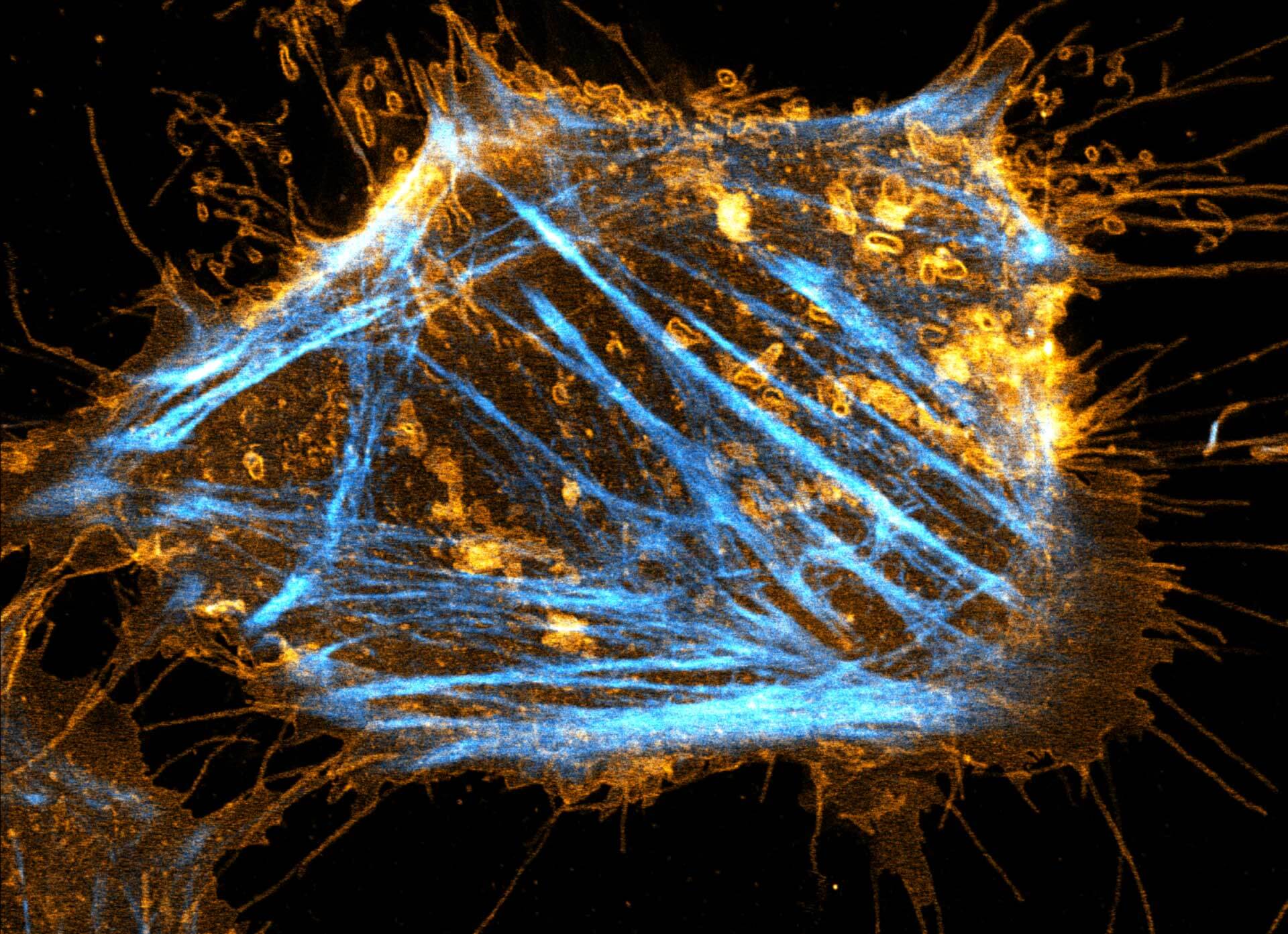
Two color live-cell STED and confocal image of a mammalian cultured cell stained with abberior STAR RED membrane (orange) and abberior LIVE 590 actin (cyan).
Description
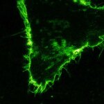
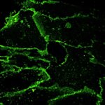
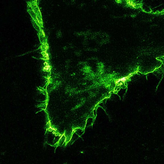
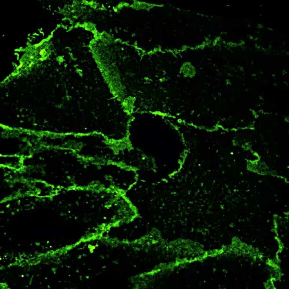
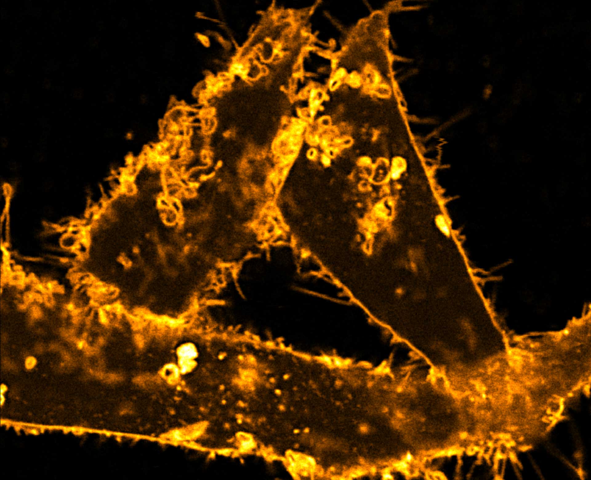
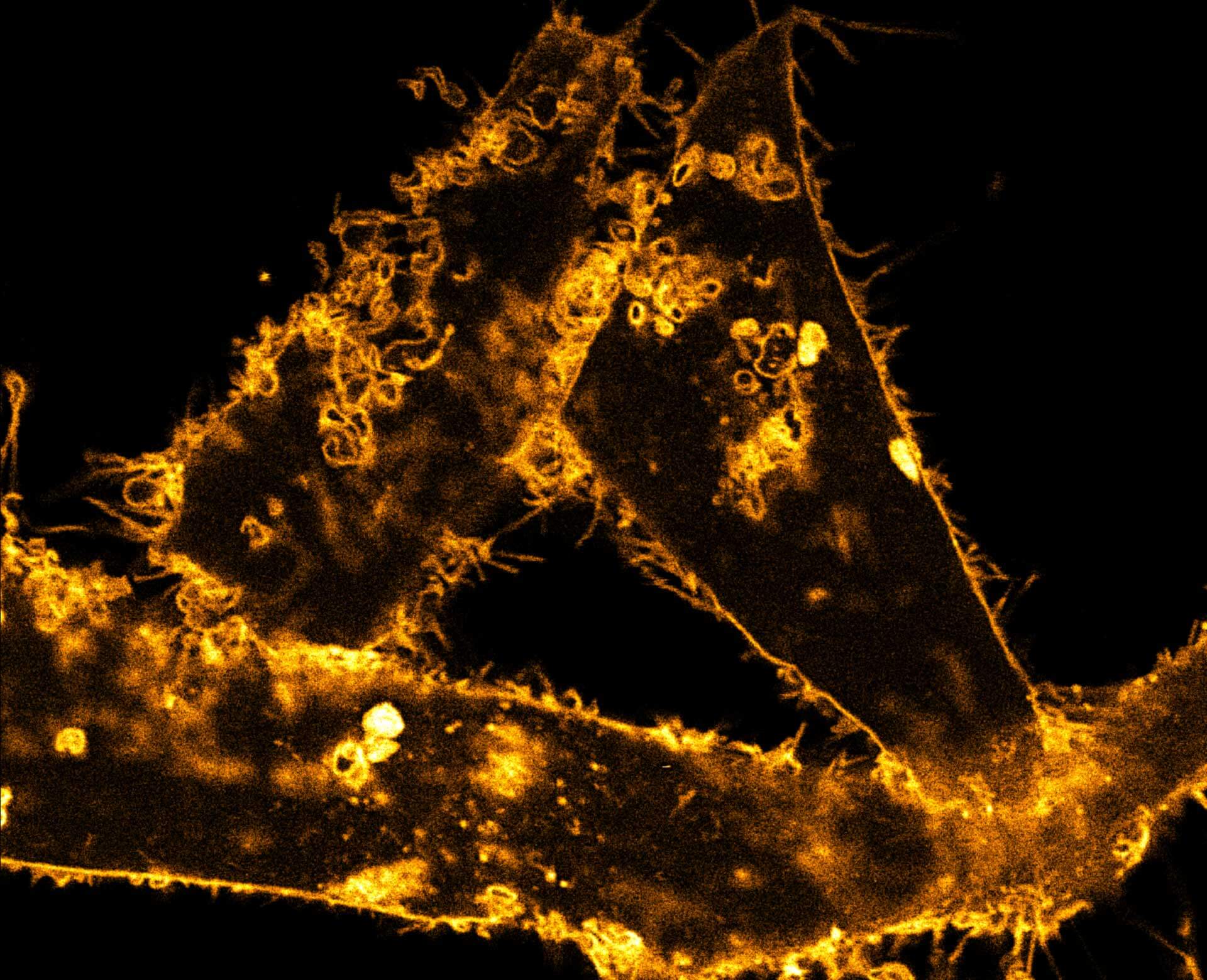
Comparison of STED and confocal of a living mammalian cell stained with abberior STAR ORANGE membrane.
Description
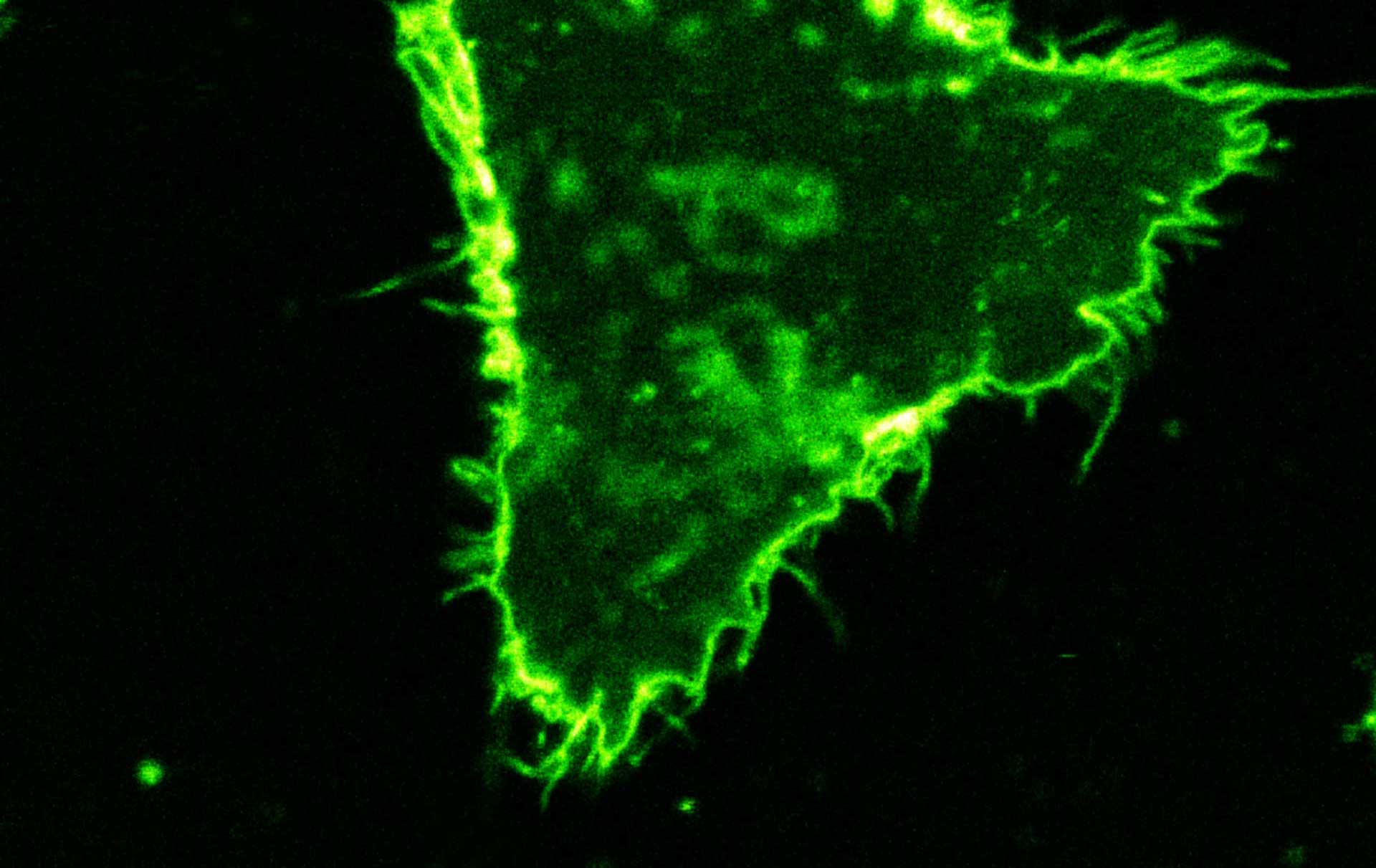
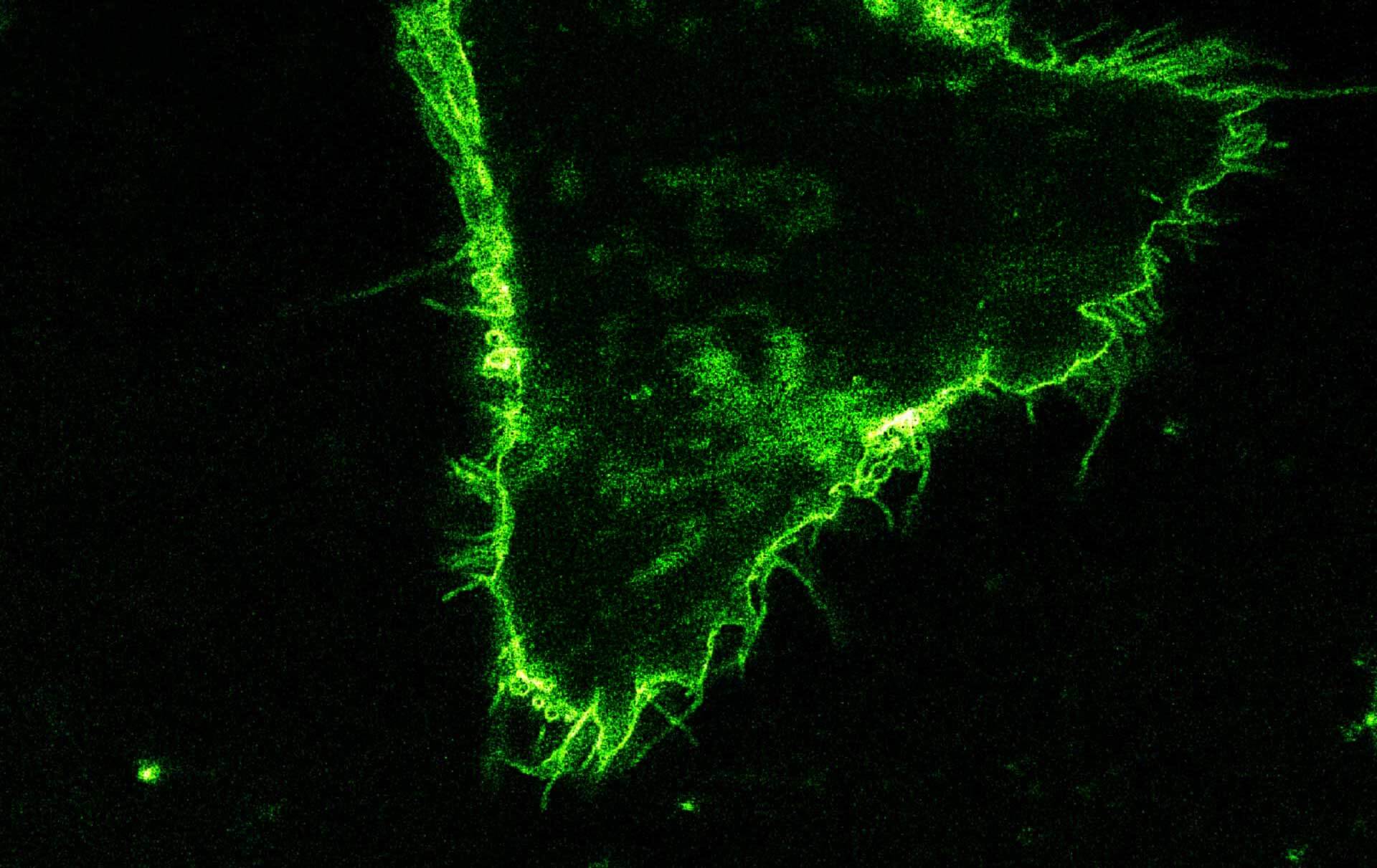
Comparison of STED and confocal of a living mammalian cell stained with abberior STAR 488 membrane. Images were acquired with our FACILITY and STED @ 595 nm.
Description
Confocal time-lapse of living mammalian cell stained with abberior STAR 488 membrane.
Description
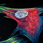
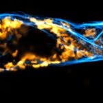
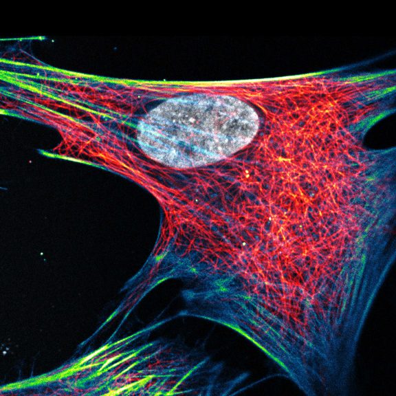
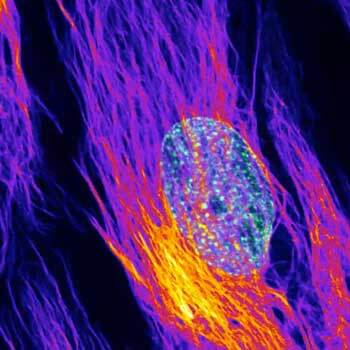
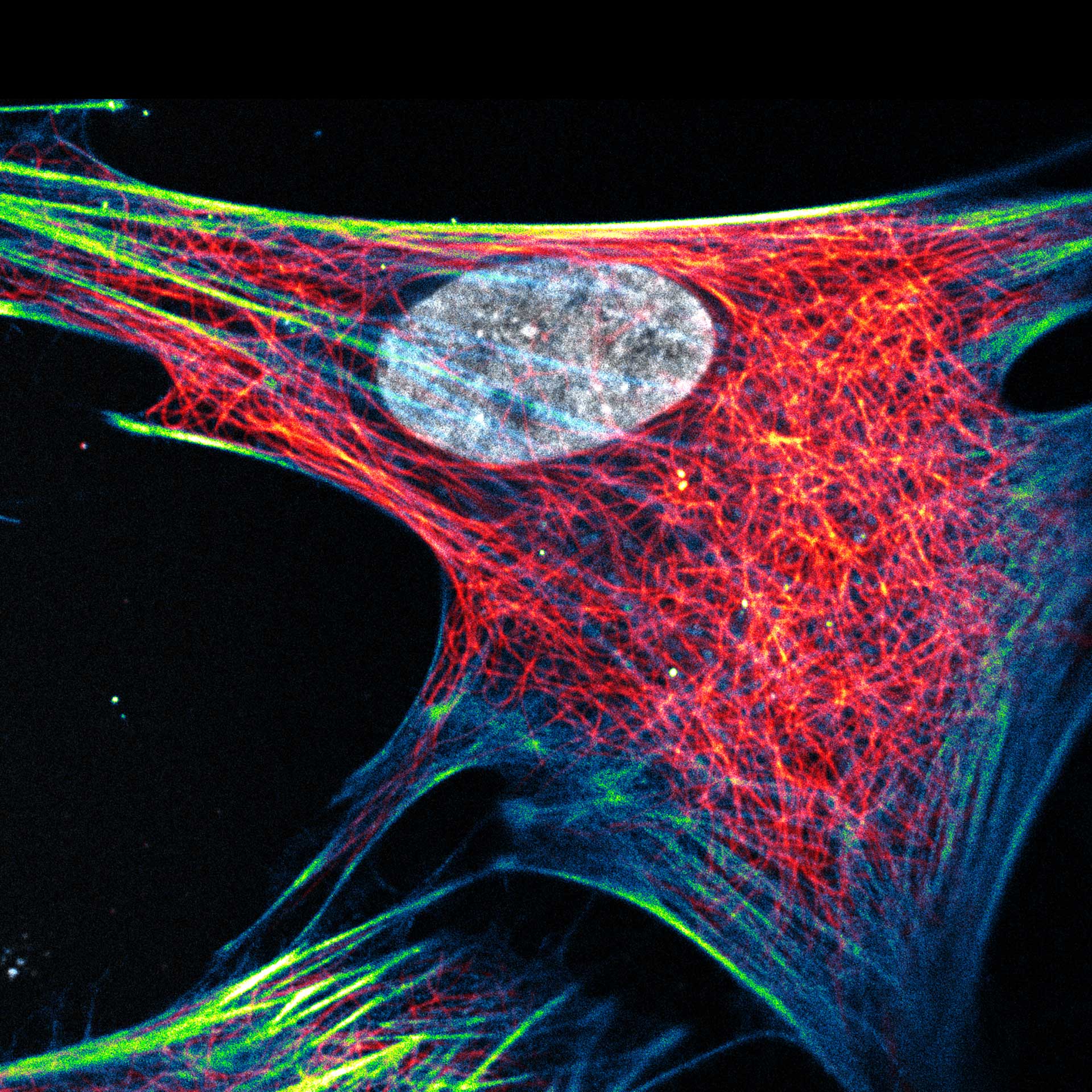
Composite of three color live-cell image of an adherent mammalian cell. This living cell was directly labelled with abberior LIVE 510 actin (blue/green), LIVE 560 DNA (gray) and LIVE 610 tubulin. This image was acquired with a STEDYCON microscope.
Description
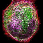
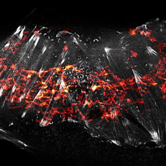
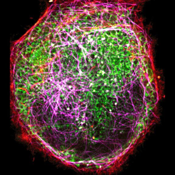
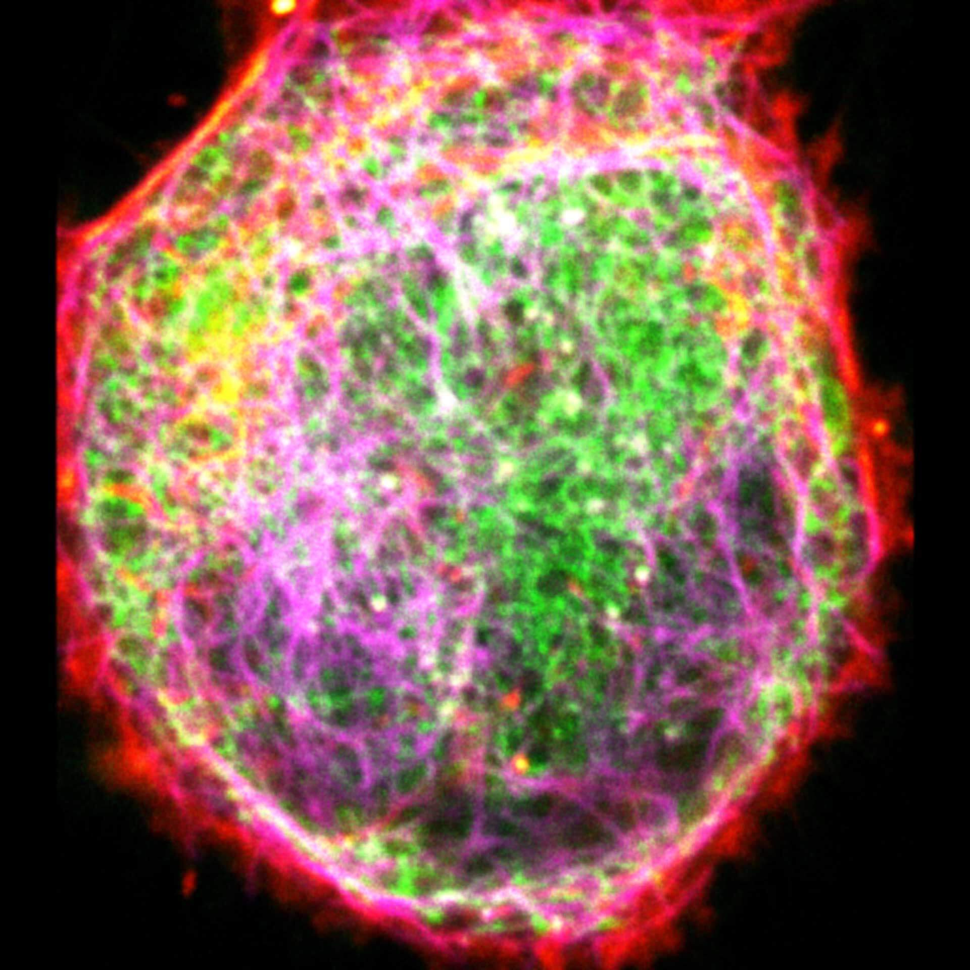
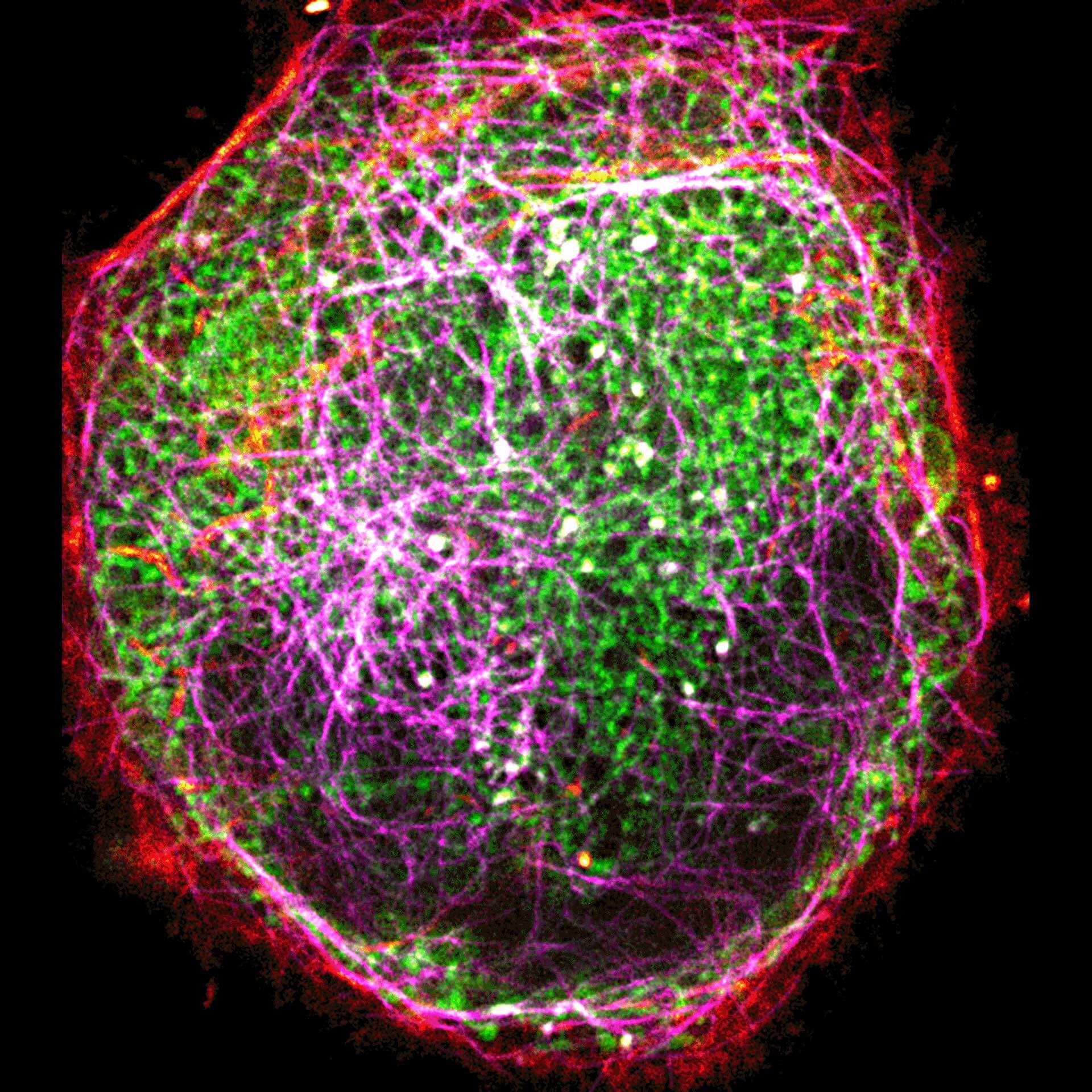
Three color live-cell STED at 775 nm: living cell labelled with abberior LIVE 460L (ER, green), LIVE 560 tubulin (magenta) and LIVE 610 actin (red).
Description
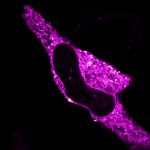
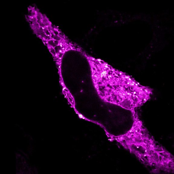
Confocal and STED time lapse of a living cell expressing SNAP-tag® fusion protein fusion protein in the endoplasmic reticulum lumen with SNAP and C-terminal tetrapeptide KDEL. SNAP-KDEL was labelled with abberior LIVE 610 SNAP ligand (benzylguanine).
The SNAP-KDEL plasmid was a gift from Francesca Bottanelli, FU Berlin.
Description
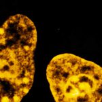
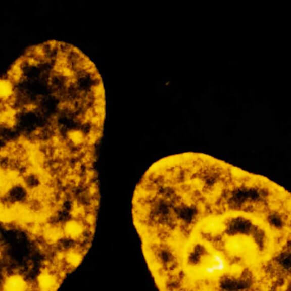
Live cell time lapse of abberior LIVE 590 DNA probe in cultured mammalian cells.
The use of very low concentrations of our abberior LIVE dyes reduces toxicity and allows long-term imaging of dynamic processes.
Image was acquired with the FACILITY microscope.

Description
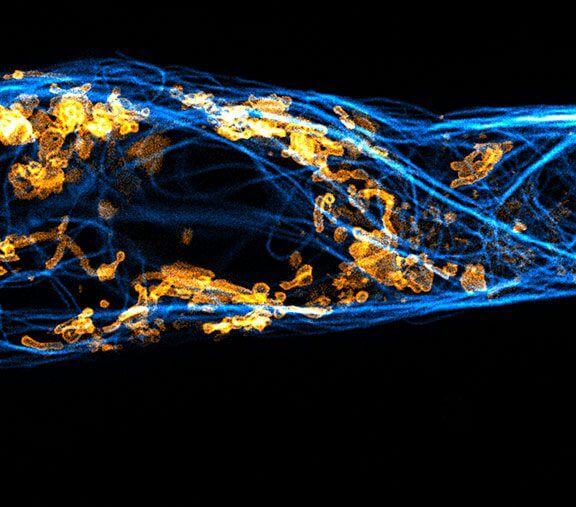
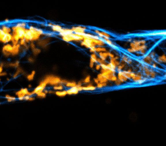
Two color live-cell confocal and STED image of a mammalian cell expressing a SNAP-tag® OMP25 fusion protein decoration the outer membrane of mitochondria. OMP25 is visualized by our new abberior LIVE 610 SNAP ligand (orange). Tubulin filaments are highlighted with abberior LIVE 550 tubulin (cyan).
The SNAP-OMP25 plasmid was a gift from Francesca Bottanelli, FU Berlin.
Description
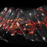
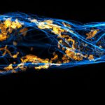
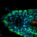
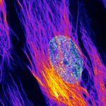
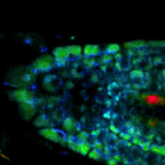
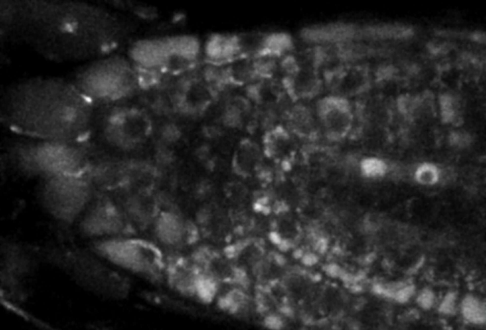
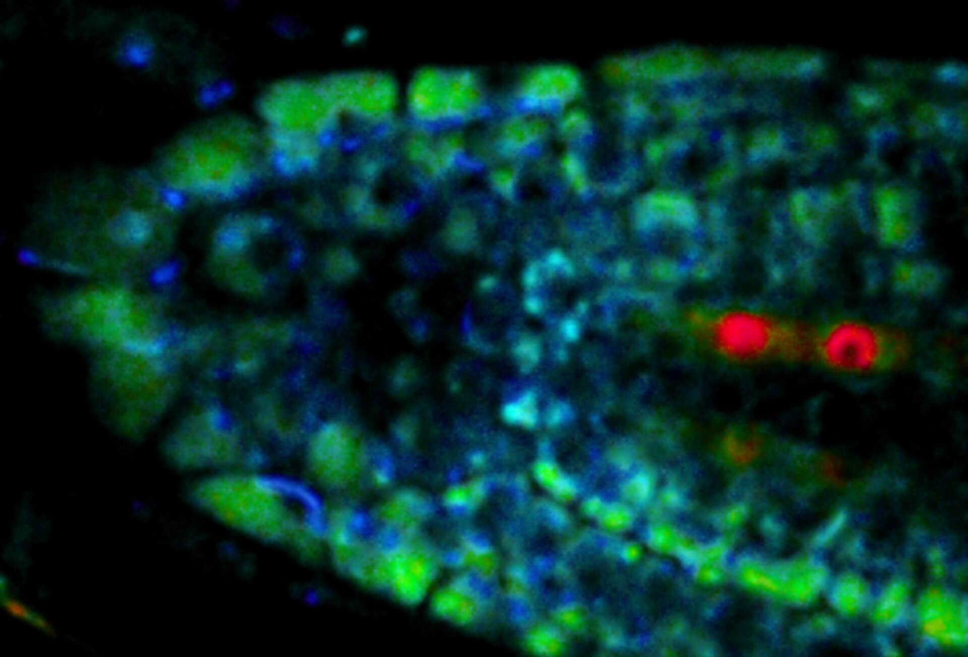
Live-cell sample of an Arabidopsis root tip suspended in water recorded with FACILITY. A subset of cells expresses a YFP construct. Lifetime imaging with TIMEBOW shows shifts in YFP fluorescence lifetime caused by the proteins nano-environment.
We thank Dr. Fábián Attila and Soós Vilmos (ATK, Brunszvik, HU) for providing this sample. Contact: soos.vilmos@atk.hu.
Modules:
Description
Video of mitochondria in living U2OS cells imaged for 1.5 hours with STED and deconvolved with TRUESHARP image boosting. Mitochondria were visualized with abberior LIVE SiR HaloX staining the outer mitochondrial membrane marker TOMM20. FLEXPOSURE adaptive illumination was used to reduce the light burden on the sample.
Modules:
Description
Living HeLa cells stained with the mitochondrial membrane marker abberior LIVE ORANGE mito, visualizing both outer and inner membranes. Confocal and STED images where deconvolved with TRUESHARP image boosting.
Modules:
Description
Two color live-cell confocal and STED image of a mammalian cell directly labeled with abberior LIVE 510 mito (cyan), and LIVE RED tubulin. This image was acquired with a STEDYCON microscope.
Description
OMP25-SNAP protein in mitochondria labeled with abberior LIVE 610 SNAP (magenta) and Actin^K118TAG labeled with abberior LIVE 550 click (green). Movie with 60 frames.
Description
Description
2-color STED image of a living cell expressing actin K118TAG incorporating TCO*A labeled with abberior LIVE 590 click (green). Plasma membrane is highlighted with abberior STAR RED membrane (magenta).
Description
2-color STED image of a living cell expressing actinK118TAG incorporating TCO*A labeled with abberior LIVE 550 click (cyan). Tubulin filaments were stained with our direct probe abberior LIVE 610 tubulin (yellow).
Description
STED time-lapse of living mammalian cell stained with abberior STAR ORANGE membrane.
Description
Two color live-cell STED and confocal image of a mammalian cultured cell stained with abberior STAR RED membrane (orange) and abberior LIVE 590 actin (cyan).
Description
Comparison of STED and confocal of a living mammalian cell stained with abberior STAR ORANGE membrane.
Description
Comparison of STED and confocal of a living mammalian cell stained with abberior STAR 488 membrane. Images were acquired with our FACILITY and STED @ 595 nm.
Description
Confocal time-lapse of living mammalian cell stained with abberior STAR 488 membrane.
Description
Composite of three color live-cell image of an adherent mammalian cell. This living cell was directly labelled with abberior LIVE 510 actin (blue/green), LIVE 560 DNA (gray) and LIVE 610 tubulin. This image was acquired with a STEDYCON microscope.
Description
Three color live-cell STED at 775 nm: living cell labelled with abberior LIVE 460L (ER, green), LIVE 560 tubulin (magenta) and LIVE 610 actin (red).
Description
Confocal and STED time lapse of a living cell expressing SNAP-tag® fusion protein fusion protein in the endoplasmic reticulum lumen with SNAP and C-terminal tetrapeptide KDEL. SNAP-KDEL was labelled with abberior LIVE 610 SNAP ligand (benzylguanine).
The SNAP-KDEL plasmid was a gift from Francesca Bottanelli, FU Berlin.
Description
Live cell time lapse of abberior LIVE 590 DNA probe in cultured mammalian cells.
The use of very low concentrations of our abberior LIVE dyes reduces toxicity and allows long-term imaging of dynamic processes.
Image was acquired with the FACILITY microscope.
Description
Two color live-cell confocal and STED image of a mammalian cell expressing a SNAP-tag® OMP25 fusion protein decoration the outer membrane of mitochondria. OMP25 is visualized by our new abberior LIVE 610 SNAP ligand (orange). Tubulin filaments are highlighted with abberior LIVE 550 tubulin (cyan).
The SNAP-OMP25 plasmid was a gift from Francesca Bottanelli, FU Berlin.
Description
Live-cell sample of an Arabidopsis root tip suspended in water recorded with FACILITY. A subset of cells expresses a YFP construct. Lifetime imaging with TIMEBOW shows shifts in YFP fluorescence lifetime caused by the proteins nano-environment.
We thank Dr. Fábián Attila and Soós Vilmos (ATK, Brunszvik, HU) for providing this sample. Contact: soos.vilmos@atk.hu.


