


TIMEBOW
Every fluorescence signal contains lifetime information that travels with the photons from the sample to the detector.TIMEBOW lifetime imaging elicits this information, allowing you to make the best use of it in multiple ways. Expand your imaging options, increase the resolution of your image with gentle STED, remove background signal… And in combination with our highly sensitive APDs, the groundbreaking MATRIX detector, and TRUESHARP deconvolution, you get the best out of every aspect of the signal! WithTIMEBOW, your research shines in perfect color.
gives time a color
TIMEBOW at a glance >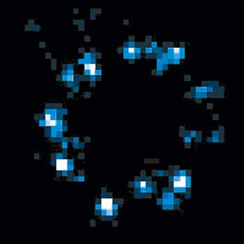
MINFLUX Module
Single-digit nanometer resolution with the MINFLUX module for our MIRAVA POLYSCOPE
MATRIX Detector
Many eyes see more than one. The MATRIX detector drastically improves signal-to-background ratio, resolution, and dynamic range.
TIMEBOW Imaging
TIMEBOW lifetime imaging for stunning results at confocal and STED super-resolution.
FLEXPOSURE Illumination
Brings down the light dose on your sample and lables dramatically. Key ingredient for volume and live-cell superresolution.
RAYSHAPE Mirror
Dynamic aberration correction with a deformable mirror over about 200 µm z-range. 140 digital actuators adjust the mirror surface within milliseconds.

Custom Solutions
We offer solutions for even the most challenging applications. Everything that can be done, we will do.
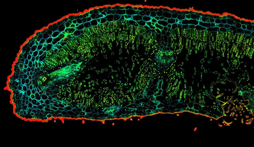
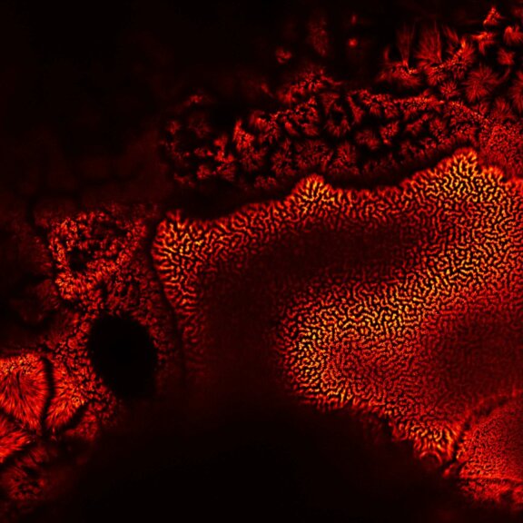
Description
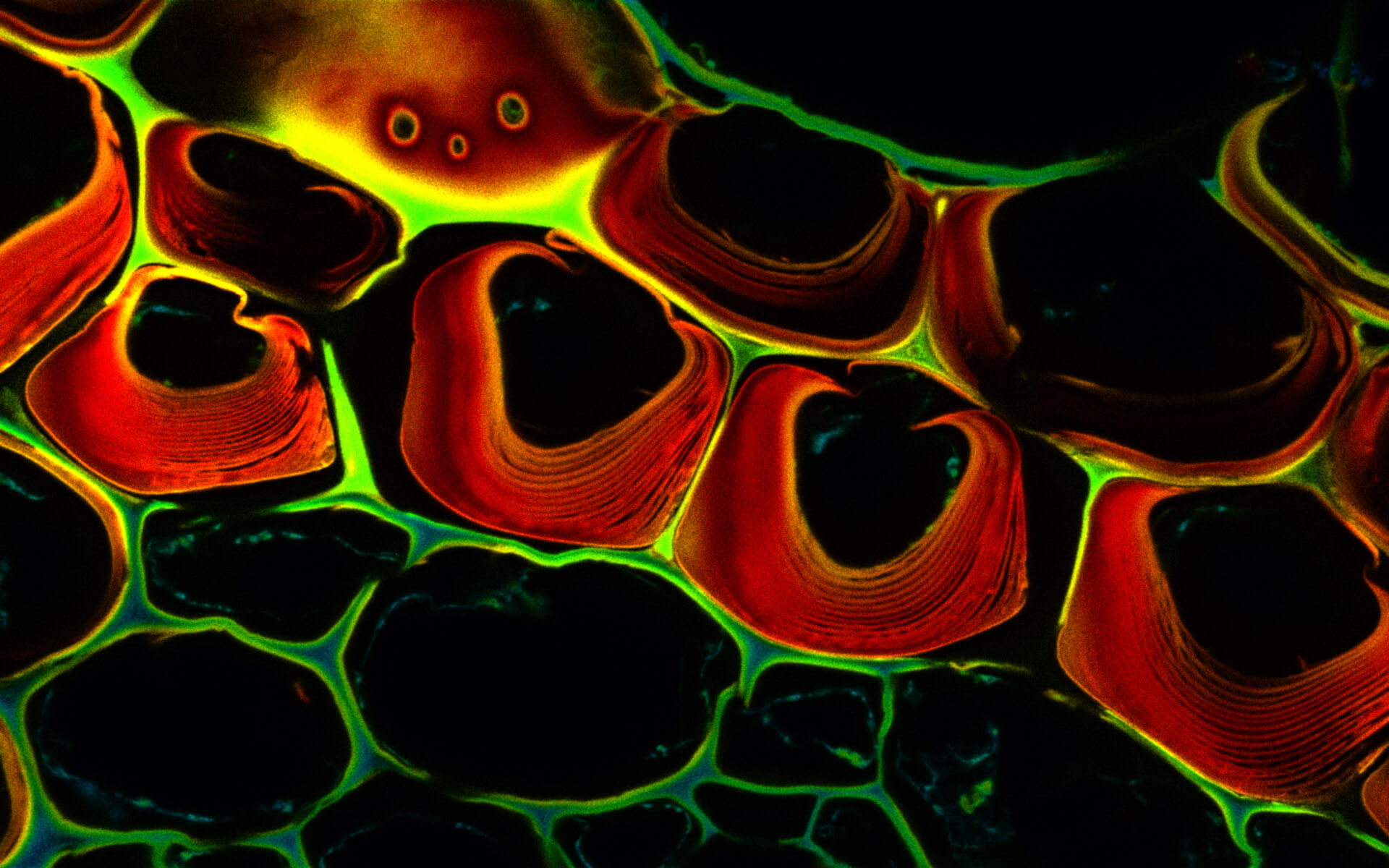
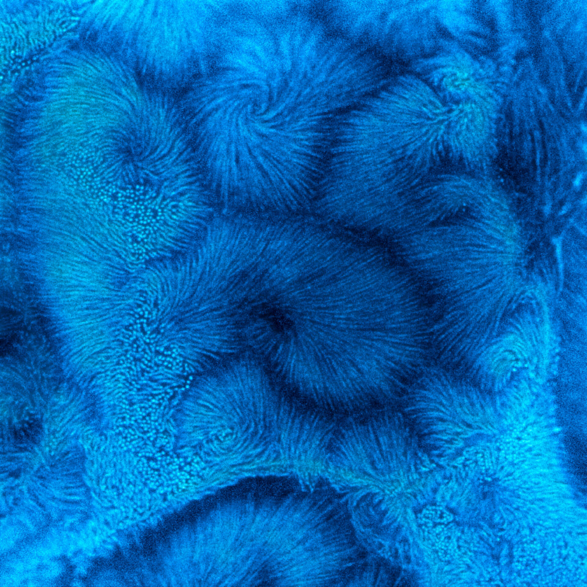
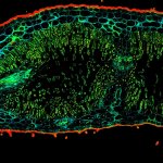
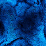
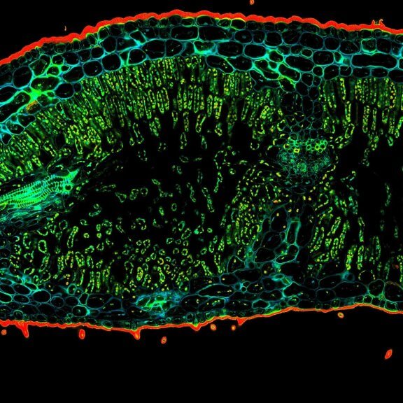
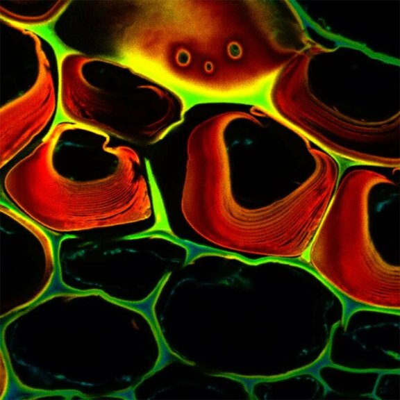
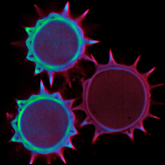
Confocal TIMEBOW acquisition of an Oleander leaf tip showing a range of autofluorescence lifetimes originating from different tissue components.
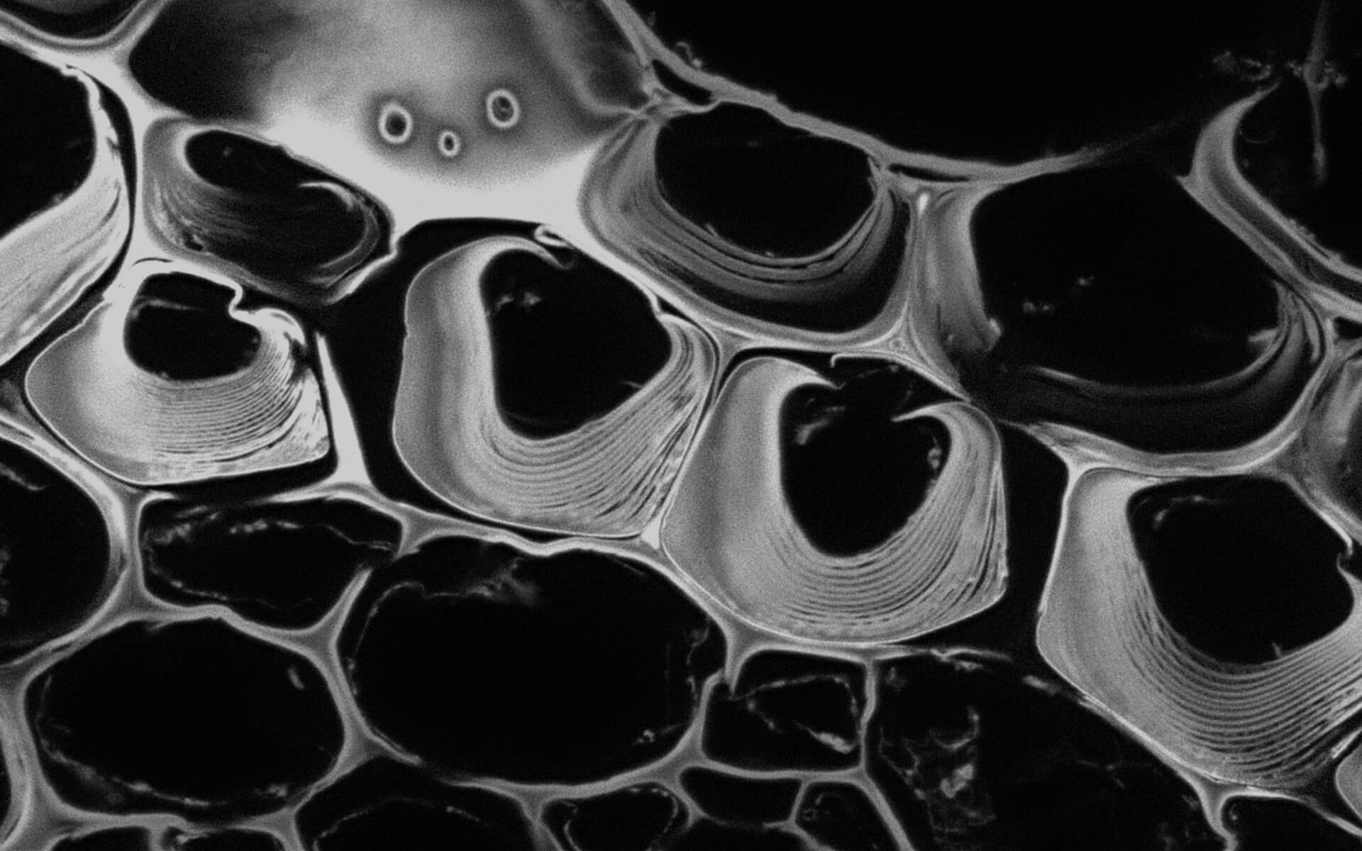
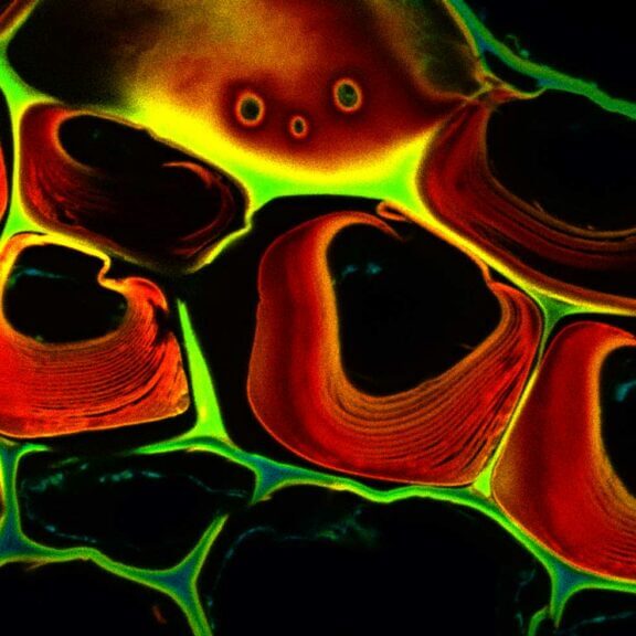
Description
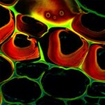
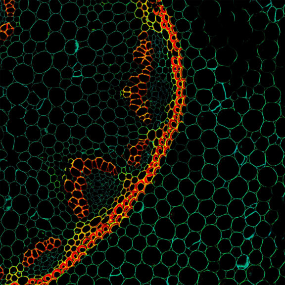
Autofluorescence in a plant stem cross section of Convallaria recorded with FACILITY. Image quality is improved by removing the background through MATRIX detection. This is combined with TIMEBOW lifetime imaging to show the differences in fluorescence lifetime due to the type of autofluorescent molecule and their nano-environment.
Modules:

Description
Autofluorescence in a plant stem cross section of Convallaria recorded with FACILITY. A large area, 10 by 10 tiles, each 125 µm by 125 µm, was imaged and stitched with image tiling in Fiji for ImageJ. This is combined with TIMEBOW lifetime imaging to show the differences in fluorescence lifetime due to the type of autofluorescent molecule and their nano-environment.
Modules:
Description
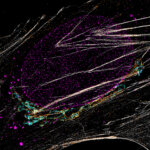
Confocal vs MATRIX + TIMEBOW STED
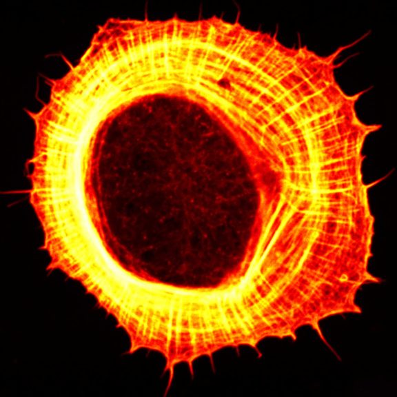
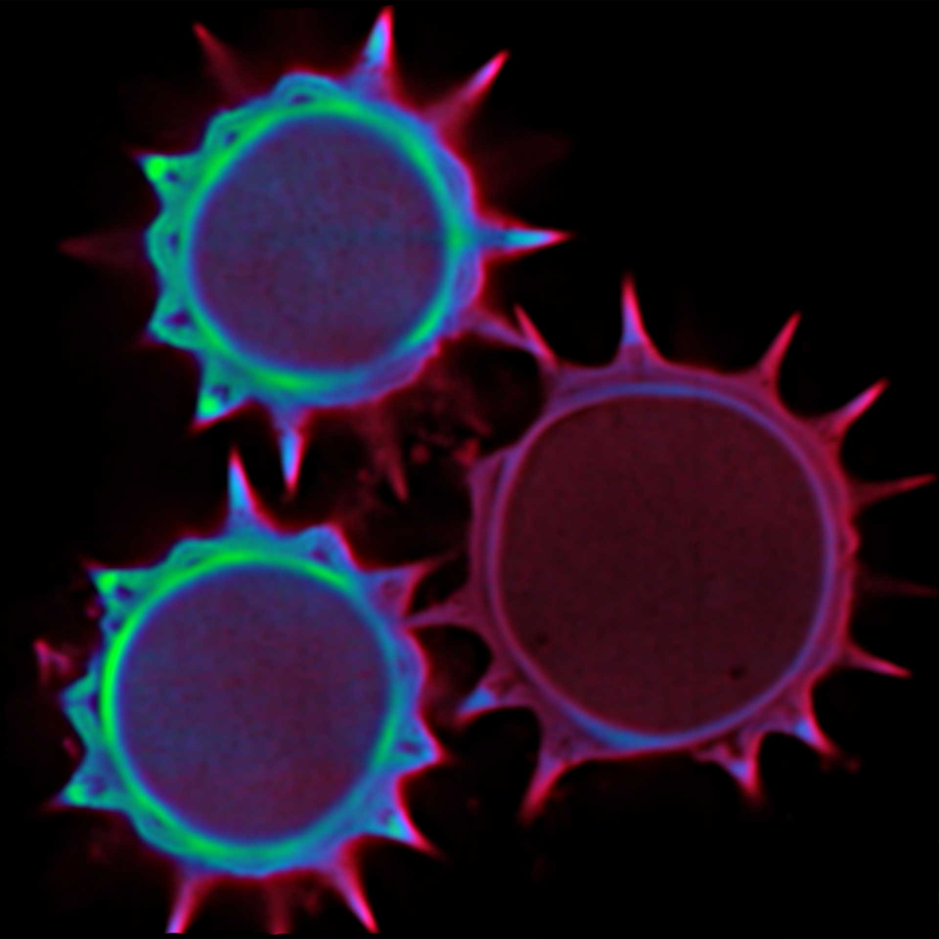
Actin in microvilli of CaCo2 cells recorded with FACILITY. Fixed cells were stained with abberior STAR RED phalloidin. Imaging of the intense-background sample is optimized by MATRIX detection by directly subtracting unfocussed data. To boost resolution TIMEBOW is used, keeping STED laser power low. Lifetime information enables the selective exclusion of STED-affected emission at the rim of each measurement.
We thank Prof. Dorothee Günzel and Jörg Piontek (Charité, Berlin, DE) for providing this sample.
Modules:
Description
TIMEBOW STED vs MATRIX + TIMEBOW STED
Actin in microvilli of CaCo2 cells recorded with FACILITY. Fixed cells were stained with abberior STAR RED phalloidin. Imaging of the intense-background sample is optimized by MATRIX detection by directly subtracting unfocussed data. To boost resolution TIMEBOW is used, keeping STED laser power low. Lifetime information enables the selective exclusion of STED-affected emission at the rim of each measurement.
We thank Prof. Dorothee Günzel and Jörg Piontek (Charité, Berlin, DE) for providing this sample.
Modules:

Description
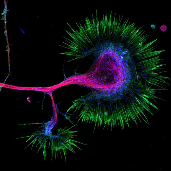
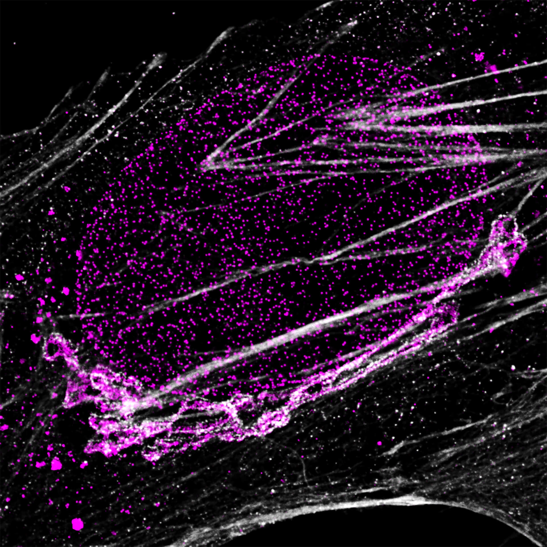
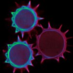
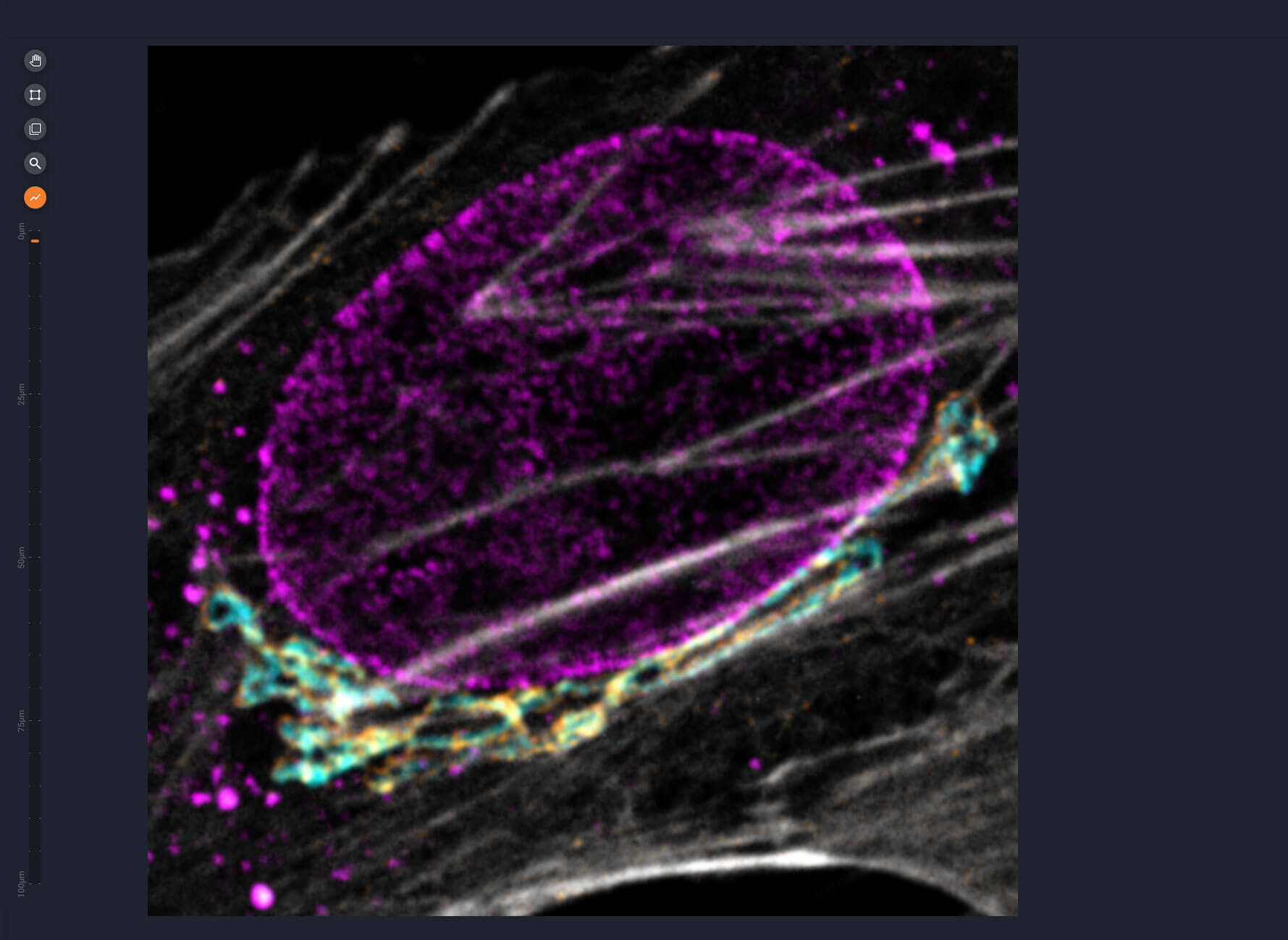
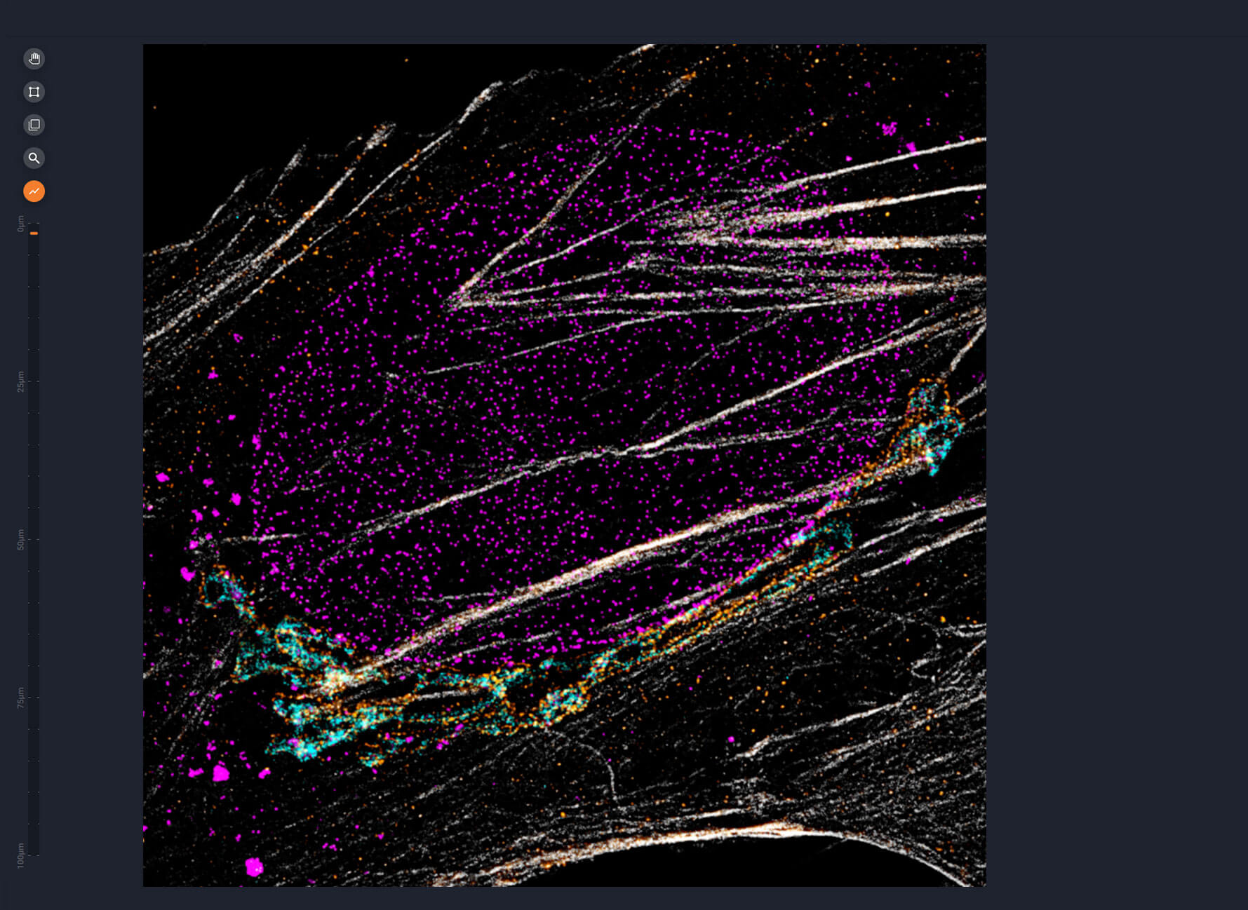
Four-color staining of fixed mammalian cells recorded with FACILITY showing nuclear pores (NUP98, STAR RED), Golgi membrane (GM130, STAR 635), Golgi medial rims (Giantin, STAR ORANGE) and actin (STAR 580 phalloidin). Shown left, just using spectral information, two red dyes (magenta) cannot be separated, because of their similar excitation properties. The same goes for STAR ORANGE and STAR 580 (grey).
With the lifetime information collected by TIMEBOW, these dyes can be distinguished, turning an image with two recording channels into a four-color image. To achieve superresolution only one STED laser at 775 nm was used.
All utilized dyes are manufactured by abberior.
Modules:
Description
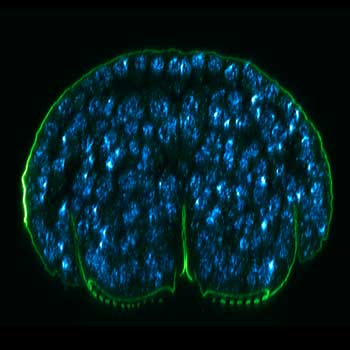
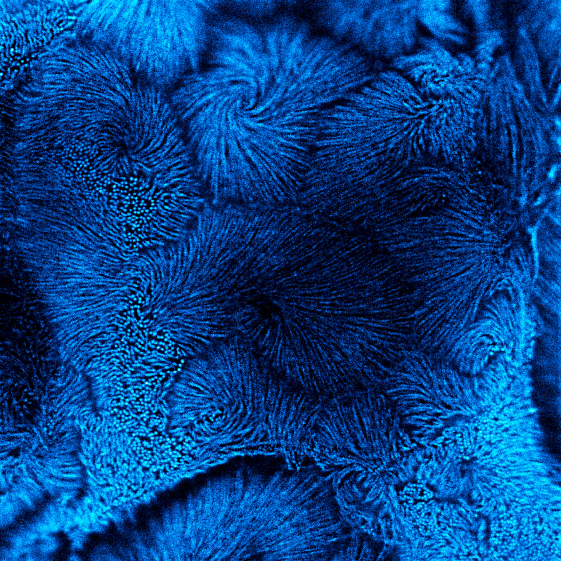
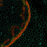
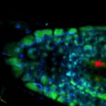
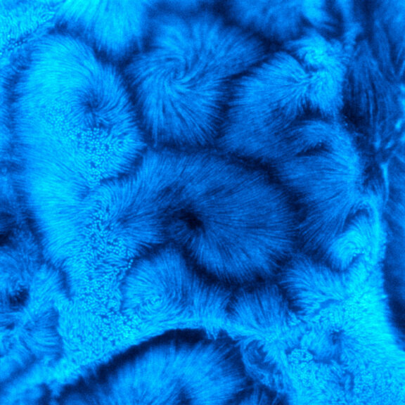
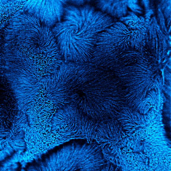
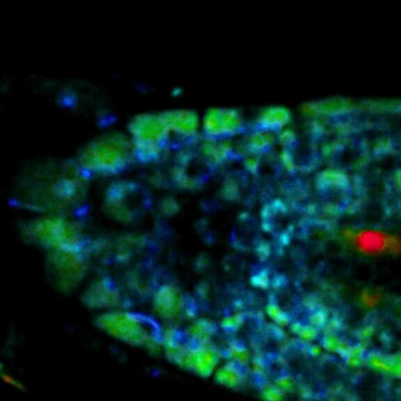
Live-cell sample of an Arabidopsis root tip suspended in water recorded with FACILITY. A subset of cells expresses a YFP construct. Lifetime imaging with TIMEBOW shows shifts in YFP fluorescence lifetime caused by the proteins nano-environment.
We thank Dr. Fábián Attila and Soós Vilmos (ATK, Brunszvik, HU) for providing this sample. Contact: soos.vilmos@atk.hu.
Modules:
Description
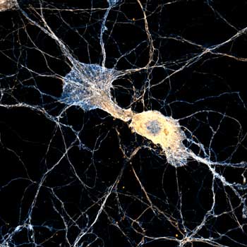
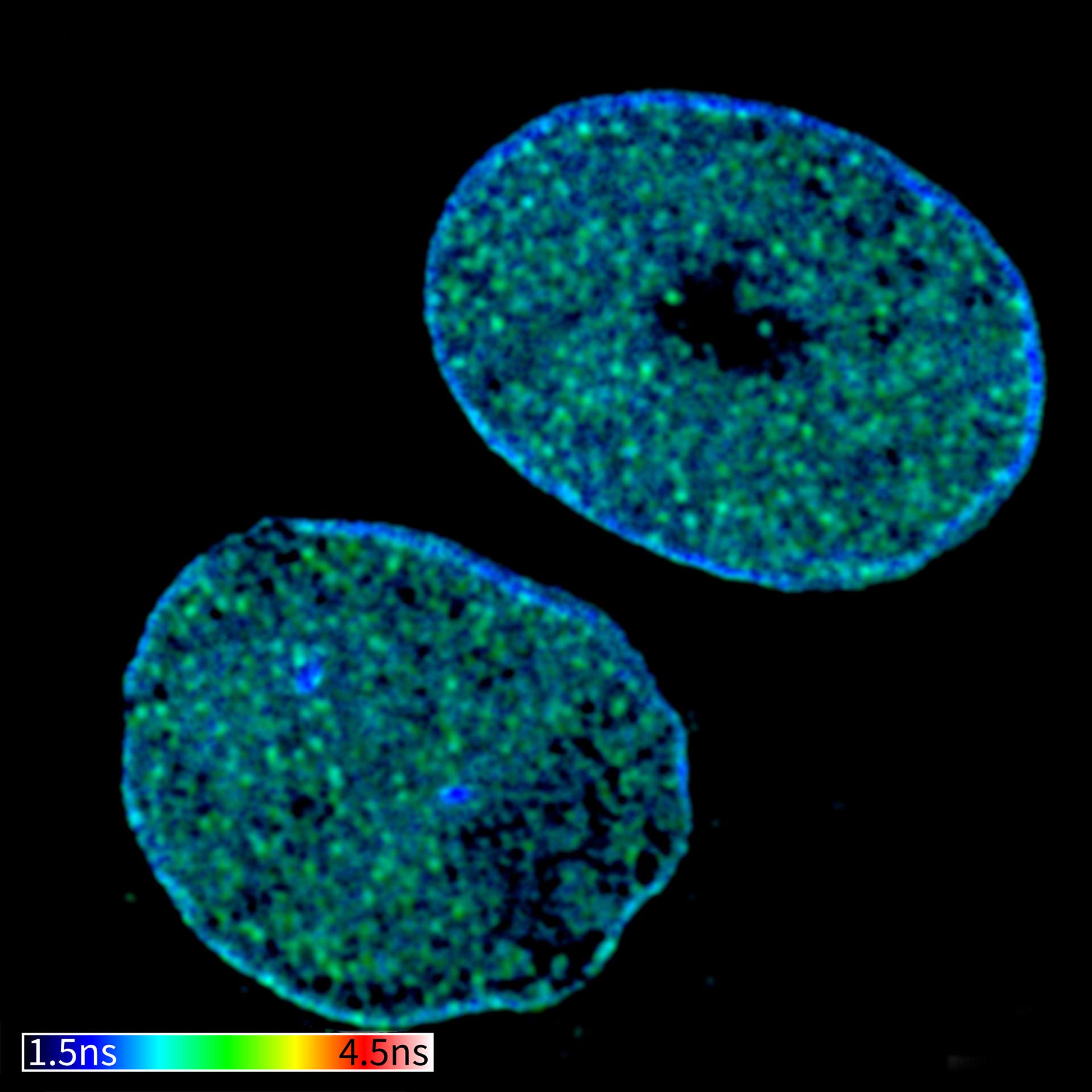
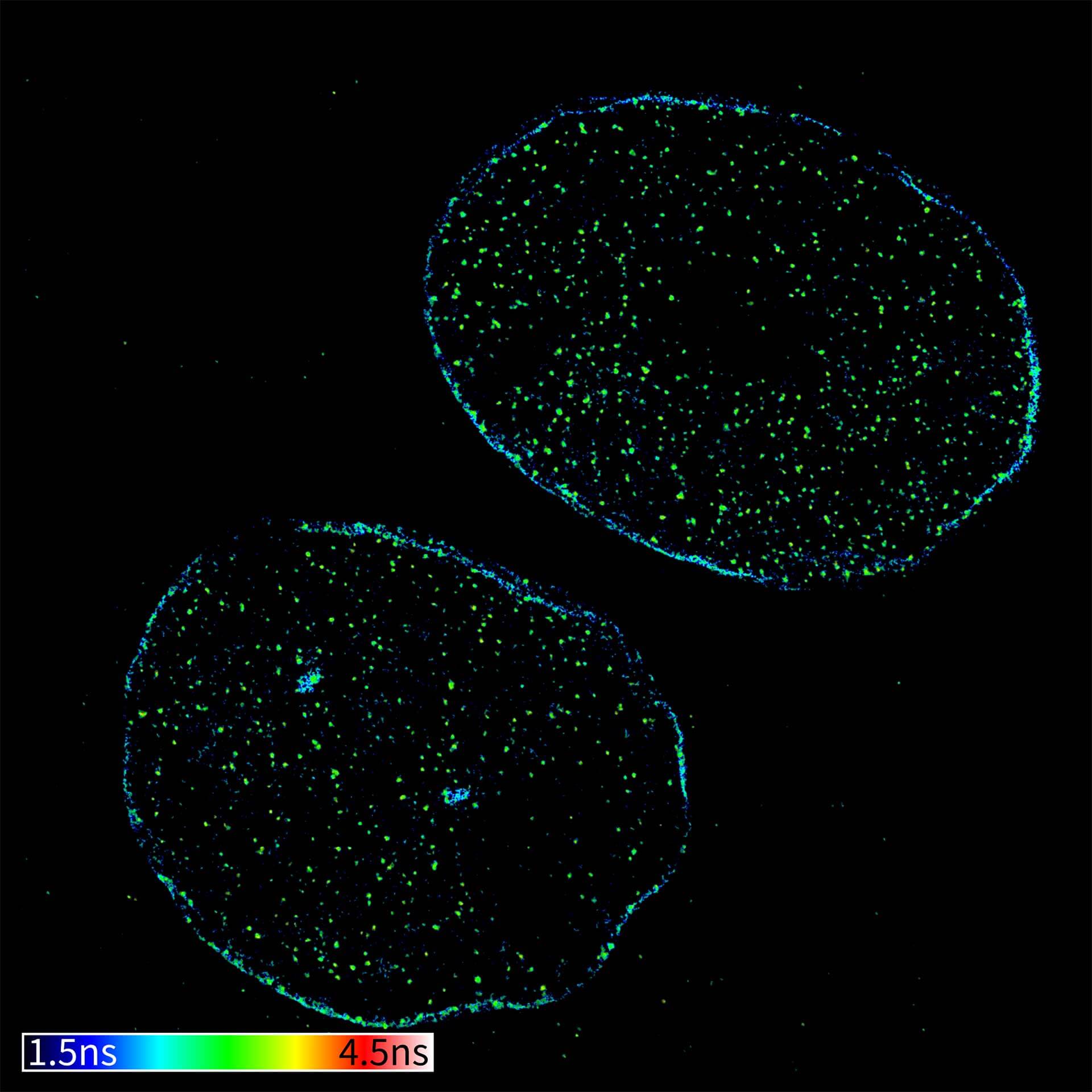
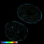
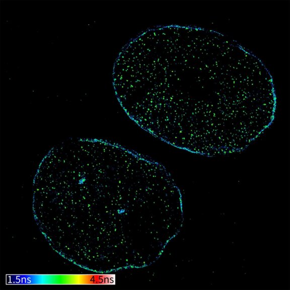
TIMEBOW confocal and TIMEBOW STED image of mammalian cells labeled with antibodies against lamin, ATTO 647N and dsDNA, abberior STAR 635P.
Modules:
Description
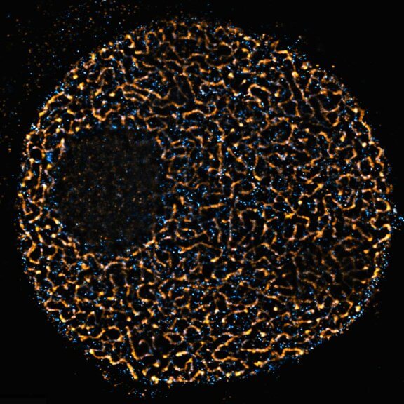
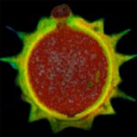
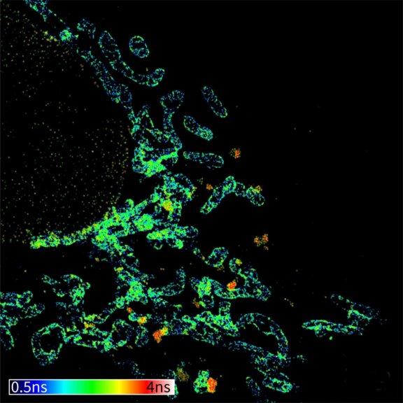
TIMEBOW image of mammalian cells labeled with antibodies against Tom20, ATTO 647N and NUP153, abberior STAR 635P. Note that nuclear pore (NUP) complex subunits appear to be localized in the cytoplasm, which is due to the NUP import pathway into the nuclear membrane.
Modules:


TIMEBOW
lifetime data evaluation at its best
Why limit yourself to just intensity and color? With TIMEBOW, you unlock an entirely new dimension of information: fluorescence lifetime. Make the most out of every photon you can get and gain insights into your sample that are invisible to traditional imaging.
Learn about the cellular microenvironment by monitoring changes in fluorescent lifetime. See subtle structural differences that are hidden in standard images. Exclude background signal or unwanted fluorescence based on lifetime signatures. Expand your imaging options by separating fluorophores emitting in the same spectral channel.
You may also use lifetime information to increase resolution by combining it with STED: gentle TIMEBOW STED gives you more resolution with less STED power while protecting your sample – for clearer, more detailed images.
TIMEBOW lifetime imaging is available both on MIRAVA POLYSCOPE and on STEDYCON 2. The direct integration into our LiGHTBOX and STEDYCON smart control software allows you to work with the data seamlessly.

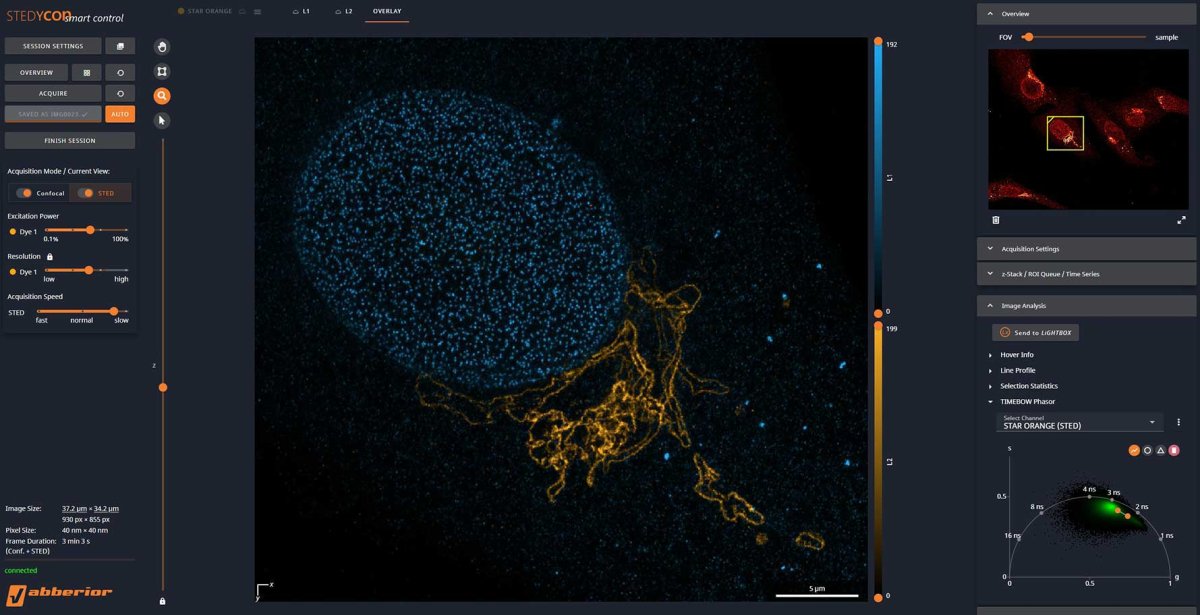
Intuitive LiGHTBOX software with TIMEBOW
Picture 1
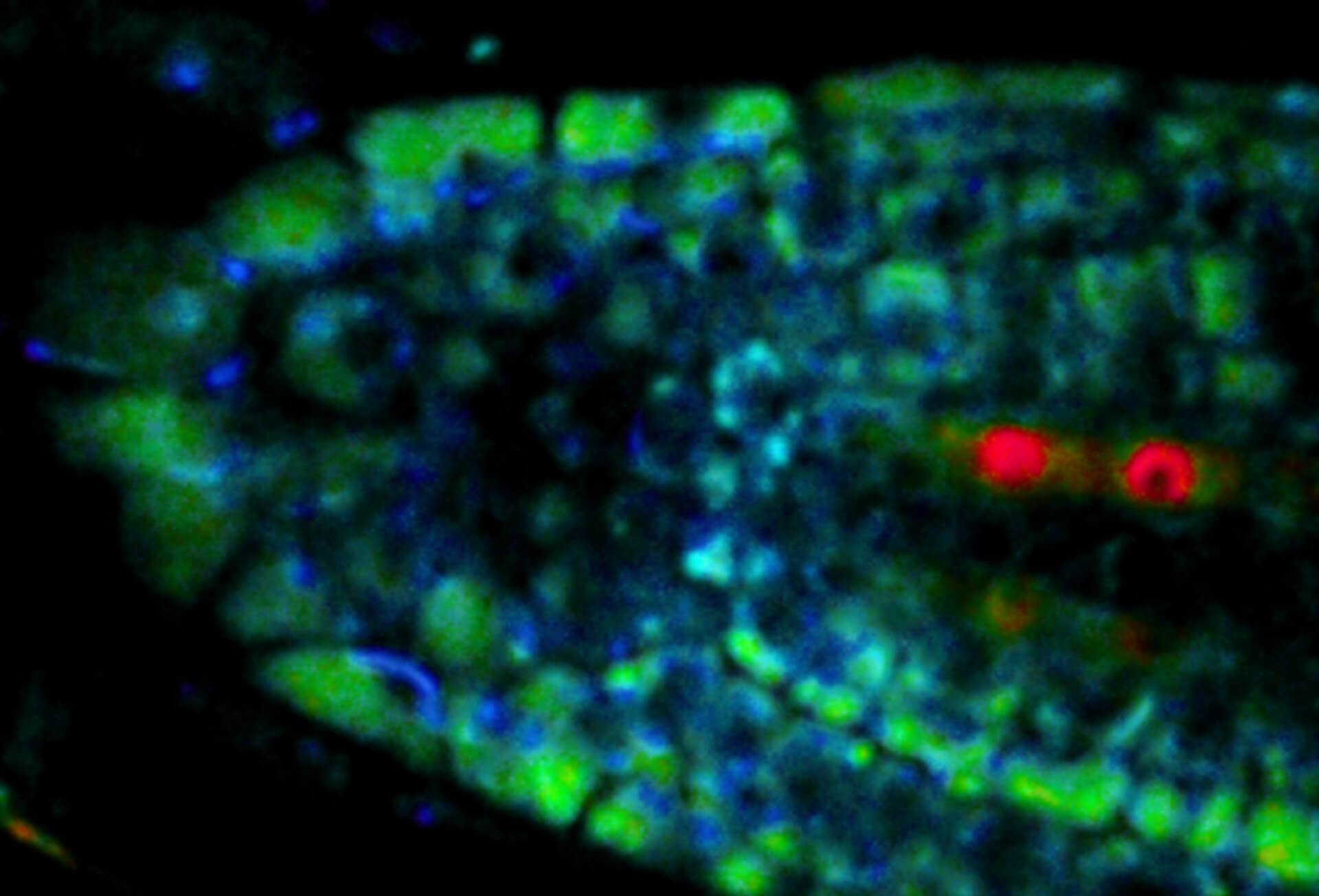
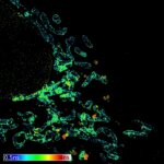
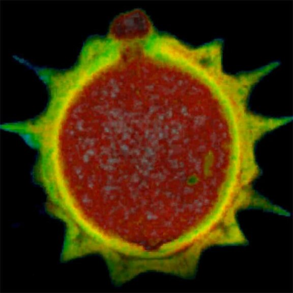
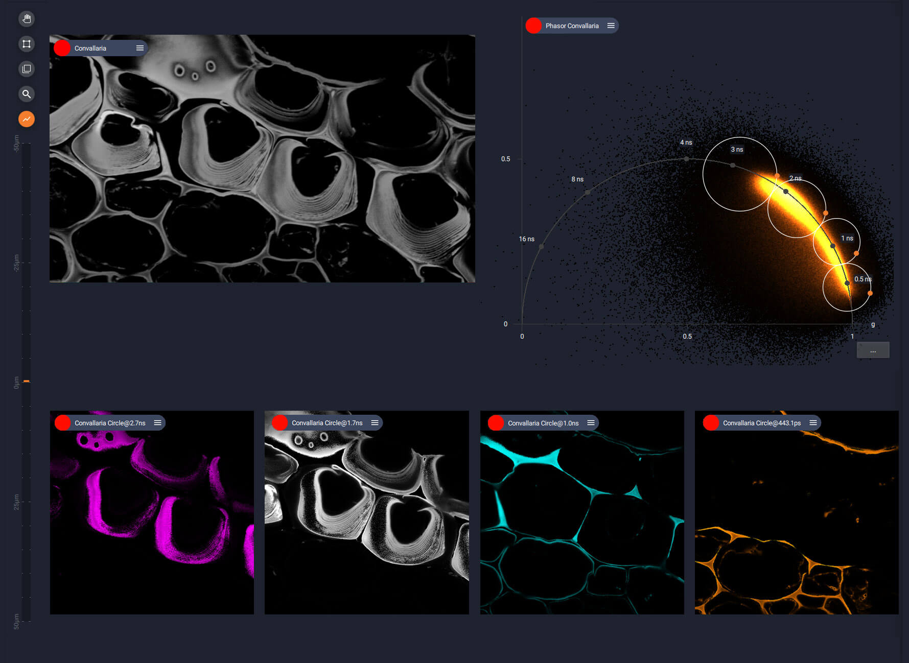
Color-coded fluorescence lifetime in an autofluorescent sample: plant stem cross section of Convallaria.
Intuitive LiGHTBOX software with TIMEBOW
Picture 2
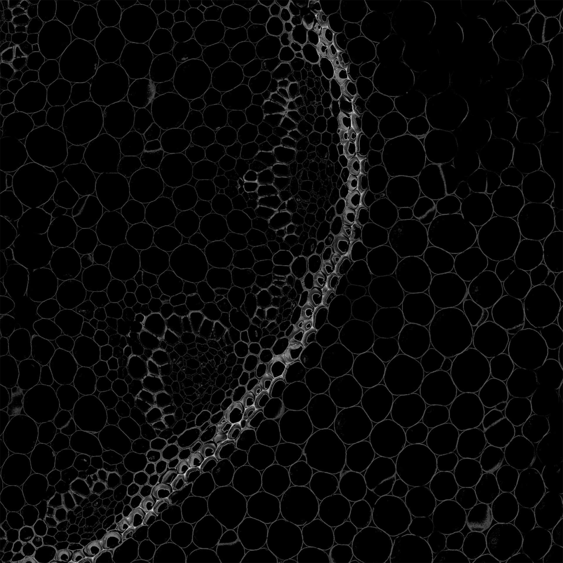
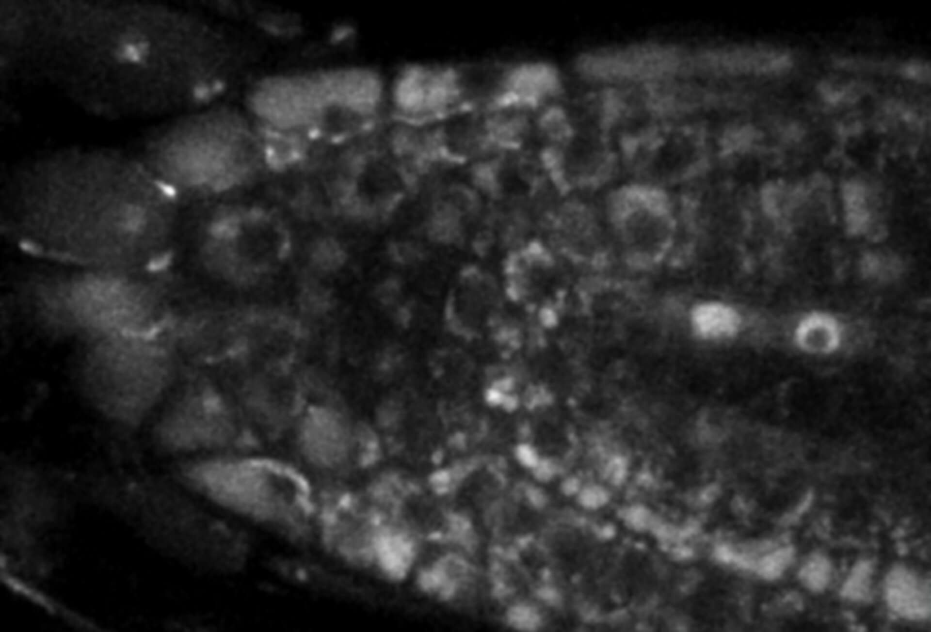
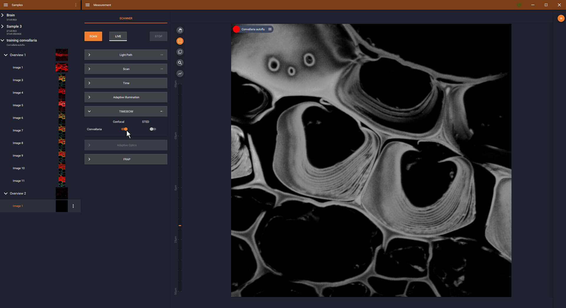
The grey intensity image without lifetime data. Image quality is already improved by removing the background with MATRIX detection.
Intuitive LiGHTBOX software with TIMEBOW
Picture 3
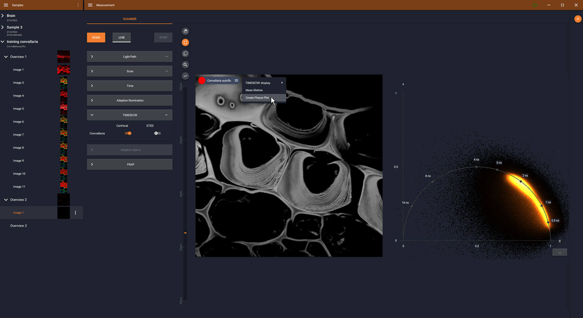
One click opens the phasor plot and gives access to lifetime data.
Intuitive LiGHTBOX software with TIMEBOW
Picture4
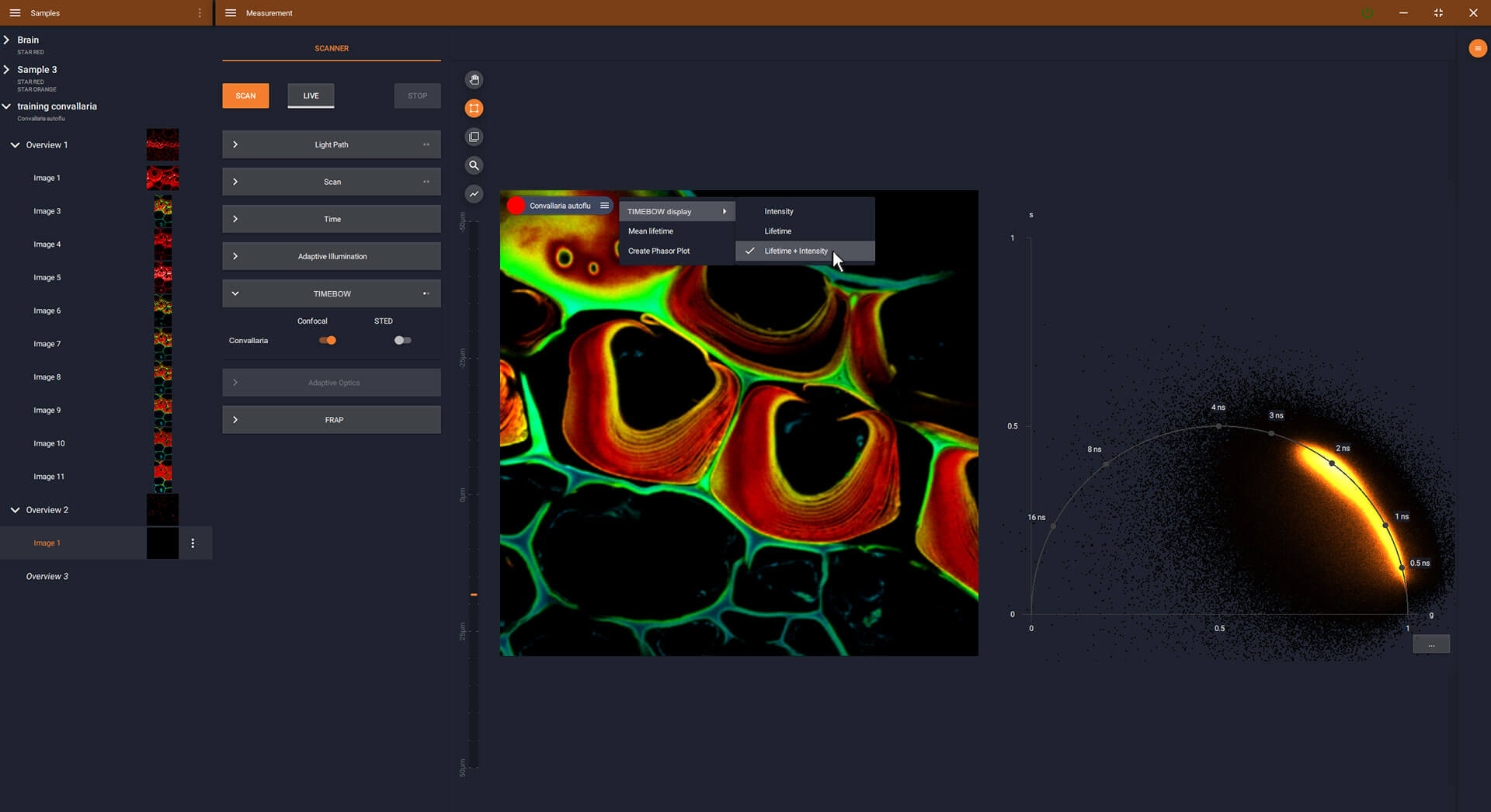
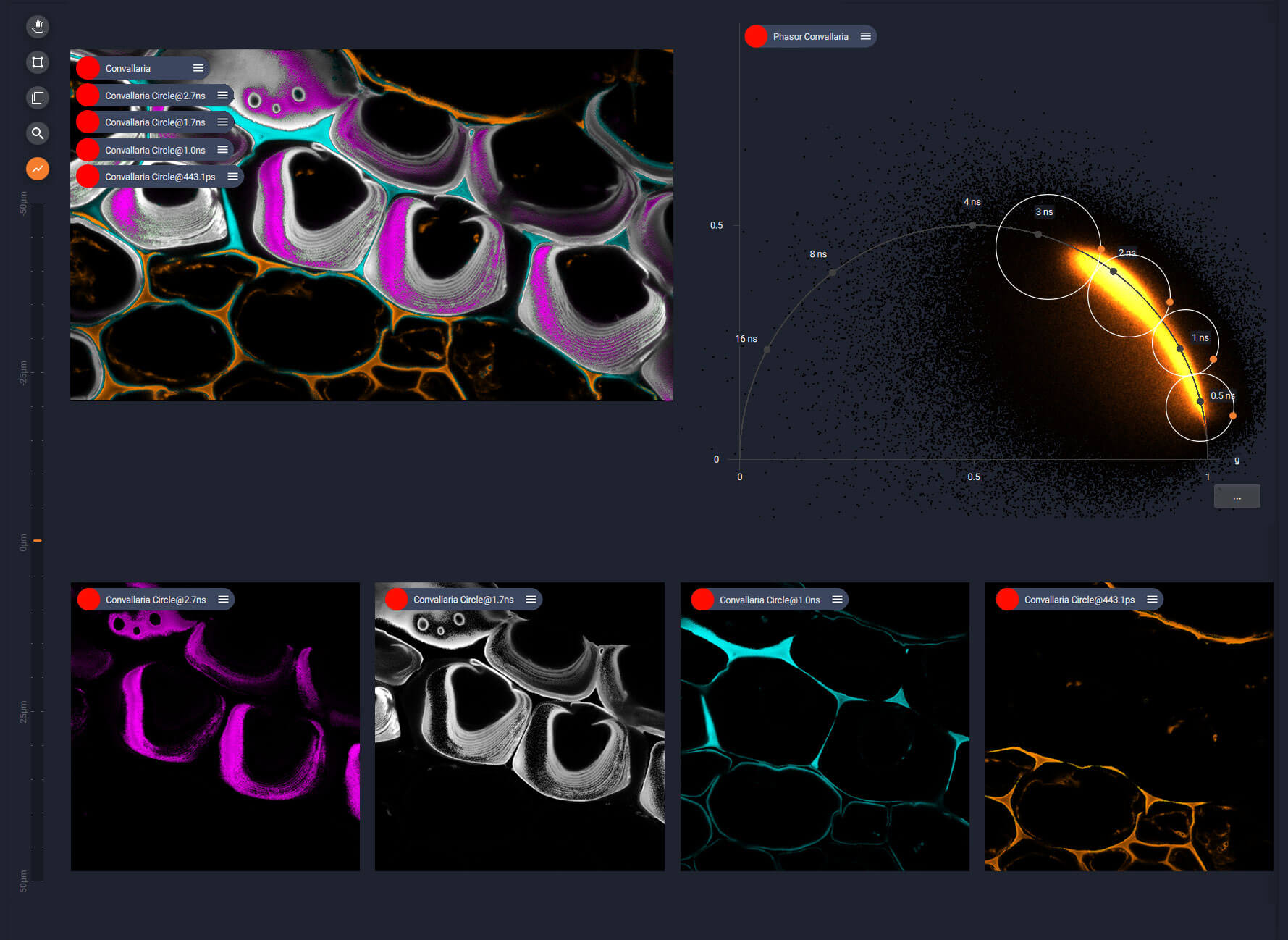
A color-coded lifetime image is just another click away.
TIMEBOW
every signal gets a lifetime tag
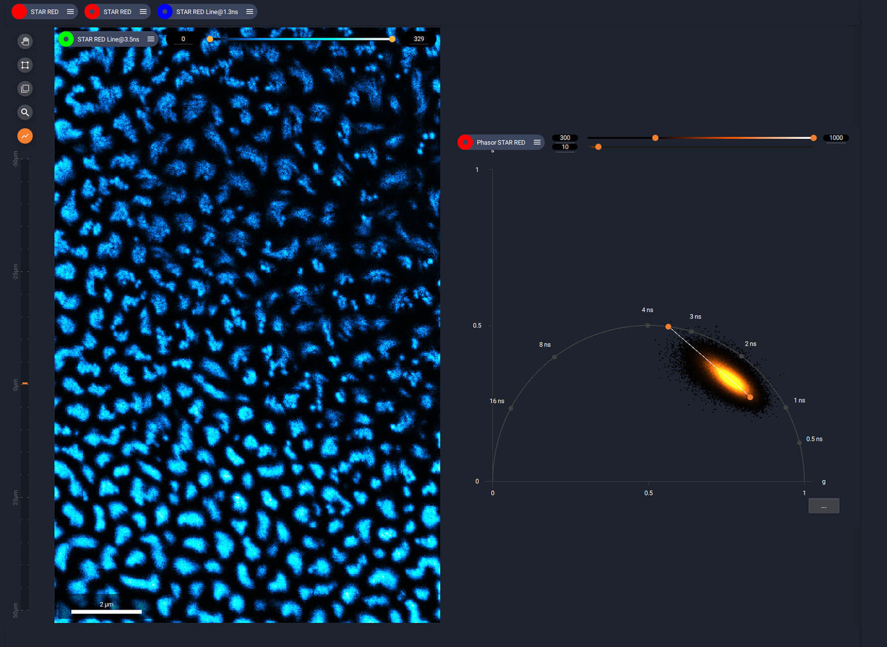
To give you the best way to work with lifetime data, TIMEBOW uses phasor analysis. This turns the temporal dynamics of fluorescence decay into a convenient plot which directly visualizes different lifetime populations. The plot is interactive and gives you the tools you need to process lifetime information within seconds.

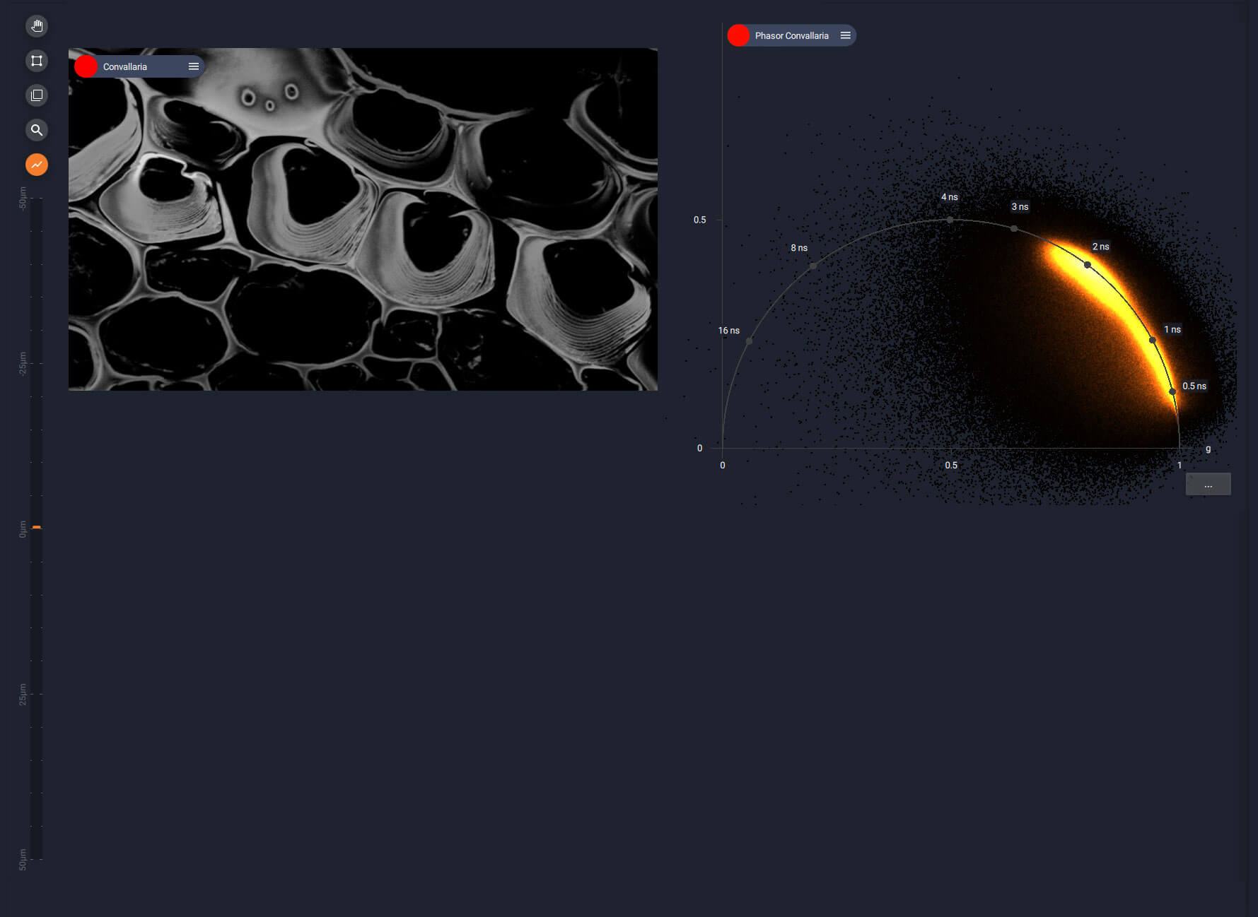
Easy separation of structures thanks to TIMEBOW
Picture 1
Lifetime separated into four species in a plant stem cross section of Convallaria.
Easy separation of structures thanks to TIMEBOW
Picture 2
Image quality is improved by removing the background with MATRIX detection. TIMEBOW gives you the interactive phasor plot to work with.
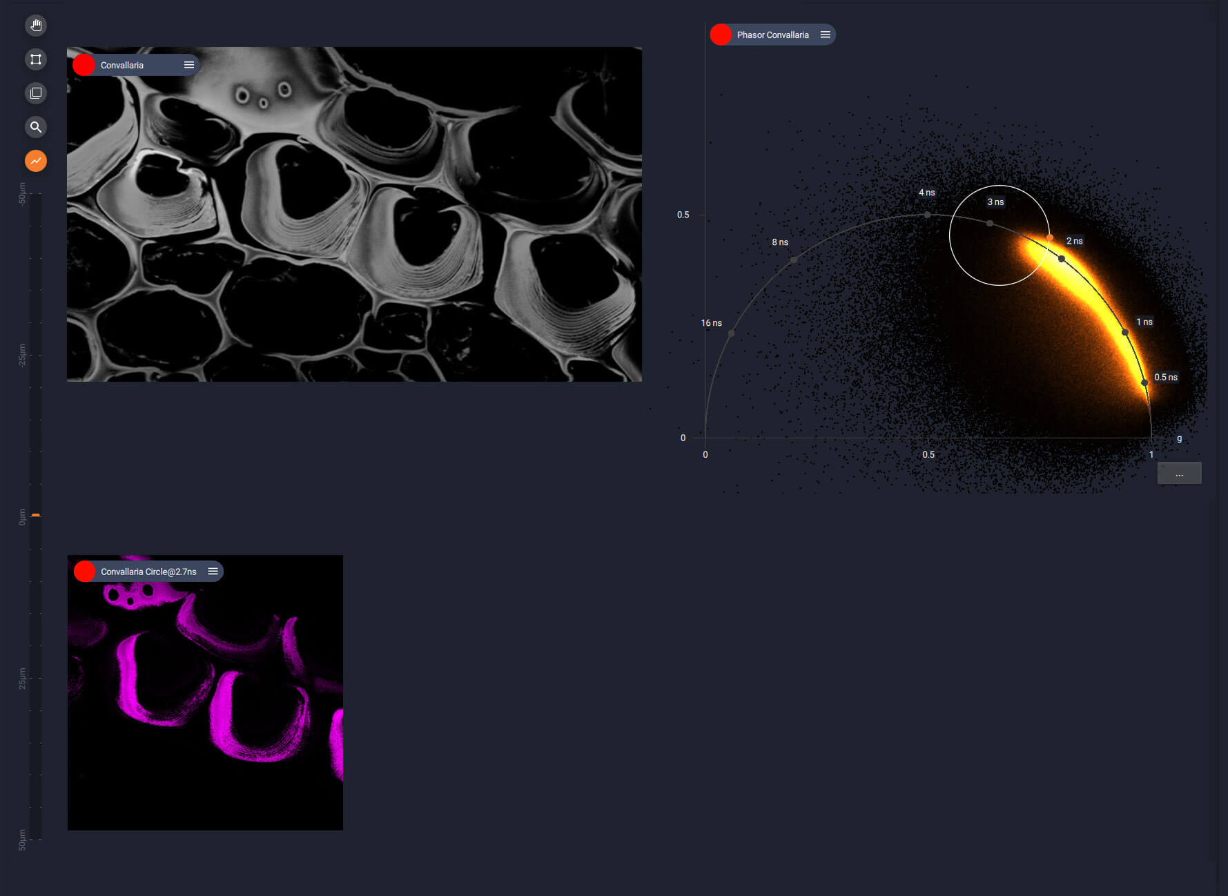
Easy separation of structures thanks to TIMEBOW
Picture 3
By simply drawing circular filters in the phasor plot, different species can be defined. Starting with the longest lifetimes …
Easy separation of structures thanks to TIMEBOW
Picture 4
… and then defining more species by adding more circles …
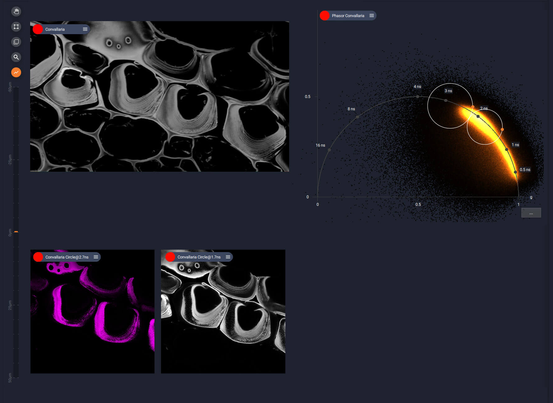
Easy separation of structures thanks to TIMEBOW
Picture 5
… and more circles … and more species …
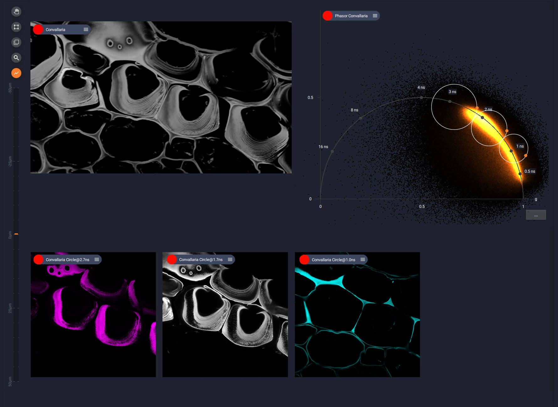
Easy separation of structures thanks to TIMEBOW
Picture 6
… until everything is separated.
Easy separation of structures thanks to TIMEBOW
Picture 7
Finally, these four species are combined, showing a clearly distinct distribution in the sample.
TIMEBOW
you will simply love it
Our superresolution imaging already produces stunning images along all spatial dimensions. Now we do the same with the dimension of time. Grey images turn into colorful sample portraits as color-coded lifetime plots reveal unseen structures. Fluorophores in the same spectral channel suddenly are clearly separated, giving you more channels and more flexibility than ever before.

Expand your imaging options with TIMEBOW
Picture 1
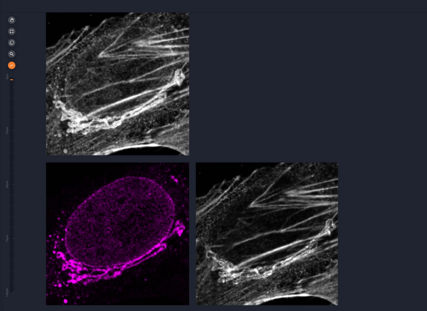
A super-resolved four-color image recorded with only one STED laser.
Expand your imaging options with TIMEBOW
Picture 2
Traditionally, one of the rules of labeling was: dyes with similar excitation spectra cannot be combined in one sample. But here, this is exactly what we do!
Expand your imaging options with TIMEBOW
Picture 3
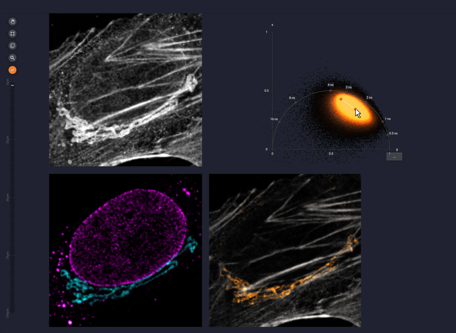
TIMEBOW allows for straightforward separation by lifetime, simply by drawing a line in the phasor plot. Now the targets can be distinguished, due to their difference in lifetime.
Expand your imaging options with TIMEBOW
Picture 4
All separated layers are combined to create a four-color image, which already looks stunning in confocal …
Expand your imaging options with TIMEBOW
Picture 5
… and WOW with superresolution.
TIMEBOW on STEDYCON 2
performance in a shoebox
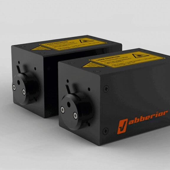
Lifetime imaging on STEDYCON 2 is powered by PicoQuant’s established MultiHarp 150 hardware. Fully integrated into the STEDYCON smart control software, command is seamless and intuitive with no need for external tools or expertise.
The interactive phasor plot assigns the image pixels to distinct populations according to their fluorescence lifetime. These populations can be unmixed into different lifetime channels.
Alternatively, you may export the lifetime data and work with it in third-party software like PicoQuant SymphoTime.
High resolution, low light dose
protect your sample with gentle TIMEBOW STED
With STED, fluorescence lifetime even encodes spatial information. As the laser power of the STED beam initially increases with the distance from its center, fluorophores away from the center are more strongly de-excited and hence display shorter lifetimes. By detecting these lifetime differences, TIMEBOW boosts STED resolution even further: gentle TIMEBOW STED gives you more resolution with less STED power while protecting your sample.
TIMEBOW + MATRIX
a perfect match only from abberior
Of course, TIMEBOW can be combined with our MATRIX array detector. This combination is even more than the sum of its parts. It brings you: higher signal-to-background ratio, better resolution and an impressive dynamic range. Every one of the 23 sensor elements of the MATRIX is used to simultaneously record both lifetime information and information about the origin of photons relative to the focal plane. With this data, we get rid of background better than a confocal pinhole ever could and we render stunning images not only richer in information, but also with exceptional contrast.
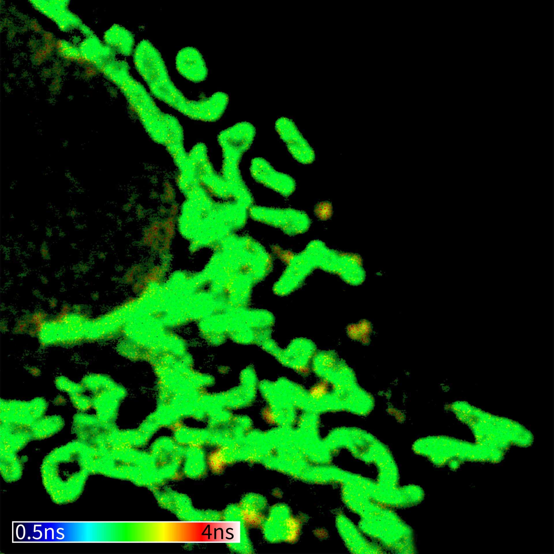
Sharpening with TIMEBOW
Picture 1
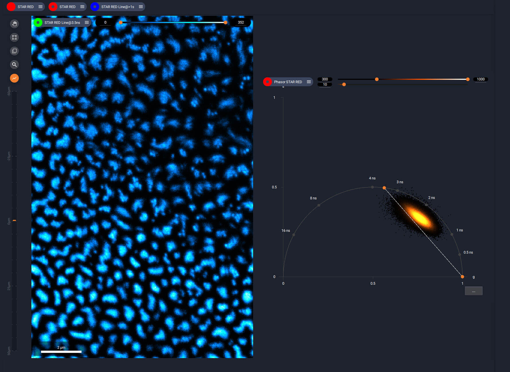
Actin in microvilli, with the background removed using MATRIX. STED imaging has a unique property, whichTIMEBOW uses to further boost the resolution…
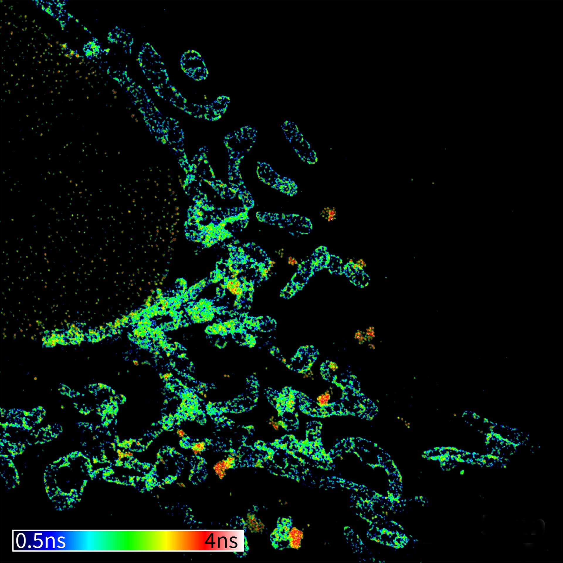
Sharpening with TIMEBOW
Picture 2
… by removing short-lived components that are not in the very center of the STED donut. This is done by simply dragging the separation line in the phasor plot.
Sample by Dorothee Günzel and Jörg Piontek, Charité, Berlin, Germany.
- FLIM, fully integrated
- Turn temporal dynamics into color-coded lifetime images
- Super-resolved four-color images with only one STED laser
- gentle TIMEBOW STED: high resolution, low light dose
- On MIRAVA POLYSCOPE, together with MATRIX: higher signal-to-background ratio, better resolution and impressive dynamic range – only available from abberior
TIMEBOW lifetime imaging for stunning results with confocal and STED superresolution
MATRIX Detector
Many eyes see more than one. The MATRIX detector drastically improves signal-to-background ratio, resolution, and dynamic range.
FLEXPOSURE Illumination
Brings down the light dose on your sample and lables dramatically. Key ingredient for volume and live-cell superresolution.