


RAYSHAPE
Ideal imaging conditions, even in turbid samples like tissue, are every microscopist’s dream. However, specimens are often inhomogeneous, and, what’s more, embedding and immersion media are hardly ever perfectly index-matched. Refractive-index mismatches compromise the focusing capabilities of your microscope and can give rise to low-resolution and poor-quality images.
abberior’s RAYSHAPE dynamic aberration correction solves this problem. Using adaptive optics with a deformable mirror, it puts diverted rays back on track, guaranteeing bright and crisp images even in complex samples, from top to bottom and regardless of embedding medium, immersion medium, or specimen. Correction happens dynamically on-the-fly and leaves nothing to desire for your results.
MINFLUX Module
Single-digit nanometer resolution with the MINFLUX module for our MIRAVA POLYSCOPE
MATRIX Detector
Many eyes see more than one. The MATRIX detector drastically improves signal-to-background ratio, resolution, and dynamic range.
TIMEBOW Imaging
TIMEBOW lifetime imaging for stunning results at confocal and STED super-resolution.
FLEXPOSURE Illumination
Brings down the light dose on your sample and lables dramatically. Key ingredient for volume and live-cell superresolution.
RAYSHAPE Mirror
Dynamic aberration correction with a deformable mirror over about 200 µm z-range. 140 digital actuators adjust the mirror surface within milliseconds.

Custom Solutions
We offer solutions for even the most challenging applications. Everything that can be done, we will do.
Description
Comparison 1/3
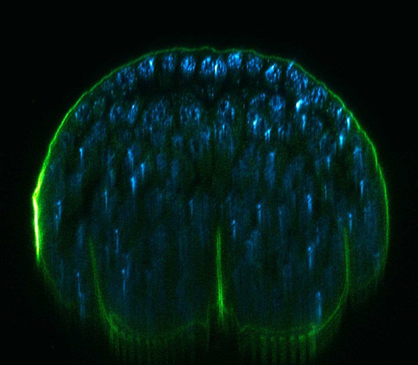
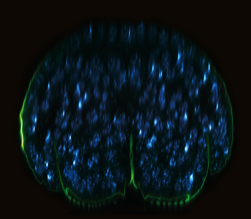
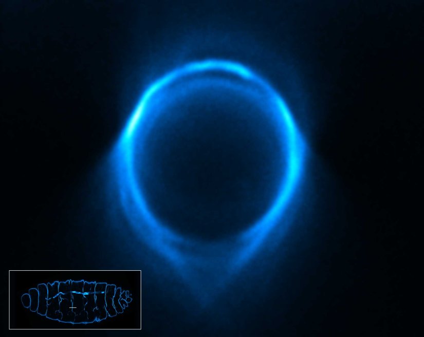
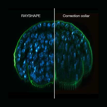
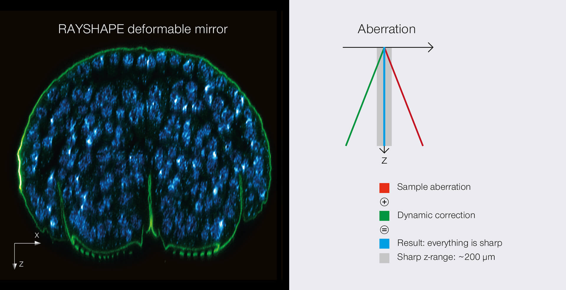
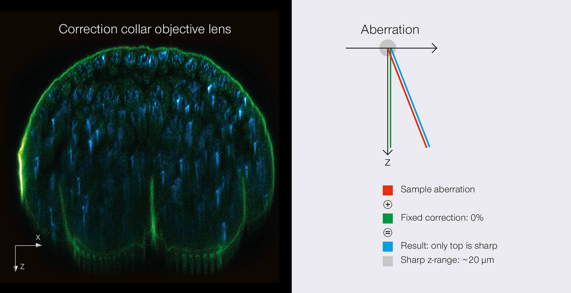
RAYSHAPE mirror versus correction collar objective lens.
RAYSHAPE dynamically corrects the entire z-range, the correction collar only a small z-range at a certain position, here close to the cover slip.
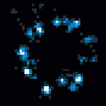
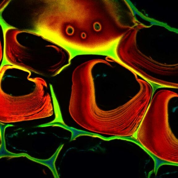
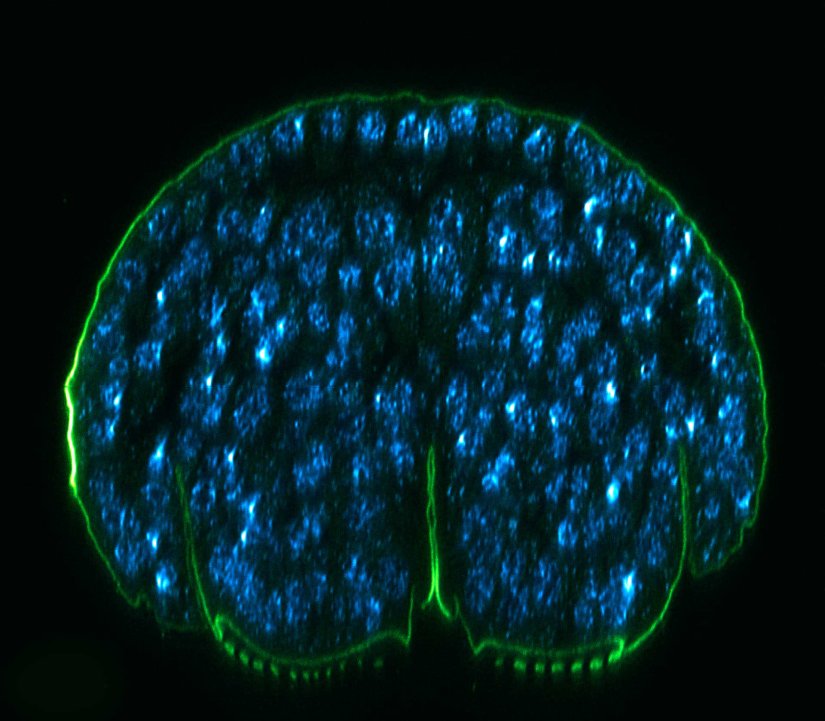
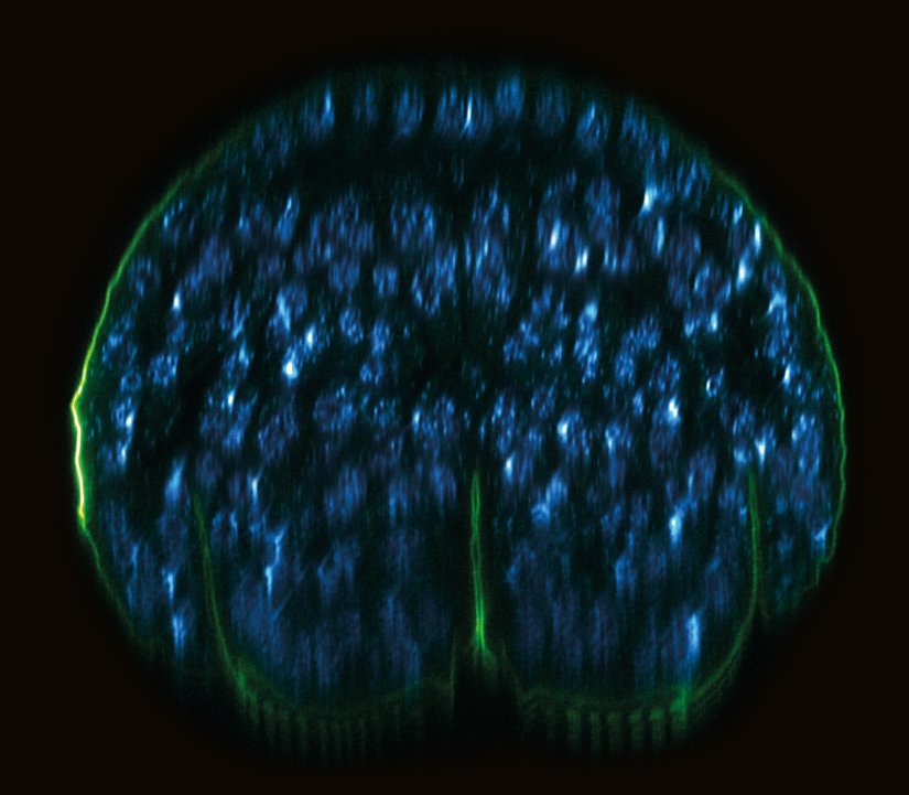
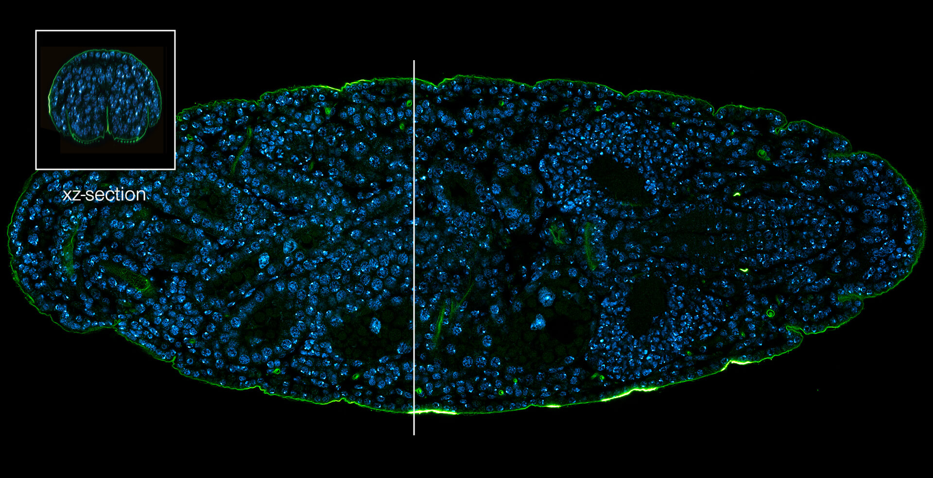
xz section of a stage 17 Drosophila embryo stained for chitin (abberior LIVE 610, green) and DNA (abberior LIVE 550, cyan).
Description
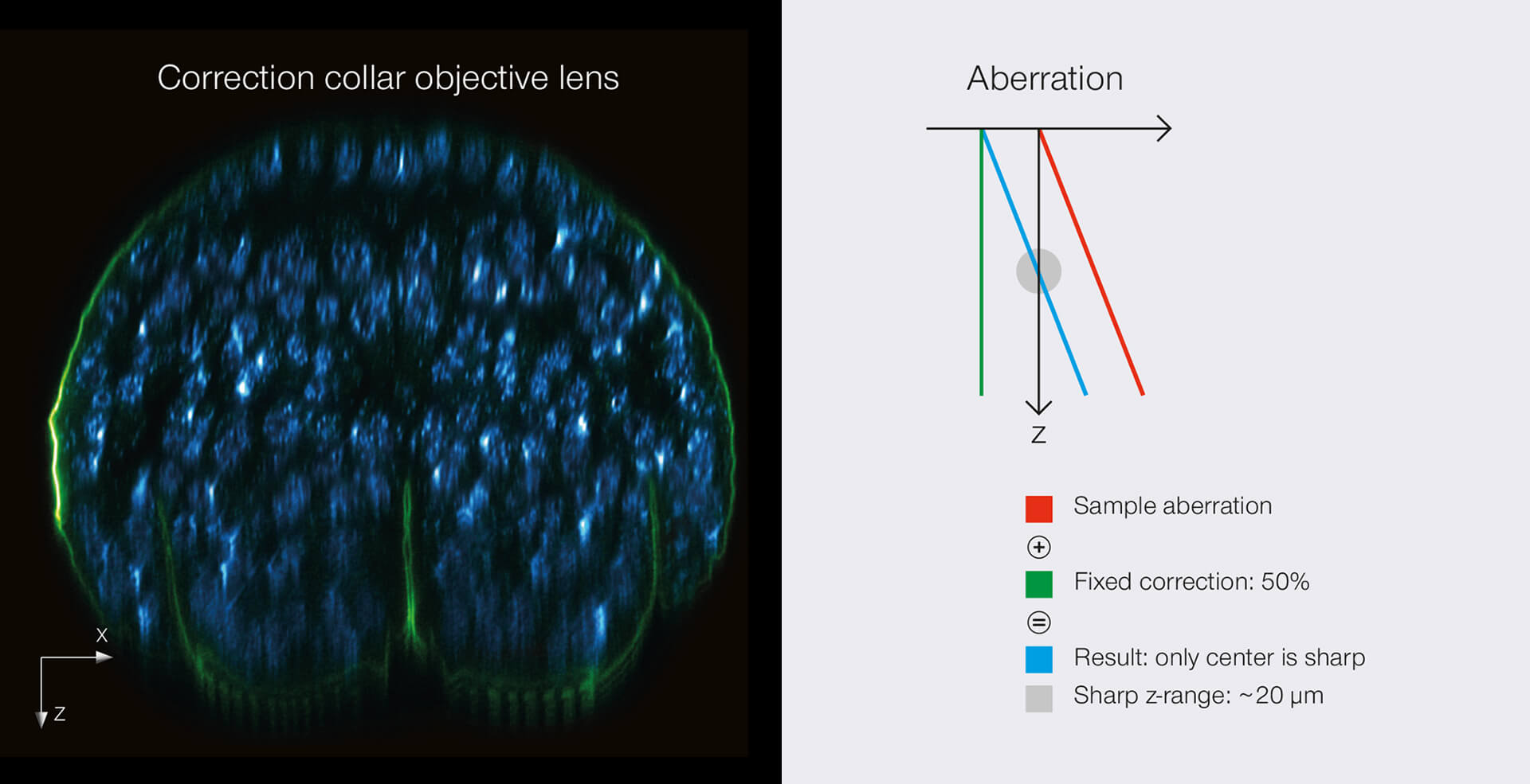
Comparison 2/3
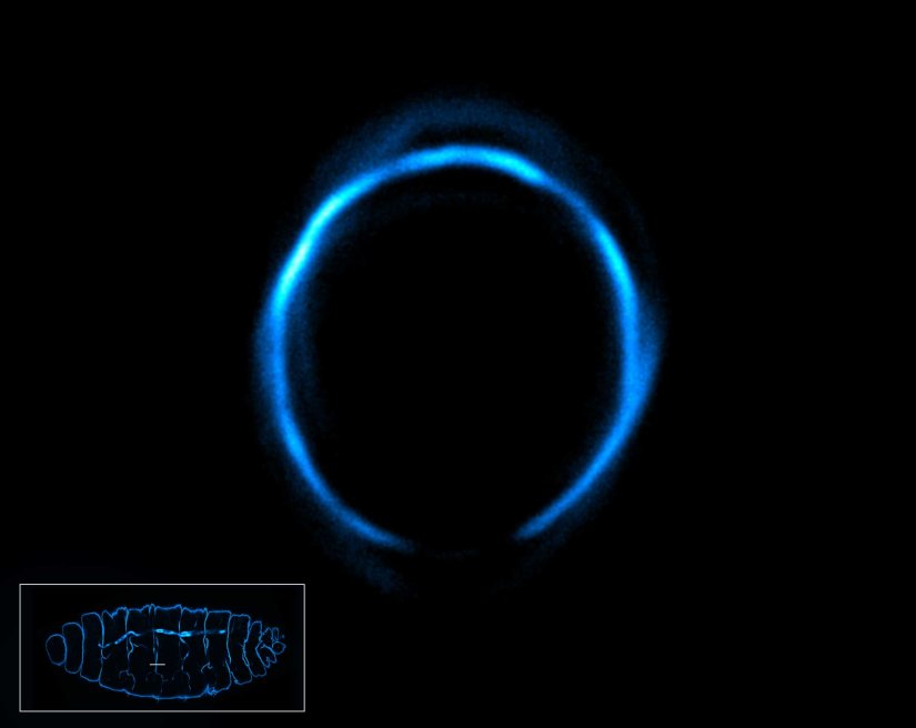
RAYSHAPE mirror versus correction collar objective lens.
RAYSHAPE dynamically corrects the entire z-range, the correction collar only a small z-range at a certain position, here at the middle of the z-range.
Description
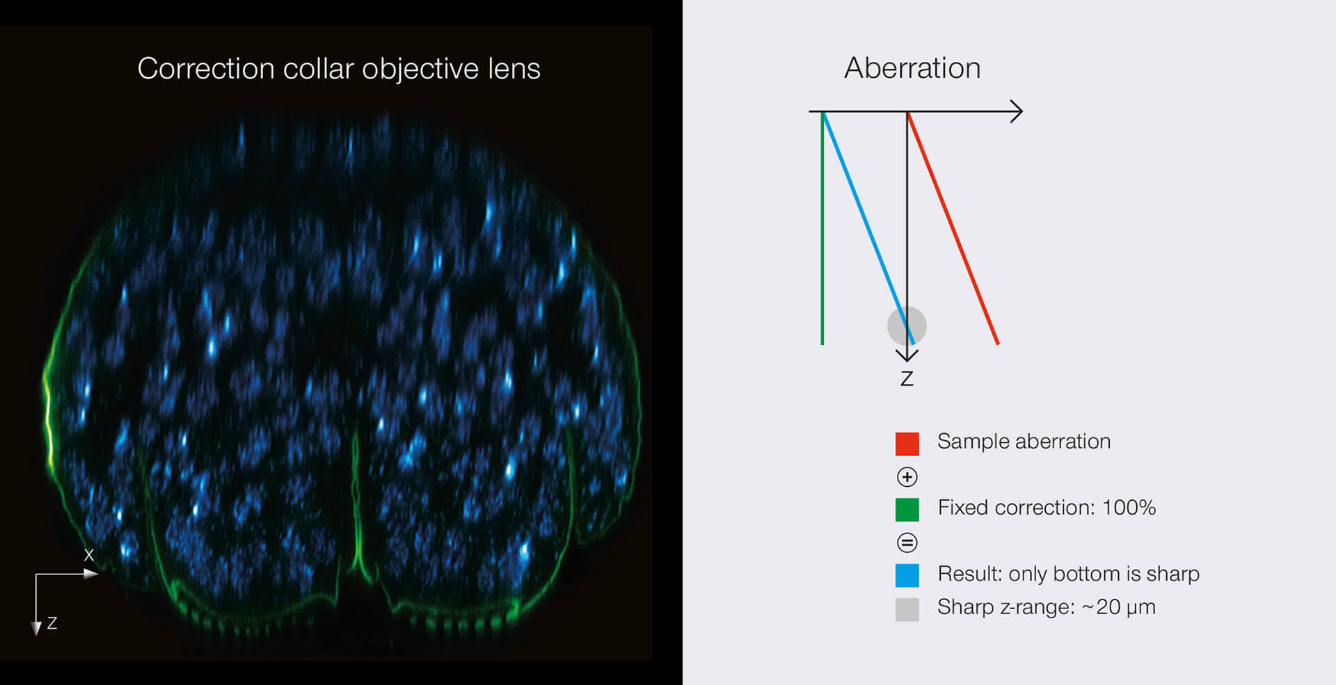
Comparison 3/3
RAYSHAPE mirror versus correction collar objective lens.
RAYSHAPE dynamically corrects the entire z-range, the correction collar only a small z-range at a certain position, here at the end of the z-range.
Description
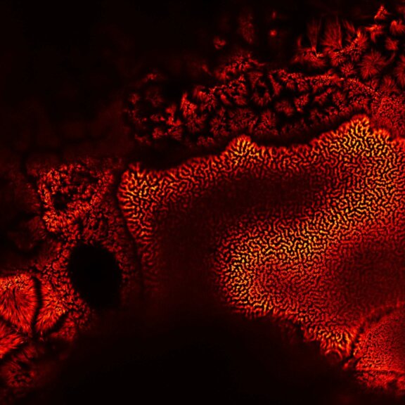
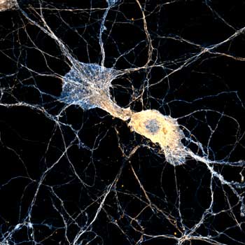
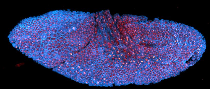
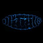
Drosophila stage 12 embryo, imaged with RAYSHAPE, stained for tubulin with abberior STAR RED and for DNA with abberior LIVE 550.
Modules:
Description
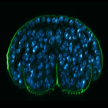
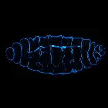
Deep tissue imaging with RAYSHAPE of a stage 17 Drosophila embryo stained for chitin with abberior LIVE 610.
Modules:
Description
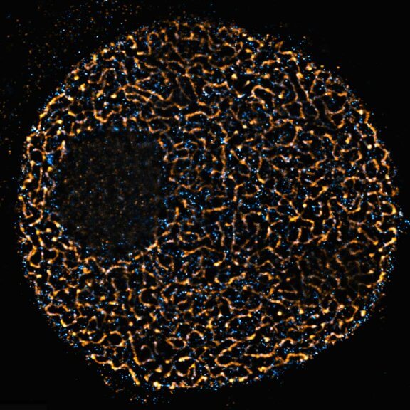
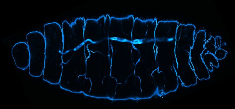
3D STED xz section of a Drosophila embryo trachea, imaged with RAYSHAPE and without abberation correction, depth 15 µm. Chitin was stained with abberior LIVE 610.
Modules:


Hello RAYSHAPE
Bye-bye, aberrations!
The deeper you dive into the sample, the more severe the aberrations become, and the more you have to correct. In order to achieve zero aberrations throughout the sample, you therefore need to permanently adjust the amount of correction as focus depth changes.
RAYSHAPE vs
correction collar
Picture 1
RAYSHAPE preserves resolution and brightness over the whole sample depth of about 200 µm by dynamically redirecting aberrated light to the right places.
In comparison, mechanical optics using a correction collar can only correct a limited z-range of approximately 20 µm …
RAYSHAPE vs
correction collar
Picture 2
In this example (Stage 17 Drosophila embryo) a correction collar objective lens is set to correct aberrations at three different z-positions: close to the cover slip …
RAYSHAPE vs
correction collar
Picture 3
… at the middle of the z-range …
RAYSHAPE vs
correction collar
Picture 4
… and at the end. None of the positions is right for the whole sample and only a small z-range looks good.
RAYSHAPE vs
correction collar
Picture 5
Overview image of this stage 17 Drosophila embryo stained for chitin (abberior LIVE 610, green) and DNA (abberior LIVE 550, cyan).
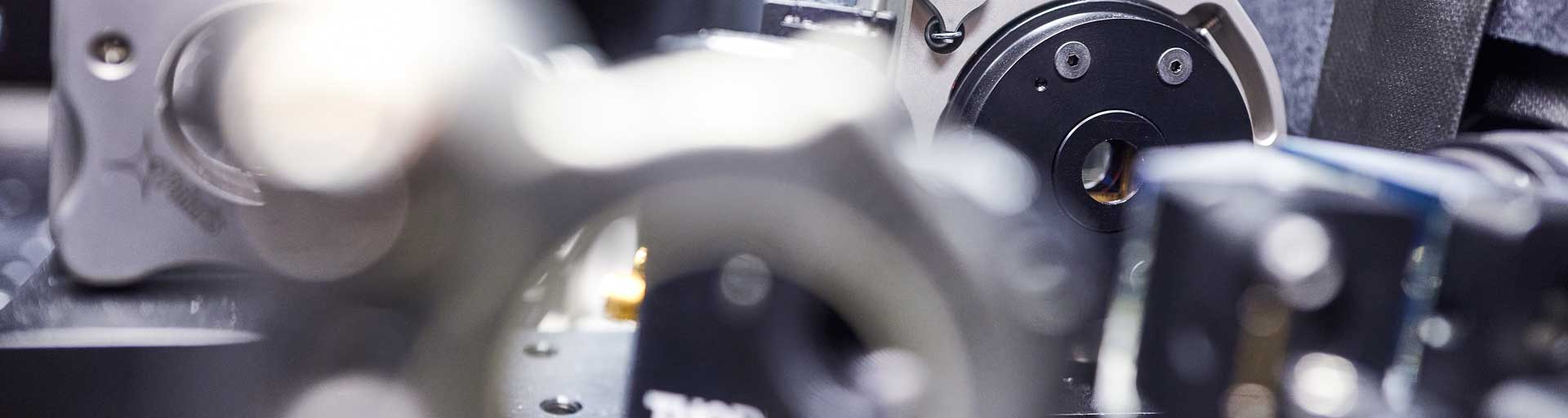
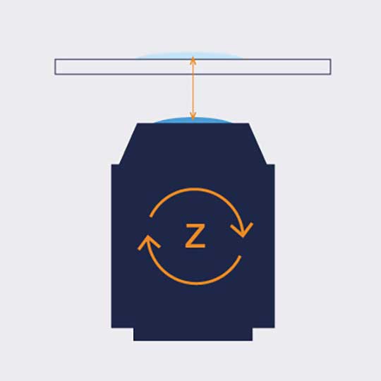
RAYSHAPE deformable mirror
only from abberior
Deformable mirrors are adaptive elements with a flexible reflective surface. 140 digitally controlled actuators adjust the mirror’s surface within milliseconds, making RAYSHAPE exceptionally precise and allowing fast z-scans with continuous adjustment of the correction to compensate for aberrations at every depth.
With RAYSHAPE, you are not forced to choose a limited region of correction while the rest of the image stays blurred, as is the case for even motorized correction collar objectives. Once adjusted to the sample, RAYSHAPE automatically follows the focus as it moves through the specimen and dynamically corrects aberrations at different depths. The focus is kept stable and image acquisition runs completely autonomously for bright, high-resolution images even very deep in the sample.
Tissue imaging has never felt better than with RAYSHAPE.
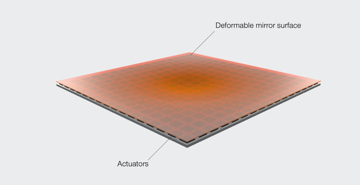
RAYSHAPE deformable mirror with 140 actuators
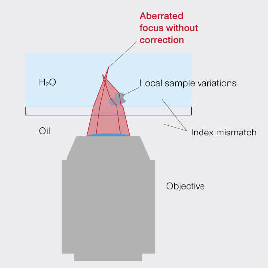
Without RAYSHAPE the focus is aberrated
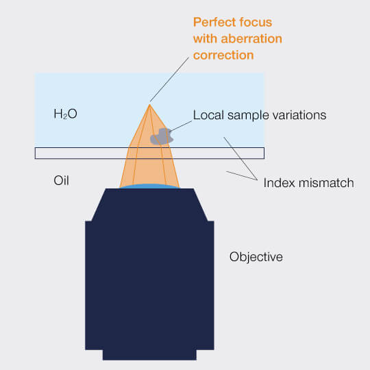
RAYSHAPE with its deformable mirror keeps the focus over the entire depth range
Mismatch between sample and the immersion medium of the objective lens results in a misshaped focal spot. To make things worse, sample inhomogeneities add to these aberrations. RAYSHAPE aberration correction compensates for these distortions by putting all rays in the right place and thus restores a perfect focus.
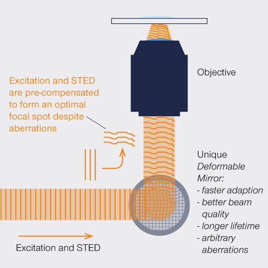
Correction of exitation STED
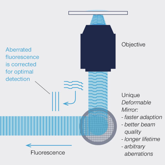
Correction of aberration fluorescence
Undistorted wavefronts entering the sample from the objective lens are normally spherical, but variations of the refractive index can distort them, leading to an imperfect focus. RAYSHAPE’s deformable mirror effectively cancels these aberrations. Applying the correct mirror shape for a specific aberration – which is a negative of the distortions introduced by the sample – achieves two things at once:
1) The wavefront of excitation and STED beam are altered to pre-compensate for aberrations. The focus is thus brought back to perfect shape.
2) The distorted wavefront of the fluorescence is also corrected before it reaches the detector.
Since RAYSHAPE can adjust the degree of correction fast and dynamically as the focus moves through the sample, the result is a clear image with strong signal and resolution from top to bottom.
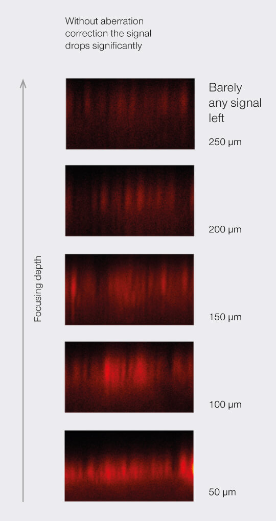
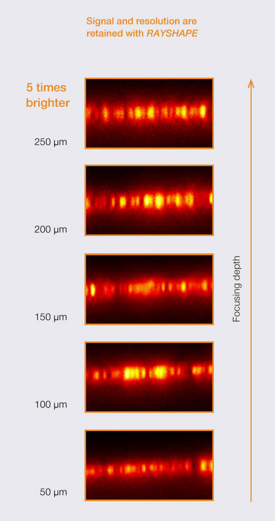
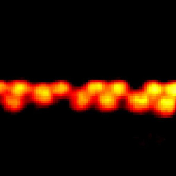
Imaging a fluorescent bead layer at different depths (63x WI objective lens, TDE embedding). The deformable mirror automatically follows when focusing depth is changed. Brightness and resolution are largely preserved up to the working distance of the objective lens.
- Up to five times higher signal in thick and complex sample sections
- Superior resolution with STED
- Often the enabler for 3D-STED imaging
- Automatic tracking of spherical aberrations
- Correction of higher order aberrations (astigmatism, coma, trefoil, …)
Super-sharp images, even in thick and complex samples

Watch Recording: Webinar on RAYSHAPE dynamic aberration correction
Hello RAYSHAPE! Bye-bye, aberrations!
MIRAVA POLYSCOPE
abberior’s MIRAVA is the first true POLYSCOPE. It unites four microscopy technologies to cover an unprecedented resolution spectrum, extending over several orders of magnitude from diffraction-limited imaging all the way…
Protocols
Not rocket science, but simple protocols that make the daily life of a fluorescence microscopist much easier.

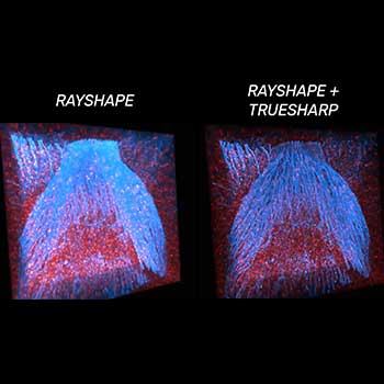
Why is TRUESHARP better at dealing with noise and background?
Learn how TRUESHARP images boosting deals with noise and background, why noise is random and how we analyze and identify signal and background separately.

How can TRUESHARP improve your everyday imaging experience?
See how TRUESHARP will strongly enhances resolution, sharpness, and brilliance of your images and why abberior’s TRUESHARP, MATRIX, and TIMEBOW technology allows you to get as close as possible to…