


Your research
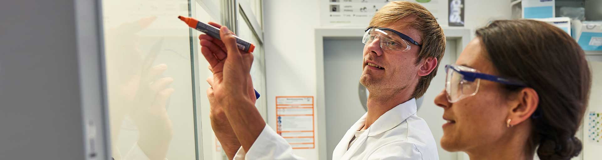
The founders and scientists at abberior not only laid the groundwork for superresolution microscopy, they also were among the first to develop its application to biological, medical, and material science. And they are always available to offer guidance should you need it. You can count on first-class advice when it comes to getting the most out of your microscope!
our solution

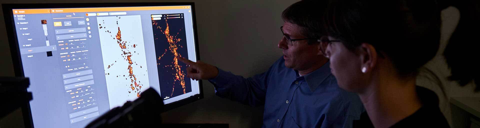
Thanks to the advances in resolution, light microscopy plays an increasingly important role in biophysics. The unique abilities of MINFLUX have been used in various studies investigating biophysical processes.
Cell biology is one of the broadest research areas within molecular biology. For some purposes, the resolution of a confocal microscope is sufficient. In other cases, you will need superresolution microscopy like STED or MINFLUX to capture relevant details beyond the diffraction limit.
Live cell imaging is used in virtually all areas of molecular biology. It places unique demands on imaging conditions and microscope. With abberior, you will find the ideal dye, a suitable microscope system, and the matching technical modules for your purpose, all with the aim of recording the best possible data while treating the sample with care.
Nowadays, 2D and 3D superresolution light microscopy techniques such as STED and MINFLUX are used more and more in material science. With them, sample preparation is comparably simple and they facilitate the investigation of the dynamic behavior of solid materials.
Light microscopy is a central tool to visualize membranes. And since membranes are highly dynamic systems, live cell imaging is of great importance. No matter what membrane process you are studying – abberior can provide the ideal equipment for your imaging experiment.
Prokaryotes, protozoa, algae, and fungal cells can be visualized with diffraction-limited microscopy. But many subcellular structures and details of host-pathogen interactions lie beyond its resolving power. Today, superresolution microscopy opens up completely new opportunities in microbiology.
Light microscopy is a valuable tool in neurobiology as it allows the analysis of the structure, function, and interaction of neuronal cells and systems in an intact and even living state deep within tissue. abberior provides microscopes for superior imaging as well as add-on modules addressing the particular challenges of neurobiological microscopy like deep tissue imaging.
Light microscopy is a key technique in physiology and is used to image specimen of vastly different length scales. Confocal, STED, and MINFLUX are well-suited for live cell imaging, which is essential to investigate physiological processes.
Imaging in botany spans several length scales, from whole organisms over tissue sections and single cells to sub-cellular structures and protein complexes. Thanks to superresolution microscopy, it is nowadays possible to image structures in unprecedented detail even in living specimen.
In virology, superresolution microscopy offers a significant advantage over electron microscopy, which has long dominated virological imaging: they can be used to image viruses and their components in living cells. It is now possible to resolve dynamic processes such as the penetration of viruses and phages into host cells and the maturation and assembly of virus particles.
Imaging spans several length scales in zoology. abberior’s microscopes are equipped with objective lenses for any magnification, and feature add-on modules tailored to tackle various imaging challenges such as intransparent structures, autofluorescence, or loss of signal in deep tissue.