Sample gallery
Fluorescence imaging, whether at confocal, STED or MINFLUX resolution, guarantees unique insights into the function and structure of life at the molecular level. Besides the scientific information content, some sample portraits provide simply beautiful images. Enjoy browsing our sample gallery.
the fine art of science
Description
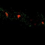
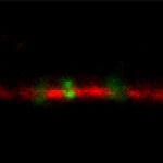
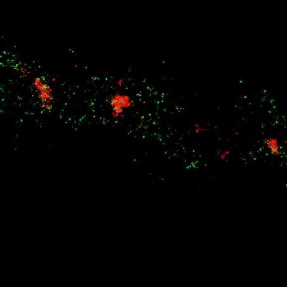
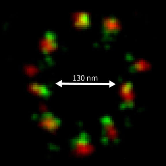
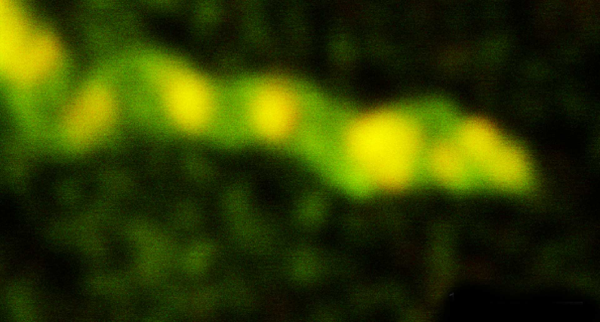
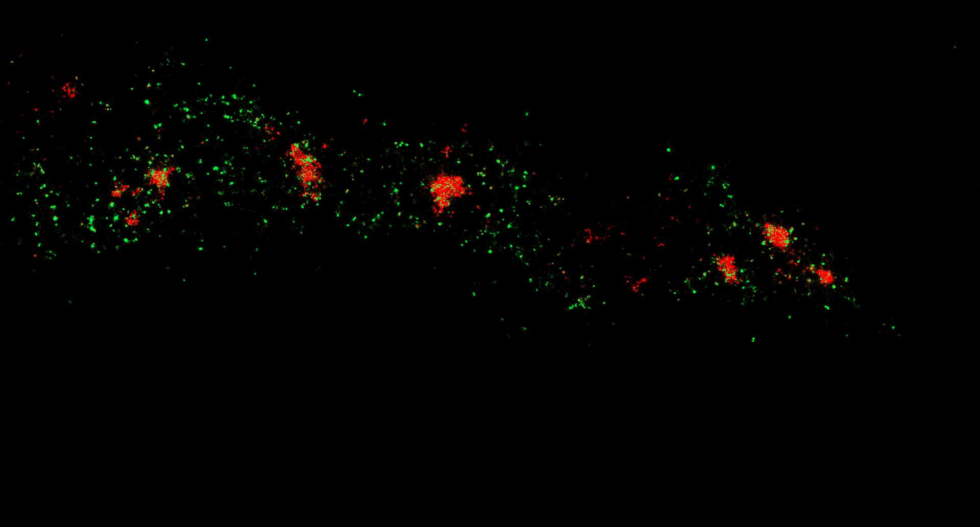
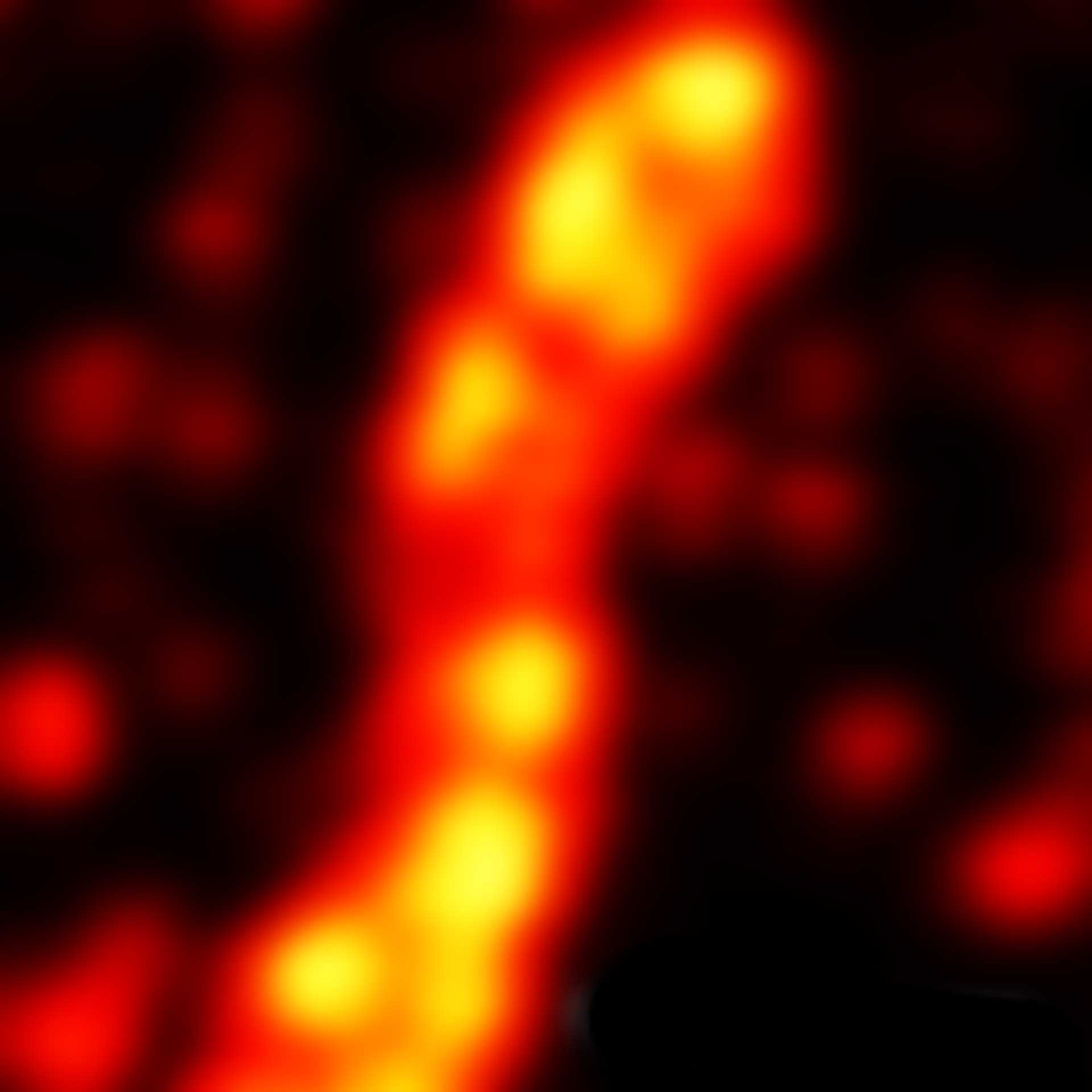
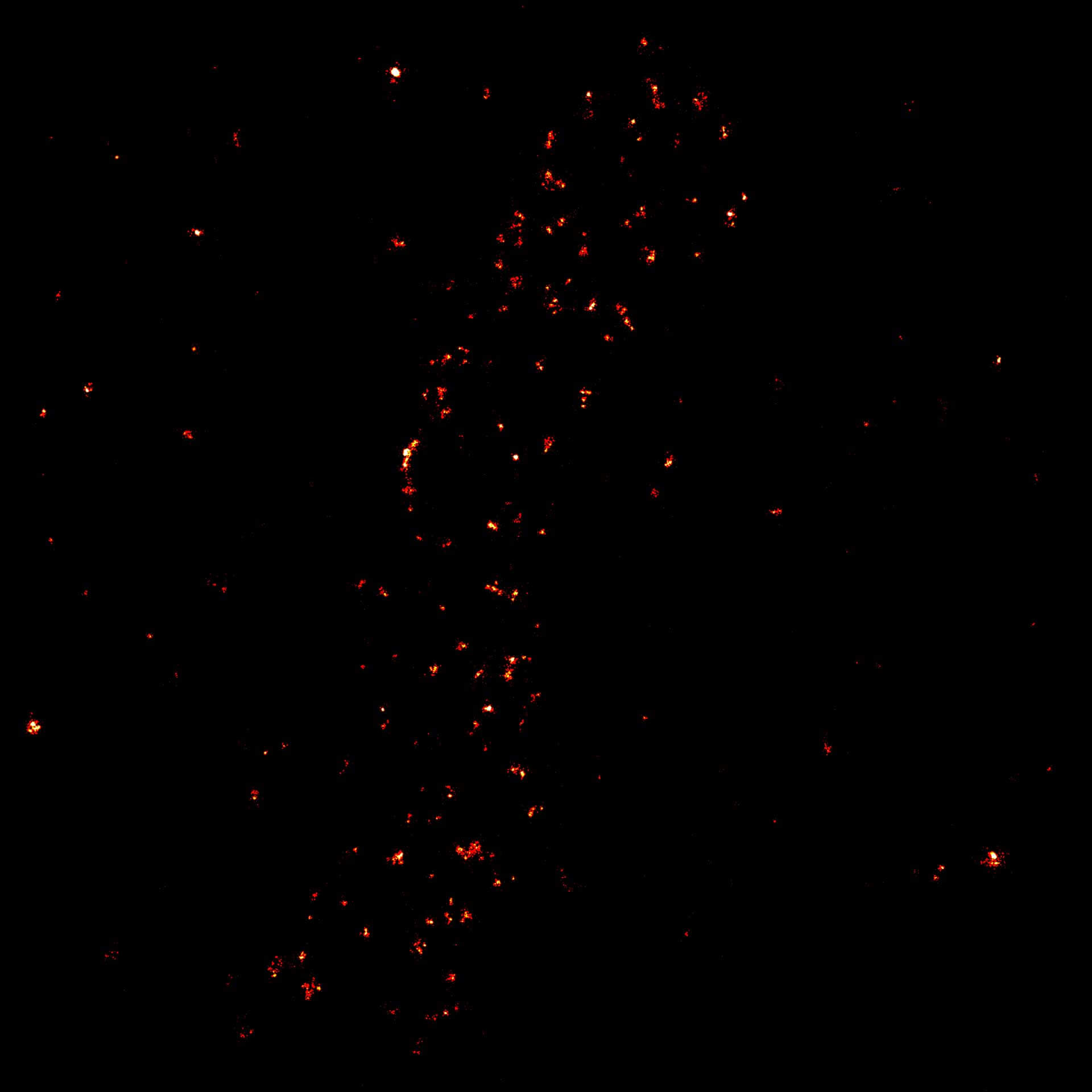
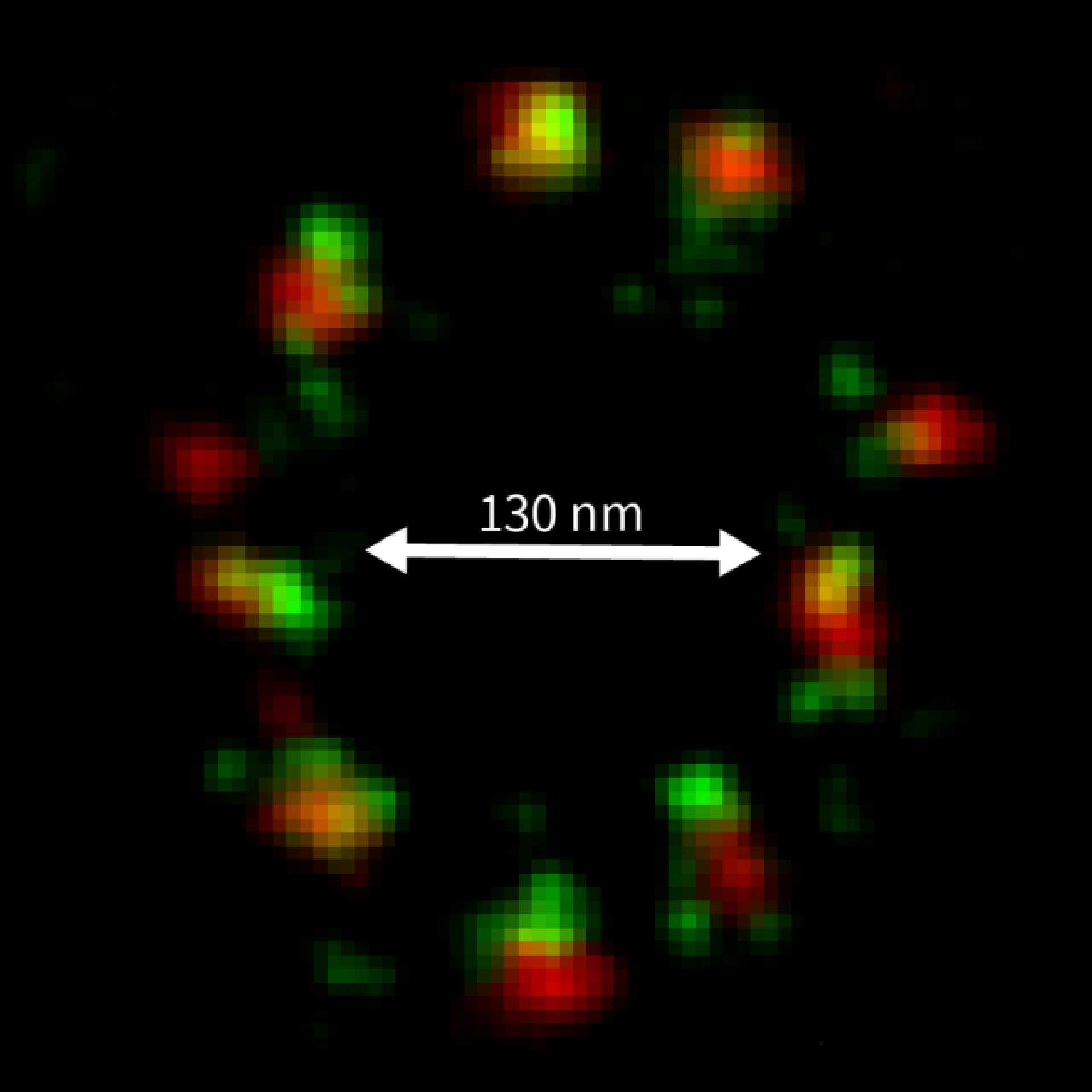
Two-color confocal and MINFLUX images of Tom20 (green) and mitochondrial DNA (red) stained with sCy5 and CF680 in mammalian cells using indirect immunolabeling. The two fluorophores were distinguished by ratiometric detection strategy. Note the dissimilar labeling density of the two imaged structures.
Description
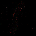
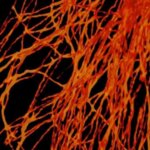
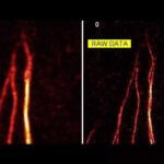
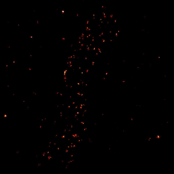
2D MINFLUX image of the mitochondrial import receptor Tom20 labeled with Alexa Fluor 647 in fixed mammalian cells using indirect immunolabeling.
Description
Modules:
Description
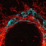
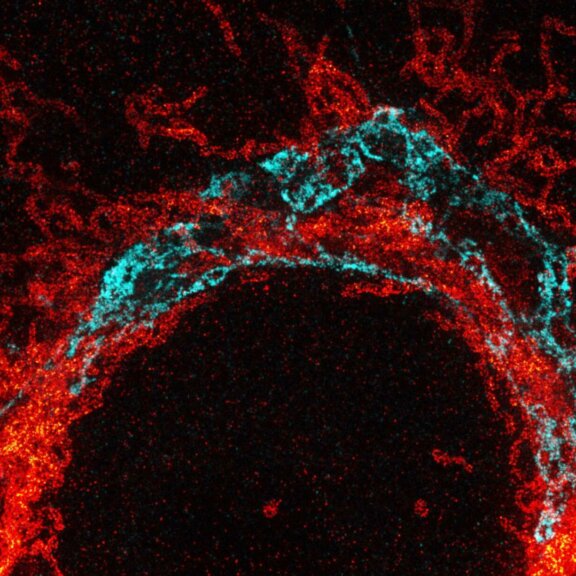
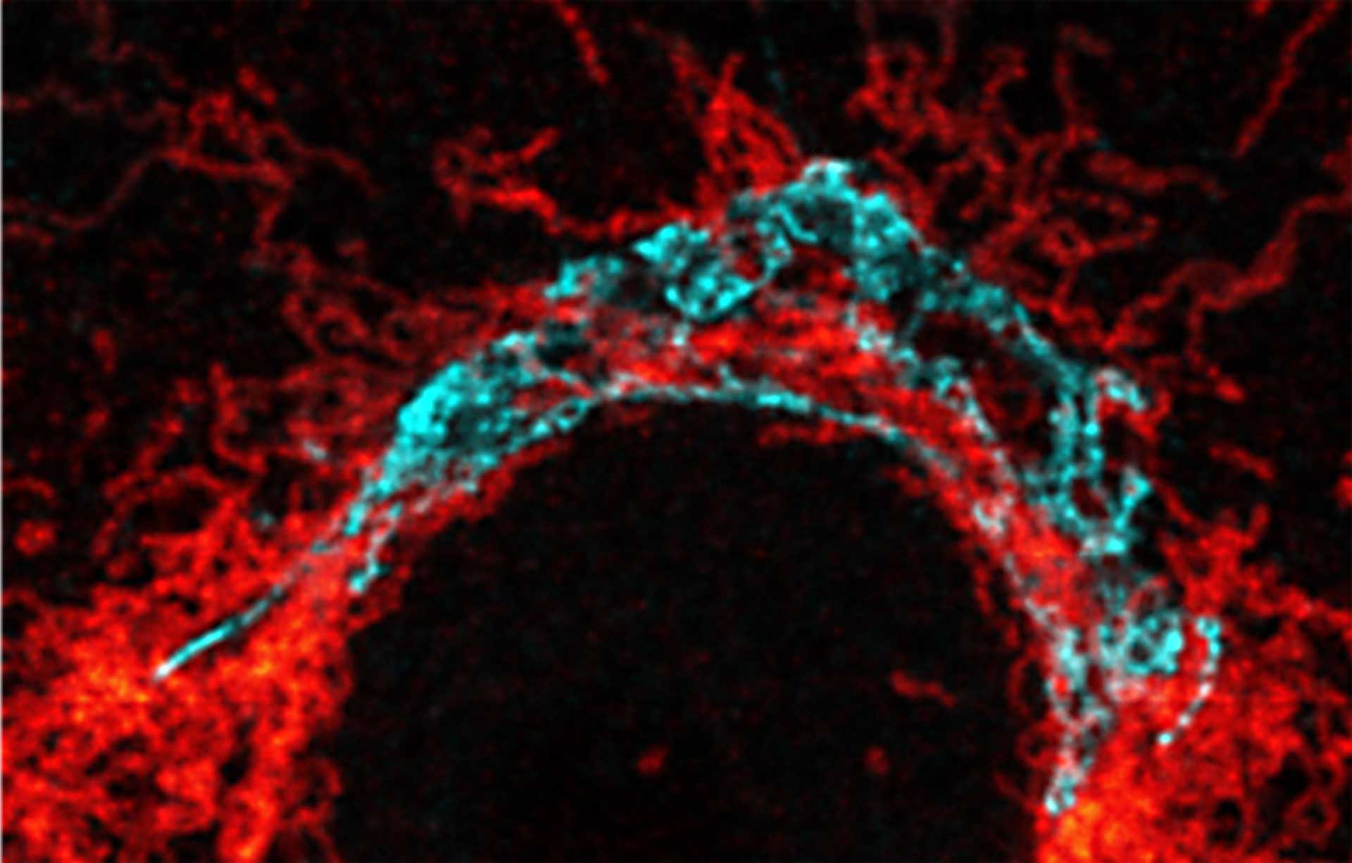
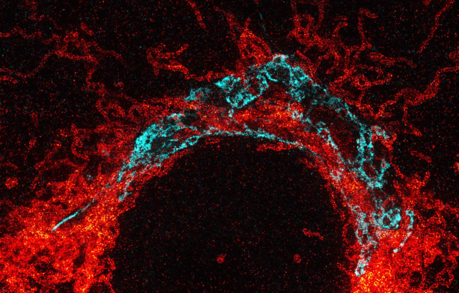
Tom20 (red) and golgi (blue) in cultured mammalian cells.
Modules:
Description
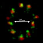
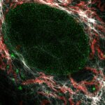
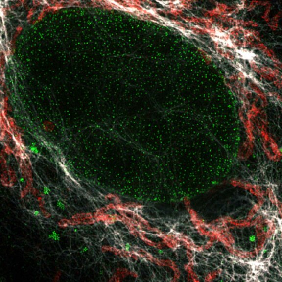
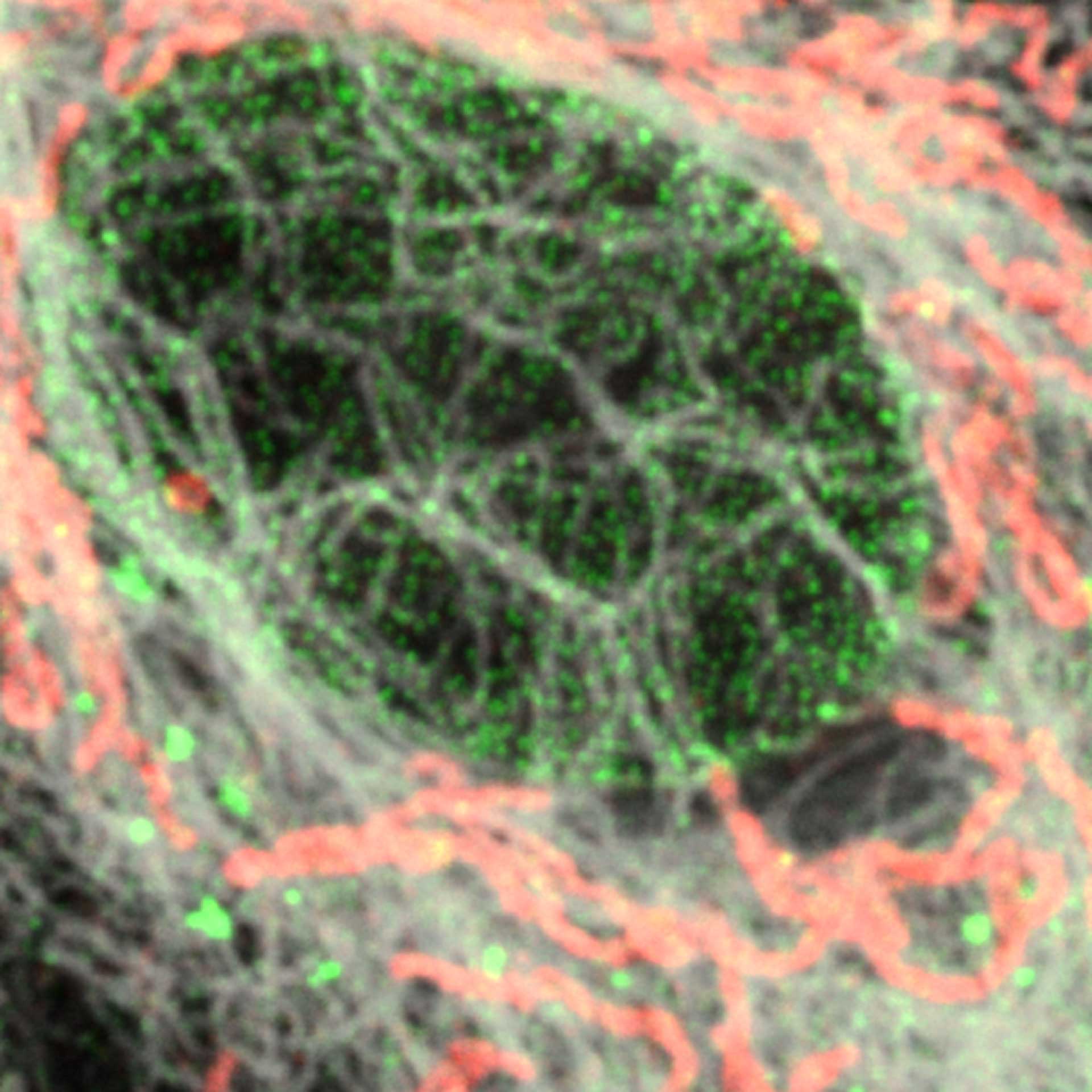
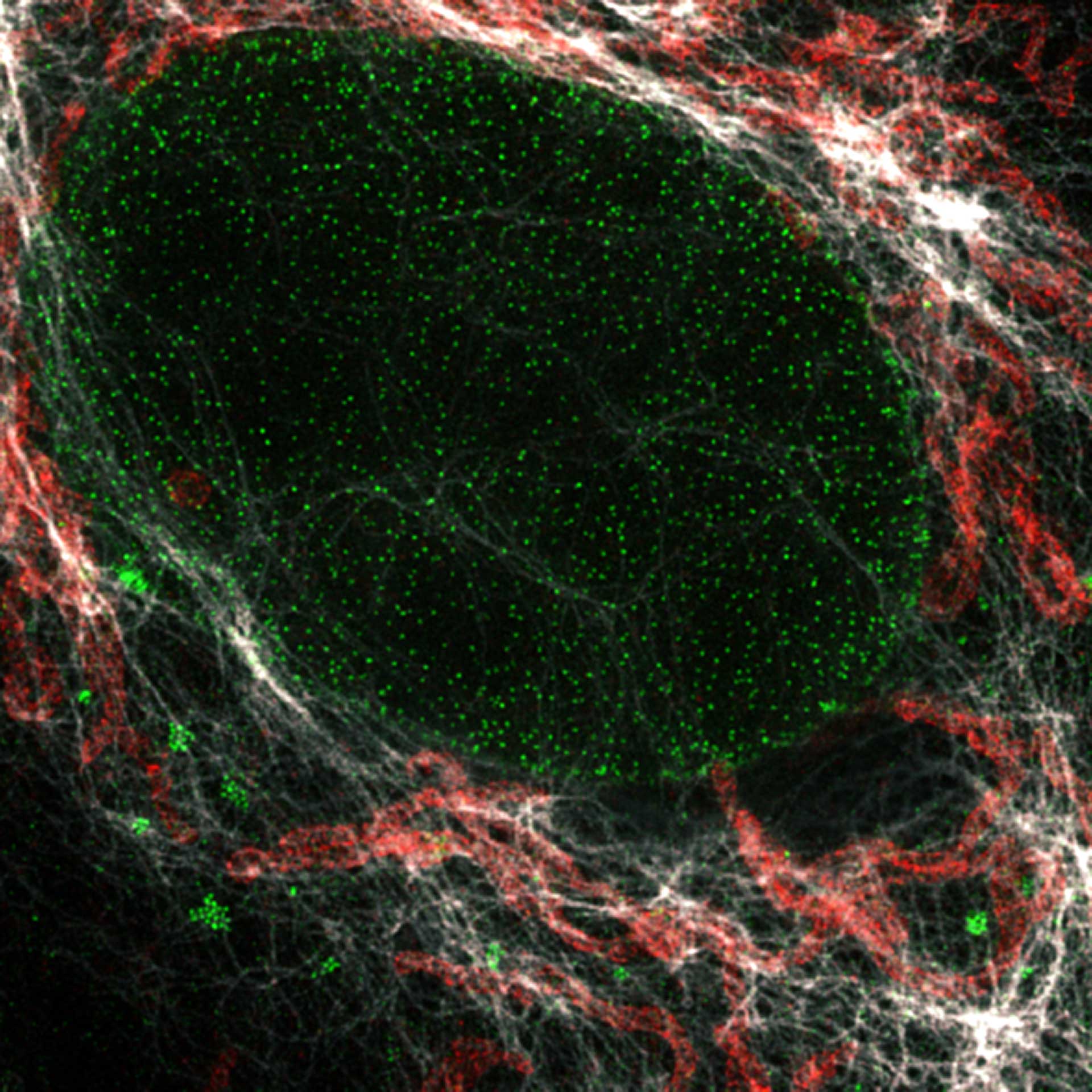
Nuclear pore complex (green), Tom20 (red), and vimentin (white) in cultured mammalian cells.
Modules:
Description
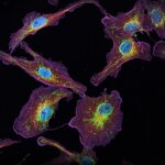
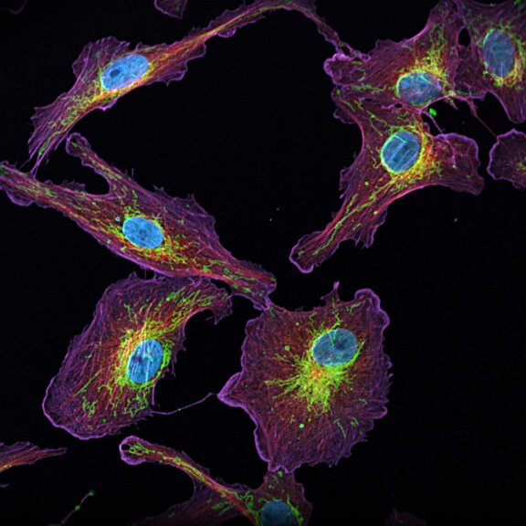
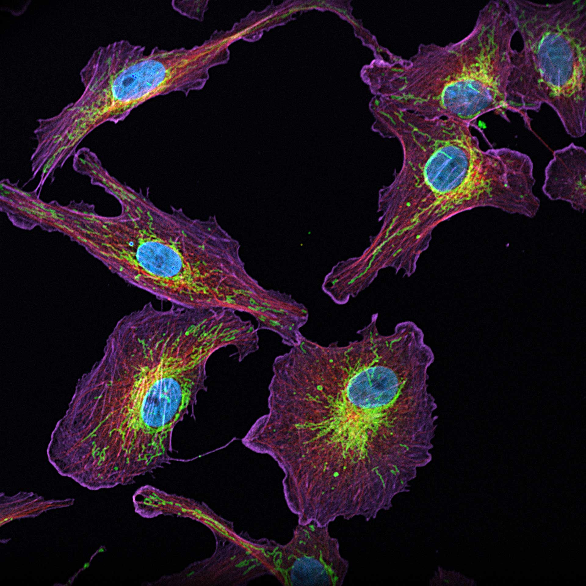
4-color confocal image of mammalian cells (DAPI, Phalloidin, Tubulin, Tom20).
Modules:


Description
Two-color confocal and MINFLUX images of Tom20 (green) and mitochondrial DNA (red) stained with sCy5 and CF680 in mammalian cells using indirect immunolabeling. The two fluorophores were distinguished by ratiometric detection strategy. Note the dissimilar labeling density of the two imaged structures.
Description
2D MINFLUX image of the mitochondrial import receptor Tom20 labeled with Alexa Fluor 647 in fixed mammalian cells using indirect immunolabeling.
Description
Modules:
Description
Nuclear pore complex (green), Tom20 (red), and vimentin (white) in cultured mammalian cells.
Modules:
Description
4-color confocal image of mammalian cells (DAPI, Phalloidin, Tubulin, Tom20).
Modules:
Description
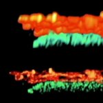
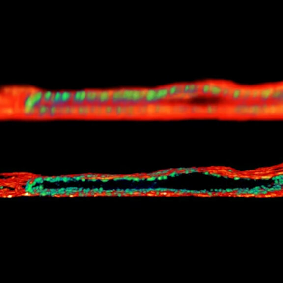
Nuclear pore complexes (NUP) and tubulin imaged with confocal and EASY3D-STED microscopy.
Modules:
Description
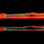
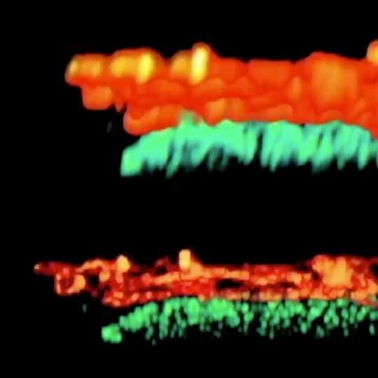
Two-color EASY3D-STED image of tubulin and giantin versus its confocal low-resolution counterpart.


