

Sample gallery
Fluorescence imaging, whether at confocal, STED or MINFLUX resolution, guarantees unique insights into the function and structure of life at the molecular level. Besides the scientific information content, some sample portraits provide simply beautiful images. Enjoy browsing our sample gallery.
the fine art of science

Description
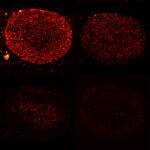
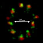
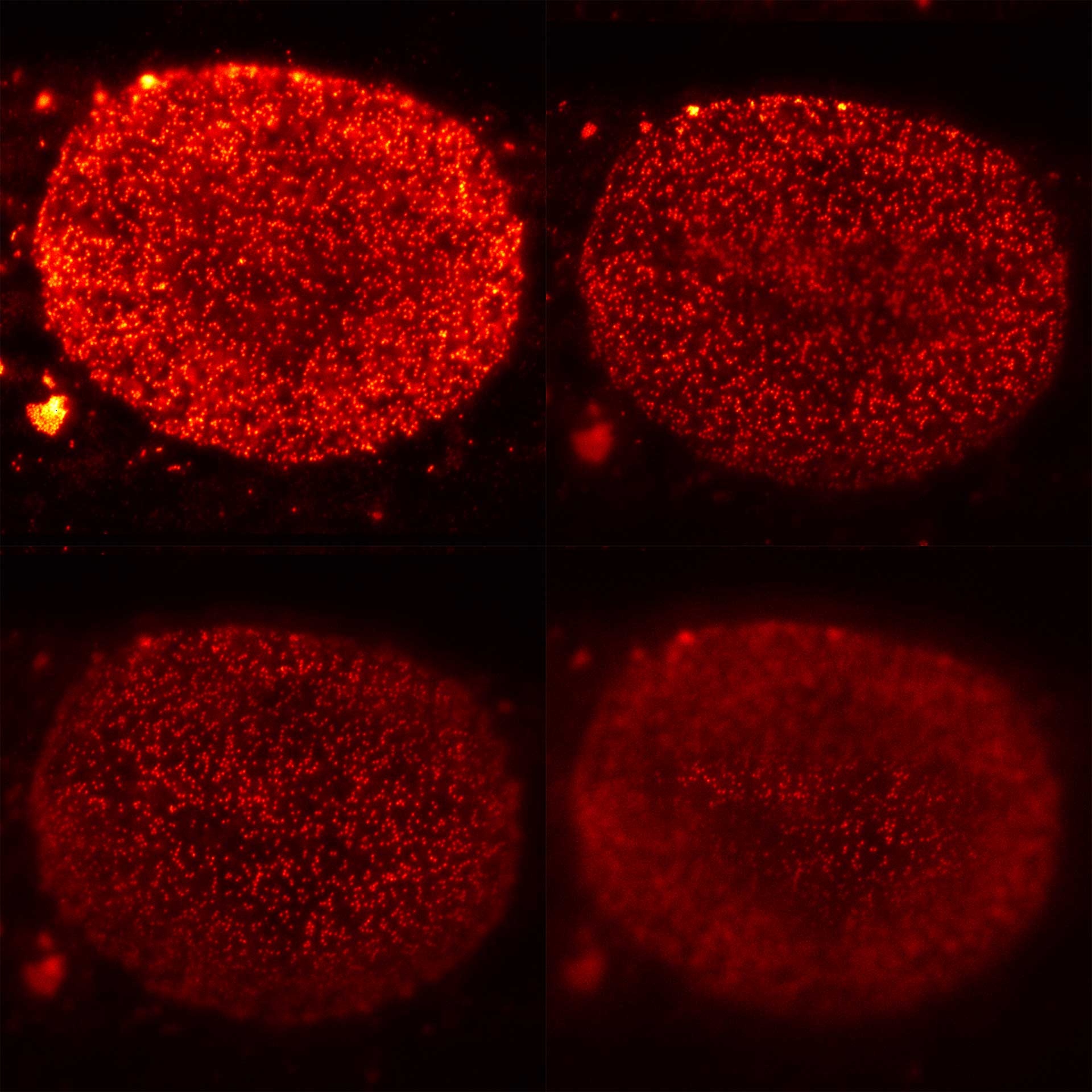
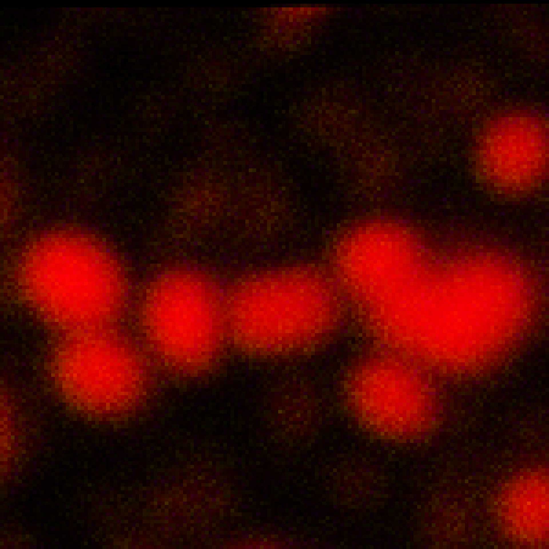
Four xy-planes from a volume stack recording of nuclear pore complexes (NUPs). With MATRIX, nuclear pores that are not in focus are effectively removed from the image.
Modules:
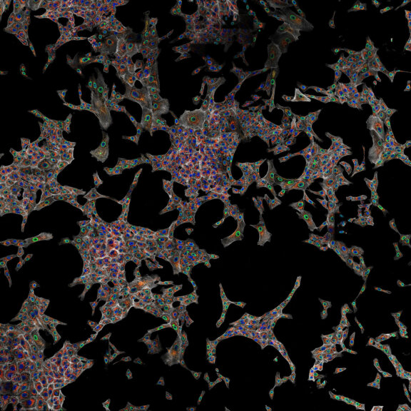
Description
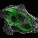
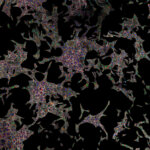
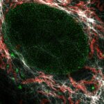
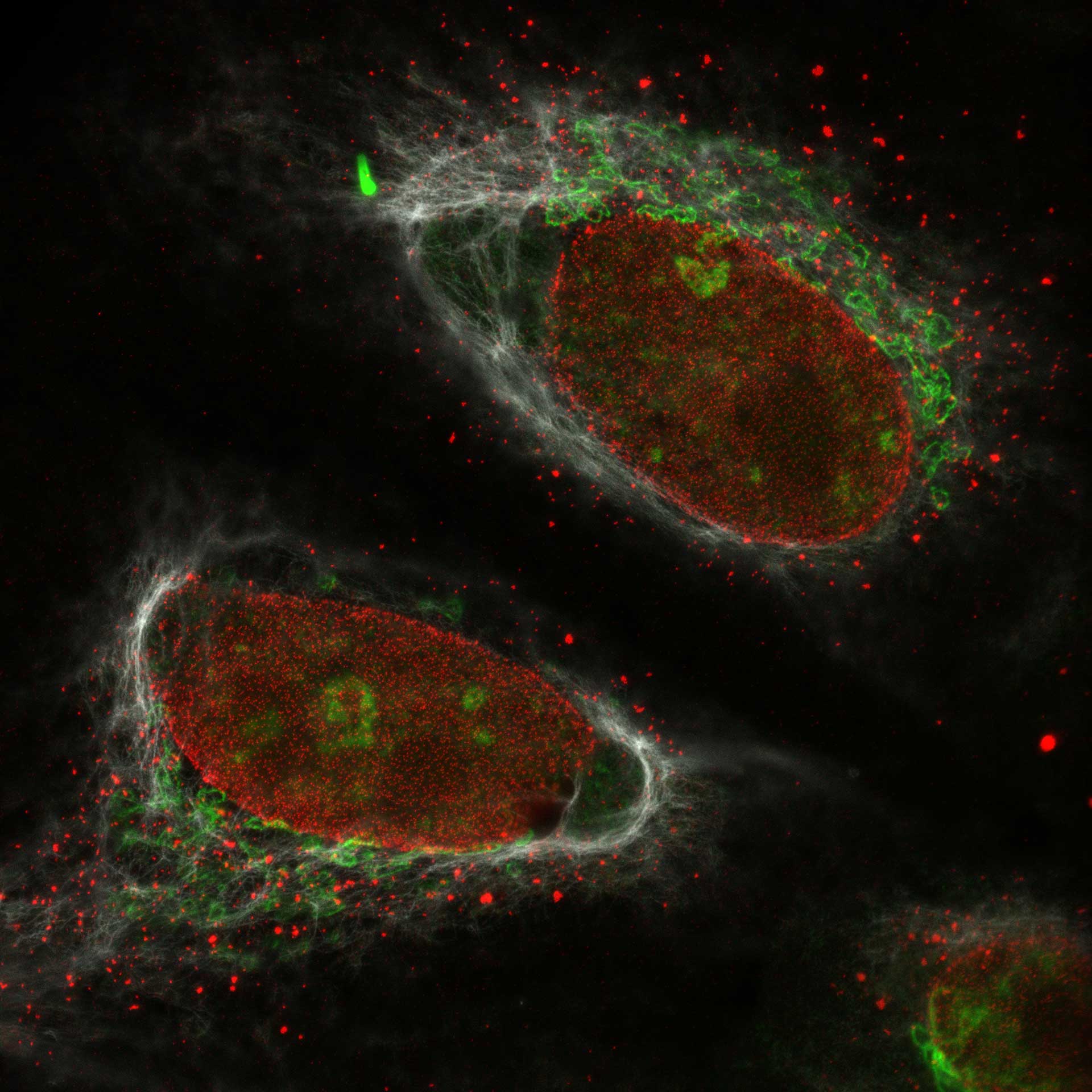
Cultured mammalian cells (NUP, Giantin, and Vimentin, labeled with abberior STAR RED, STAR ORANGE, STAR GREEN). Out-of-focus contributions of all three types of structures is effectively removed and section is improved.
Modules:
Description
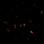
Two proteins in the Golgi apparatus were immunolabelled using primary antibodies specific for GM130 and Giantin and secondary antibodies coupled to abberior STAR 580 and abberior STAR 635P. Shown is RAW DATA. Images were acquired using a STEDYCON attached to a Zeiss Axio Imager Z2.
Description
Vero cells, Vimentin (green, abberior STAR Red) and Phalloidin (white, abberior STAR Orange)
Modules:

Description
Centrosome linker. U2OS cells in which the centrosome-linker-protein rootletin was immunolabelled using secondary antibodies coupled to abberior STAR RED. The sample was prepared by R. Vlijm at MPI for Medical Research, Heidelberg, Germany. Imaged with abberior Instruments’ STEDYCON and deconvolved with SVI Huygens optimized for STEDYCON.
Description
Description
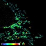
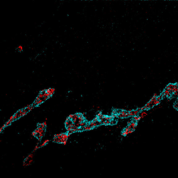
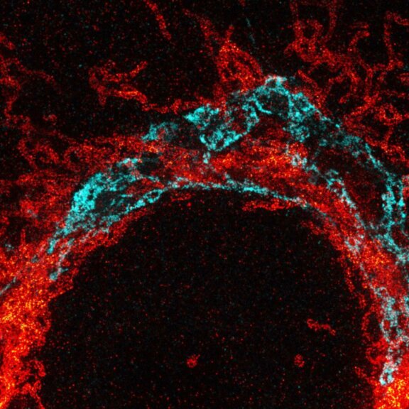
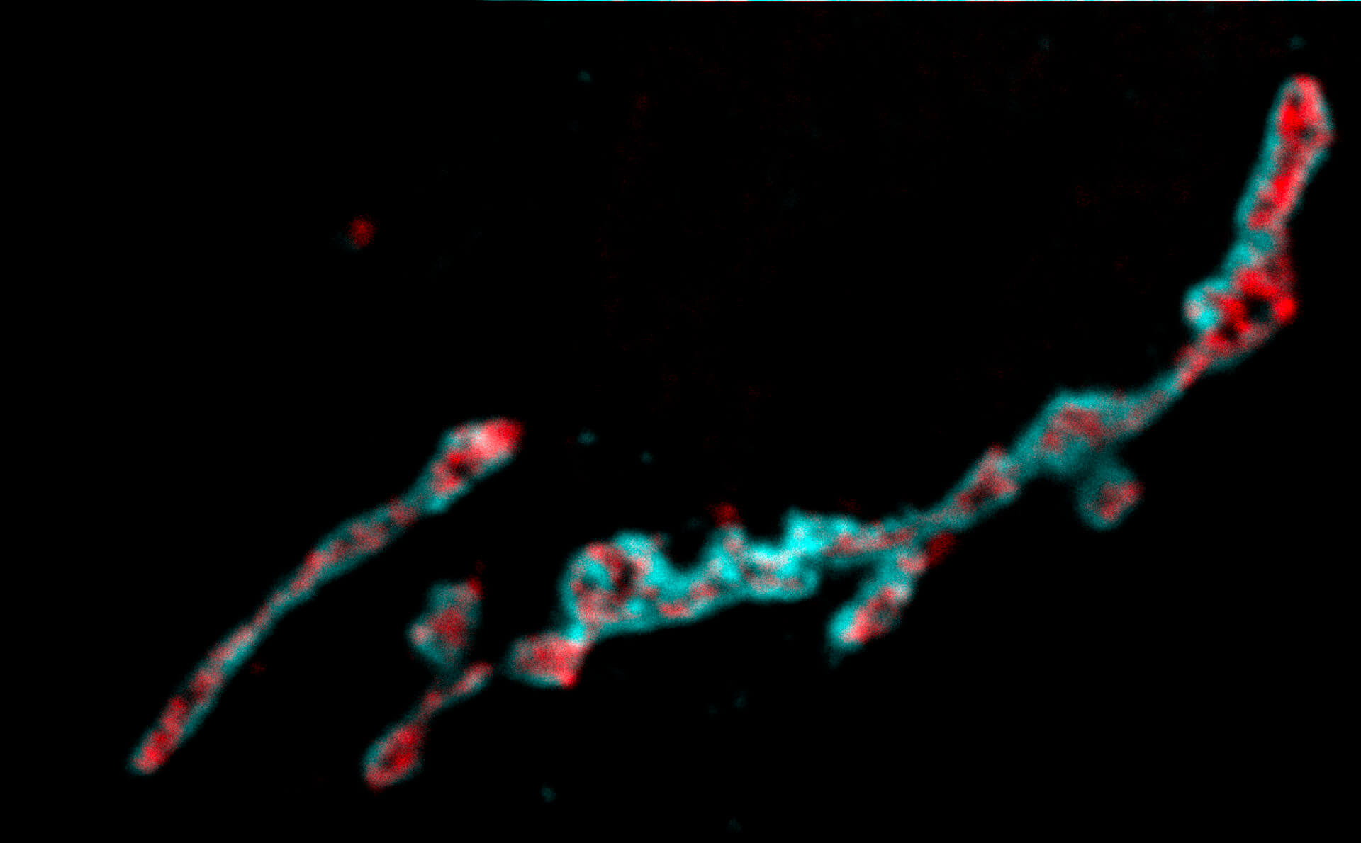
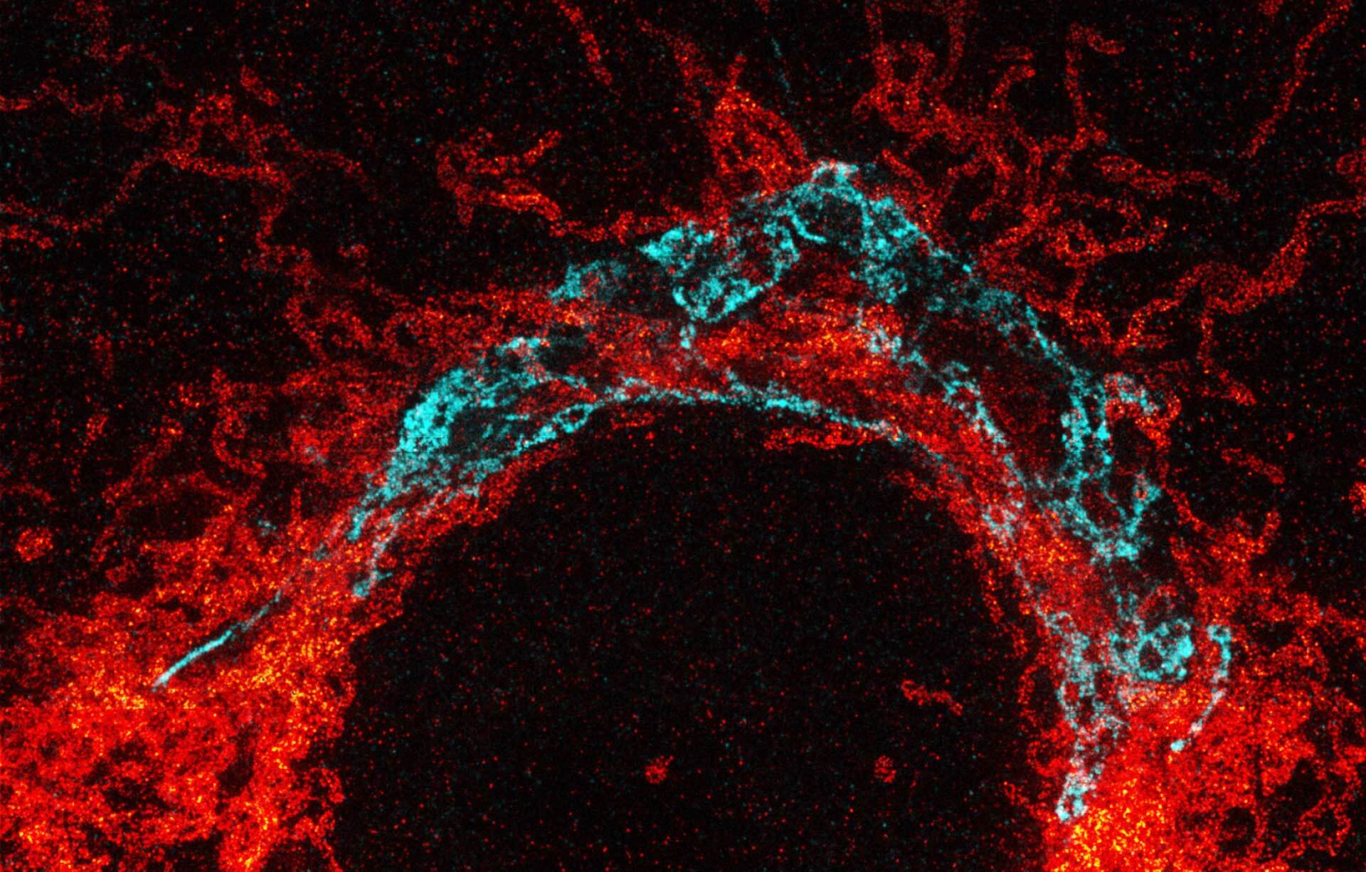
Maximum intensity projection of a z-stack showing nuclear pore complex protein (red, abberior STAR RED) and peroxisomes (cyan, Alexa 594) in mammalian cells. Imaged with abberior Instruments’ STEDYCON and deconvolved with SVI Huygens optimized for STEDYCON.
Description
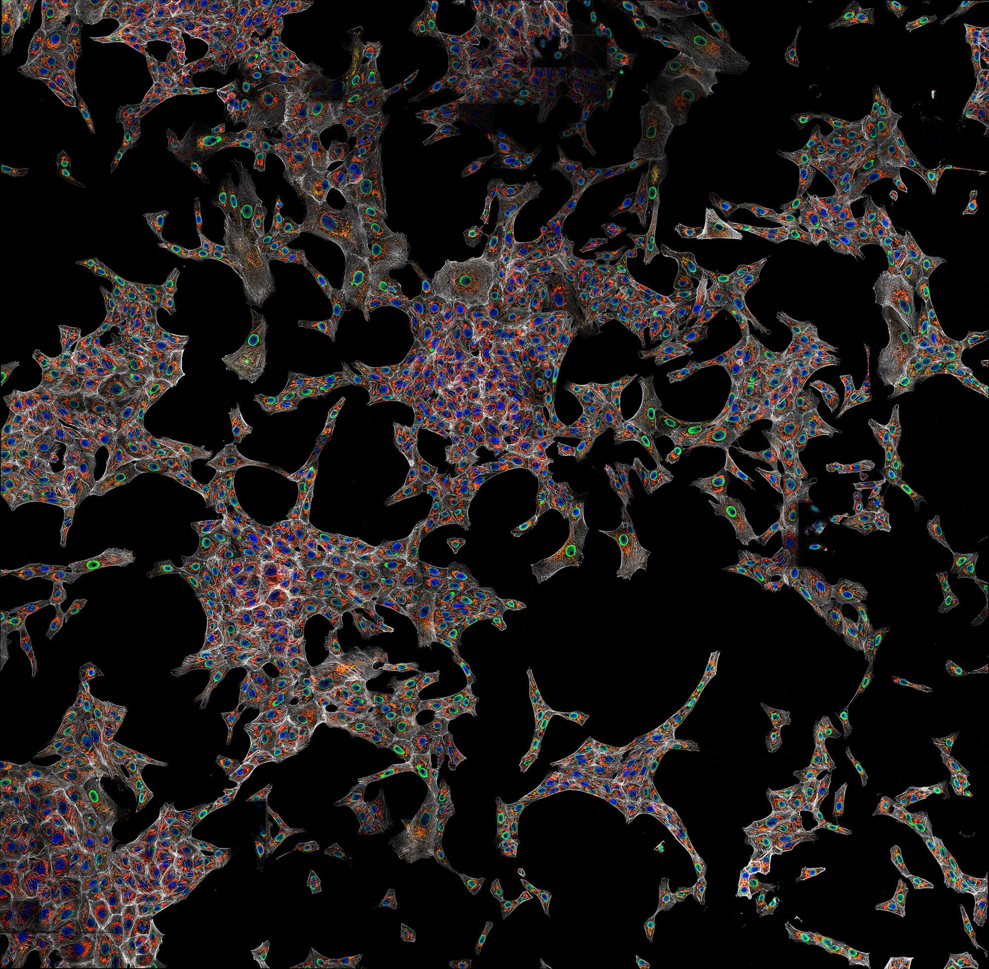
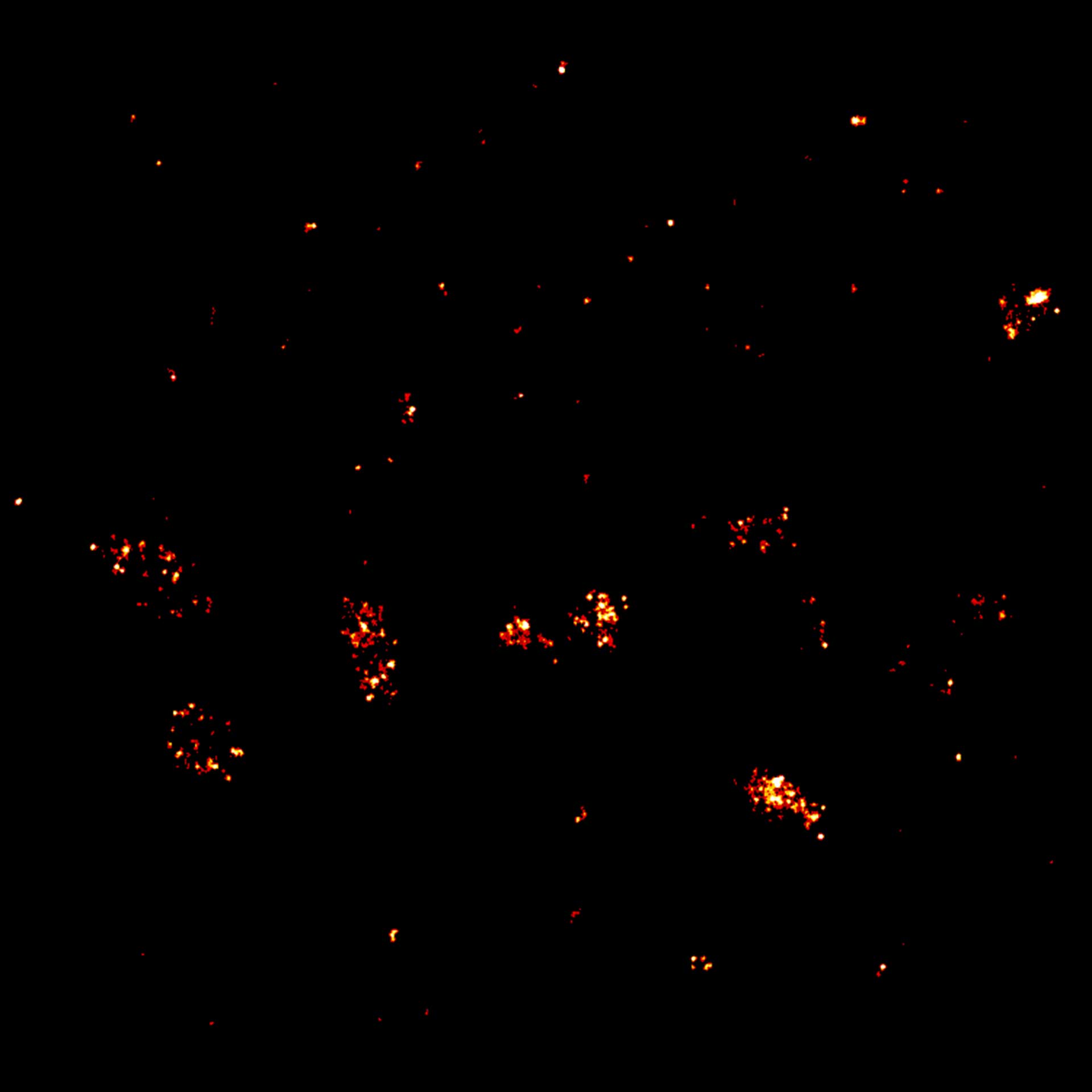
Confocal imaging of a large sample region. Shown is a 2.8 mm x 2.5 mm region, acquired using 35 by 29 tiles using the STEADYFOCUS and stitched with SVI Huygens. Sample: mammalian cells immunolabelled for the mitochondrial protein TOM20 (abberior STAR RED, red), double-stranded DNA to visualize mitochondrial DNA (abberior STAR ORANGE, green), phalloidin to label F-actin (abberior STAR GREEN, gray), and DAPI to label nuclei. Sample preparation by abberior GmbH.
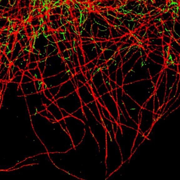
Description
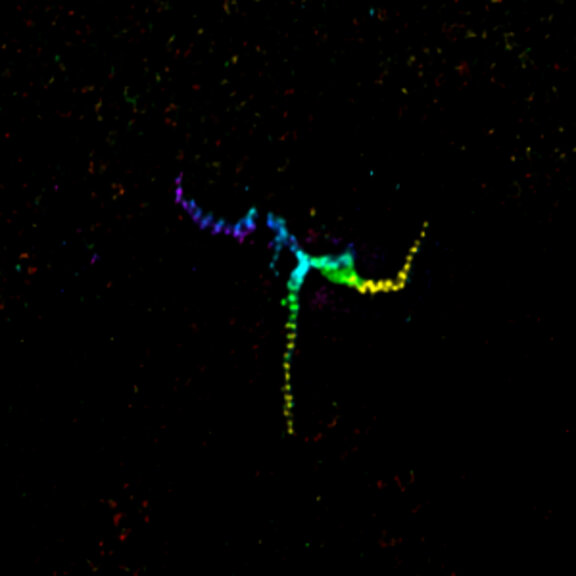
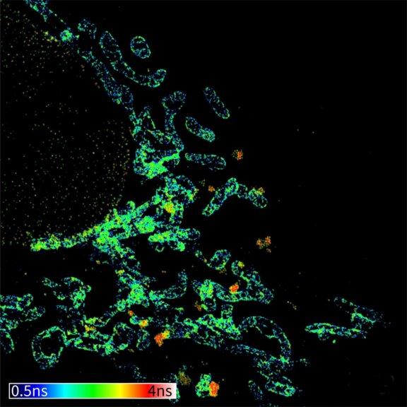
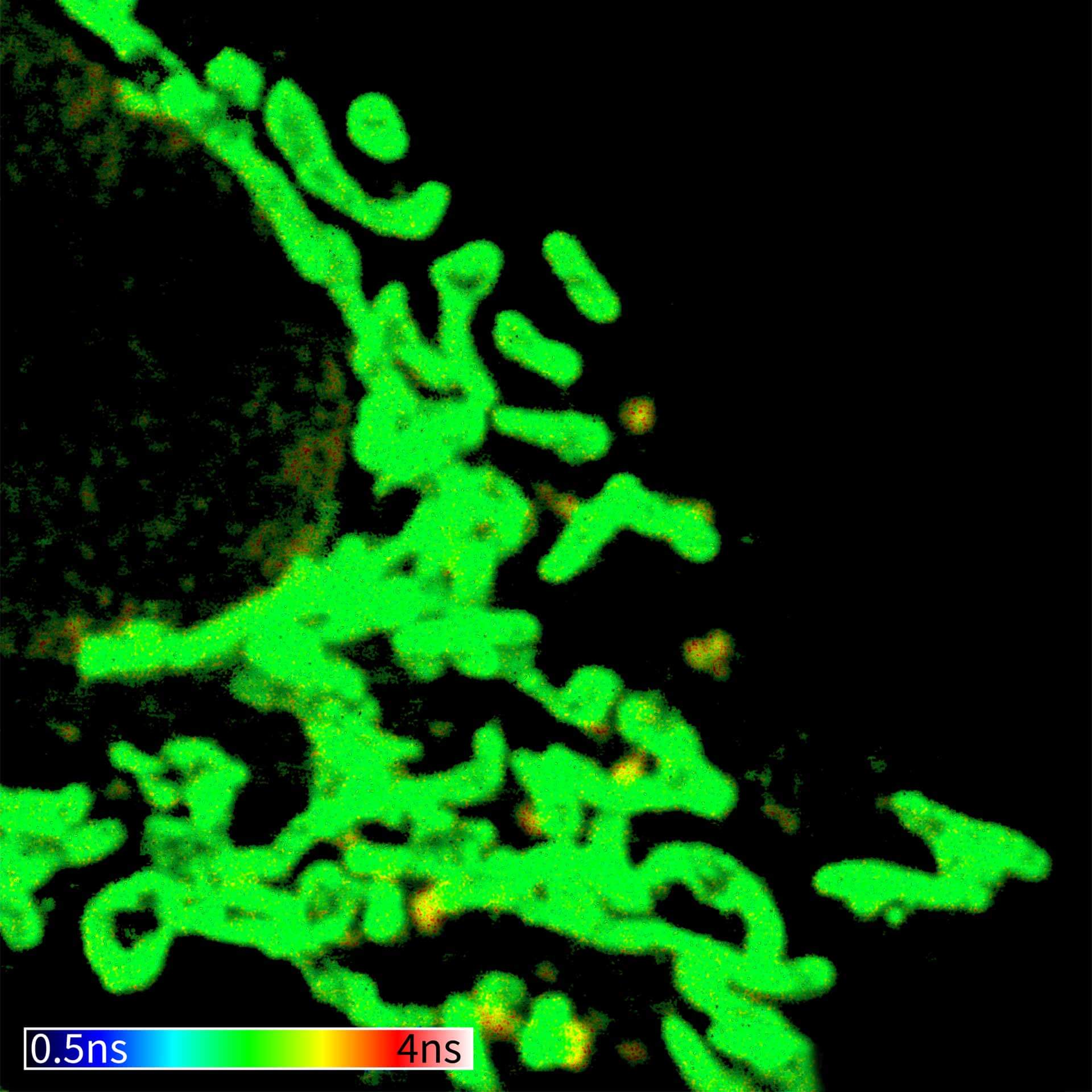
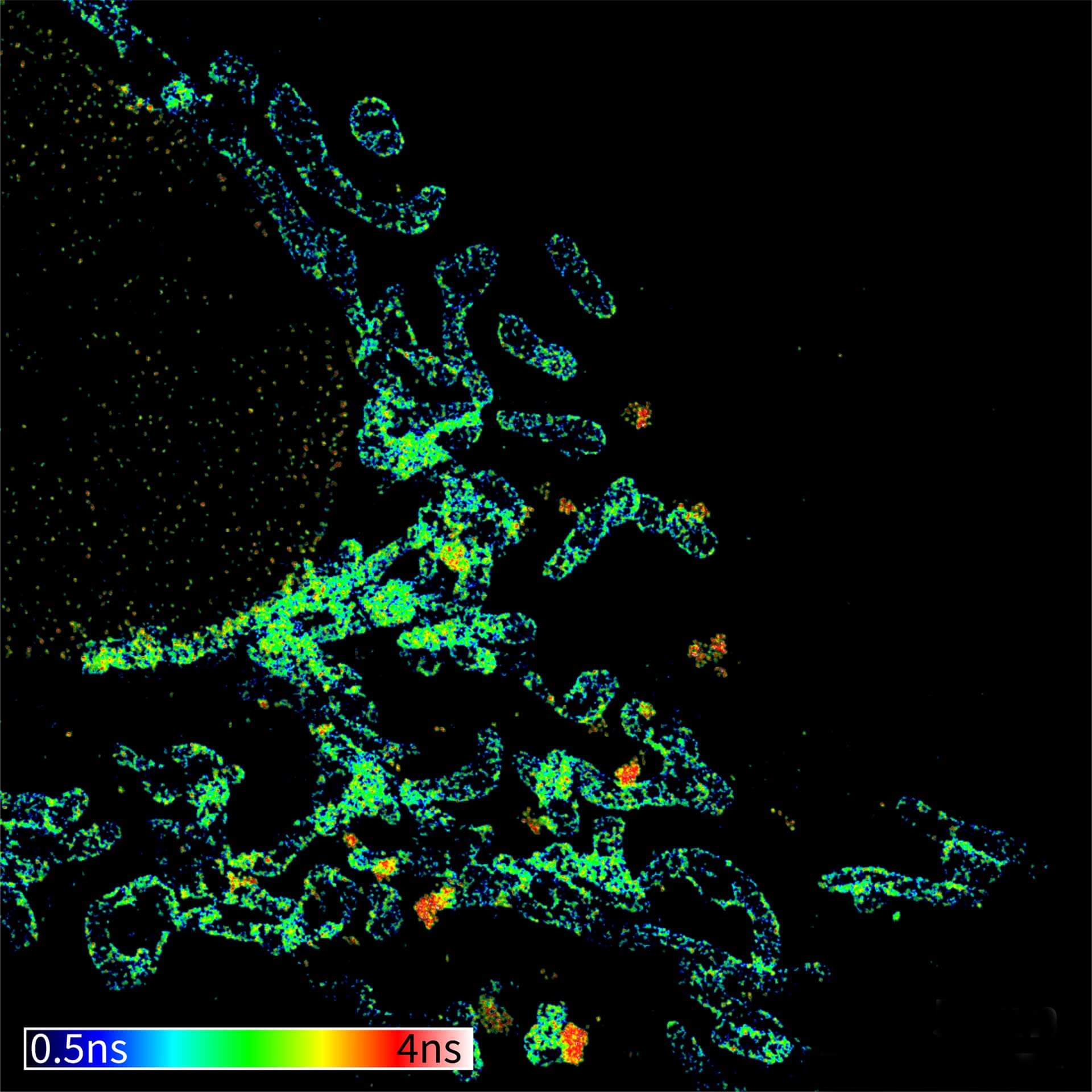
TIMEBOW image of tubulin labeled with Atto647N and vimentin labeled with abberior STAR 635P. The two labels (Atto647N and abberior STAR 635P) have the same excitation spectra, but different lifetimes. Tubulin (red, short lifetime) and vimentin (green, long lifetime) can successfully be separated in the TIMEBOW STED recording via their lifetimes.
Modules:
Description
TIMEBOW image of mammalian cells labeled with antibodies against Tom20, ATTO 647N and NUP153, abberior STAR 635P. Note that nuclear pore (NUP) complex subunits appear to be localized in the cytoplasm, which is due to the NUP import pathway into the nuclear membrane.
Modules:
Description
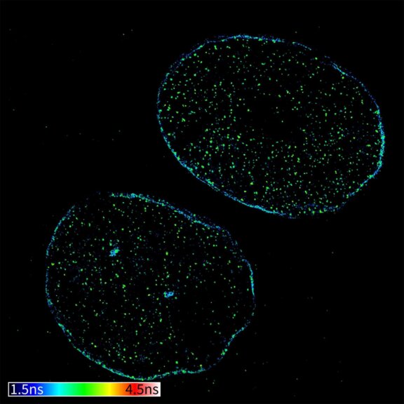
TIMEBOW confocal and TIMEBOW STED image of mammalian cells labeled with antibodies against lamin, ATTO 647N and dsDNA, abberior STAR 635P.
Modules:

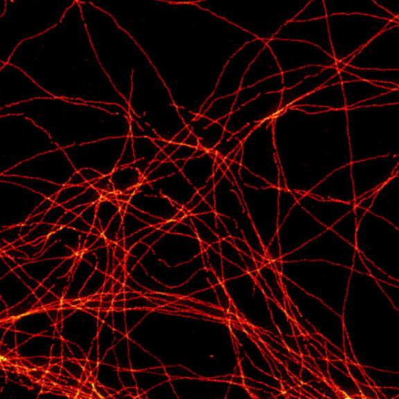
Description
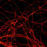
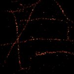
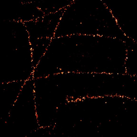
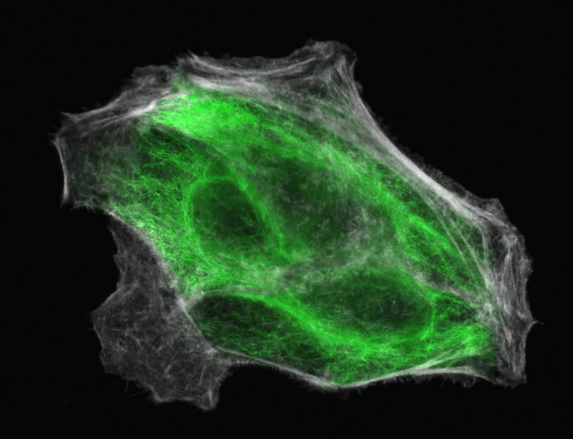
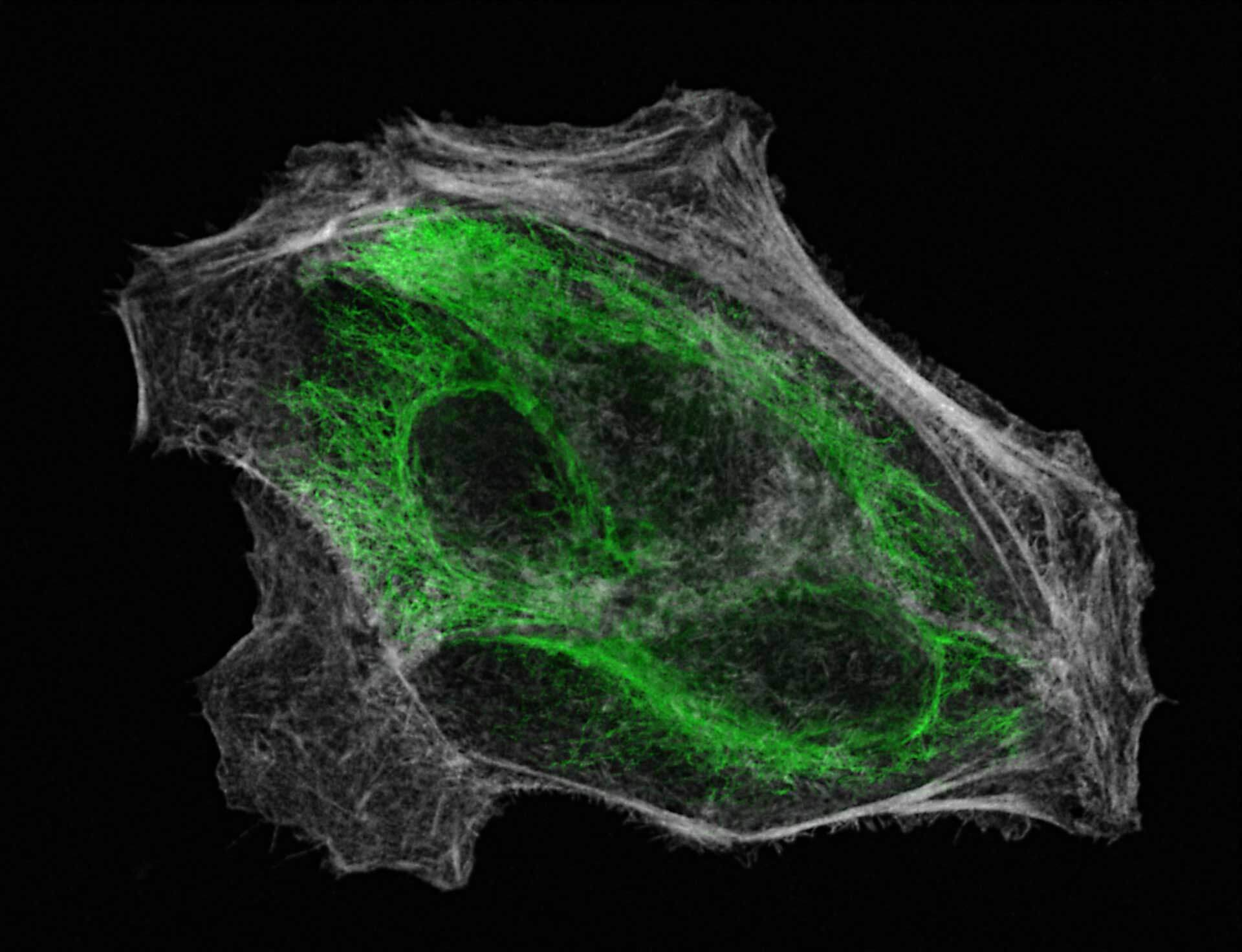
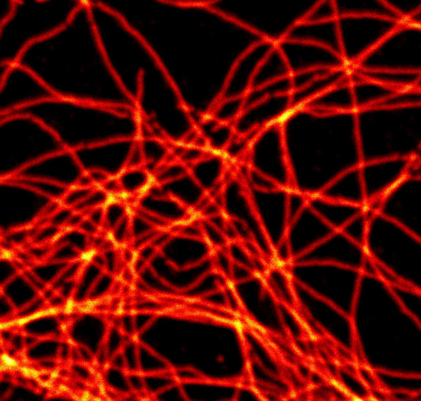
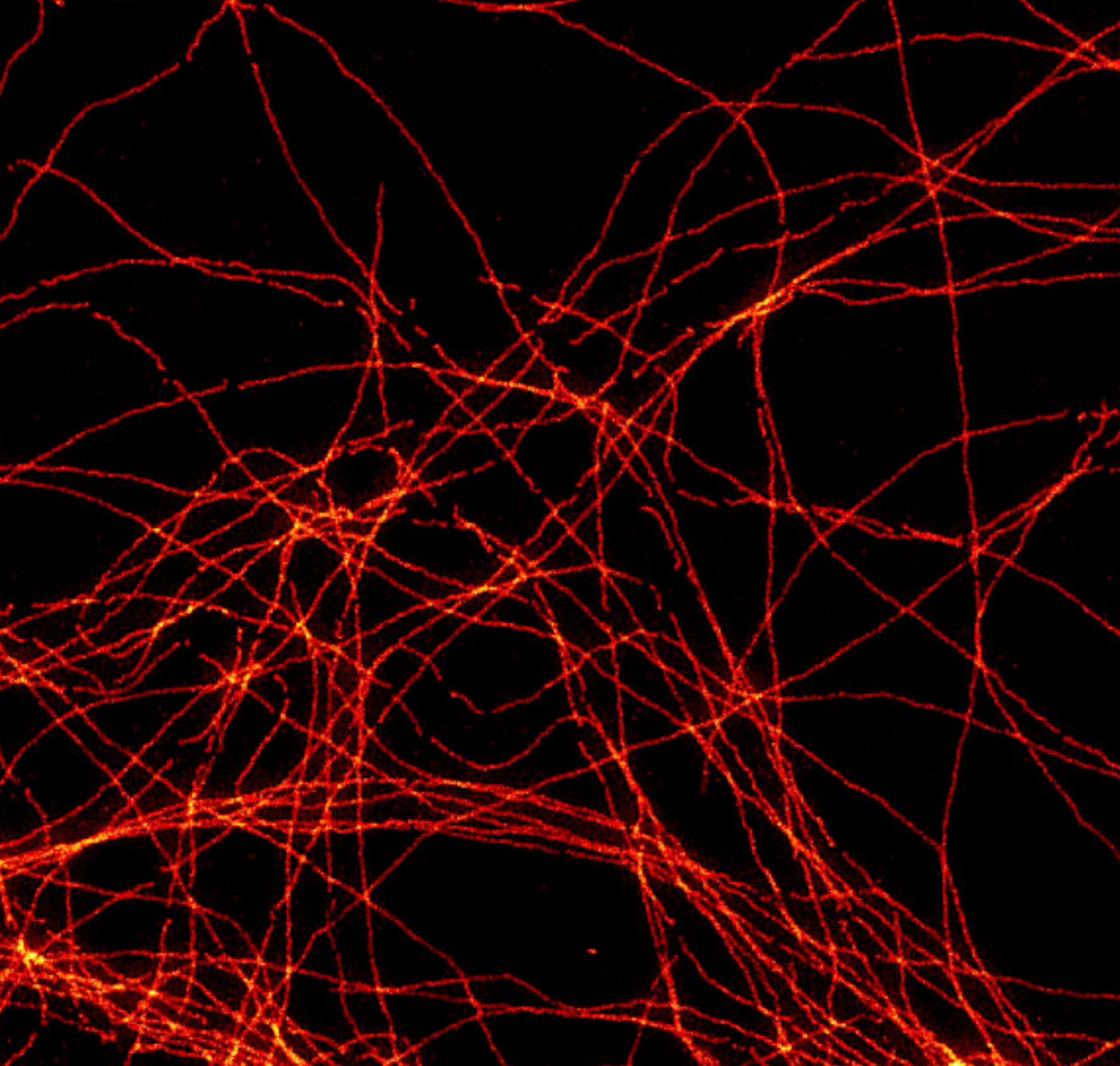
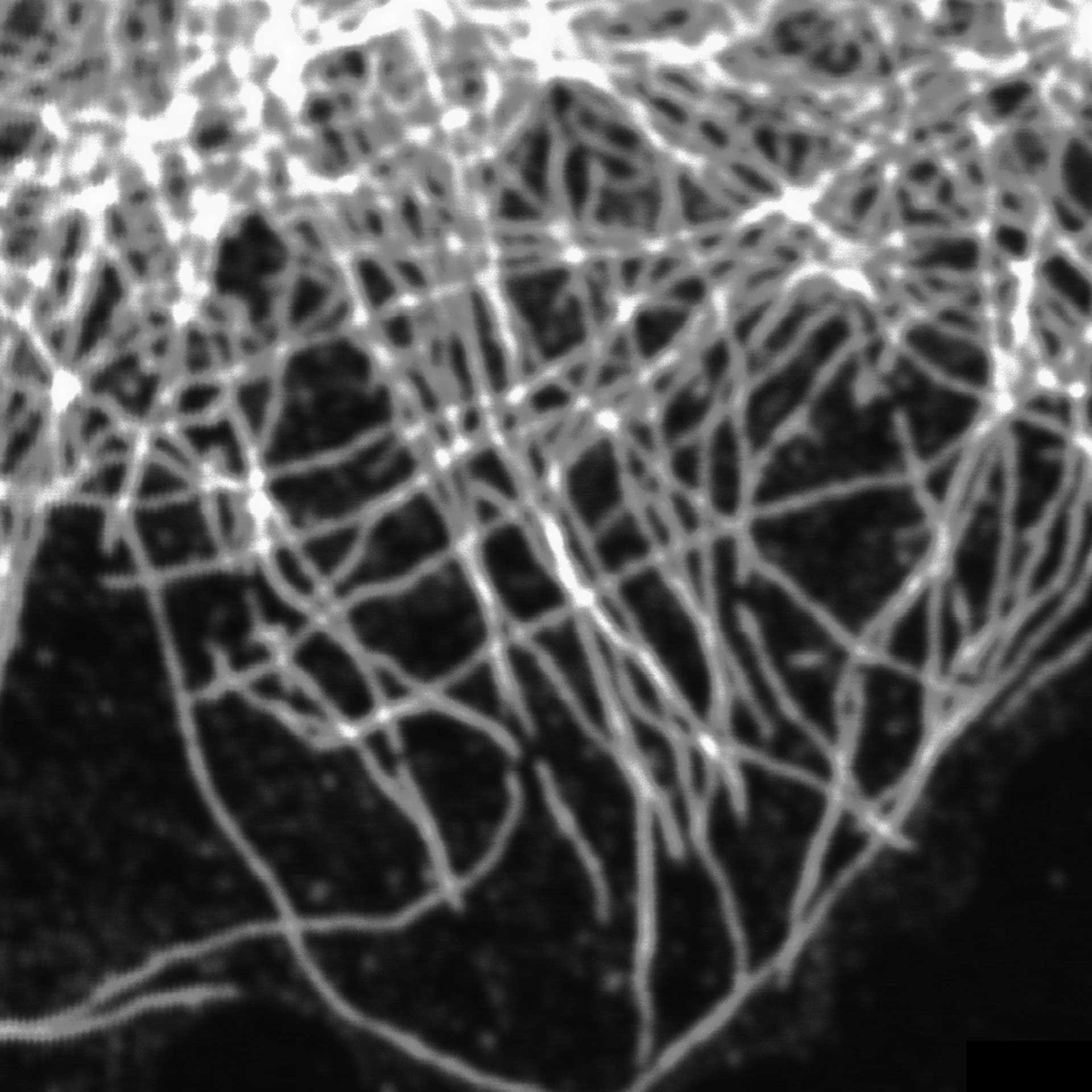
2D MINFLUX imaging of the cytoskeletal protein vimentin. Vimentin was labeled with Alexa Fluor 647 in fixed mammalian cells using indirect immunofluorescence. Note the individual filaments at intersections are invisible in the confocal image.
Description
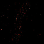
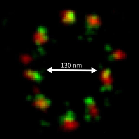
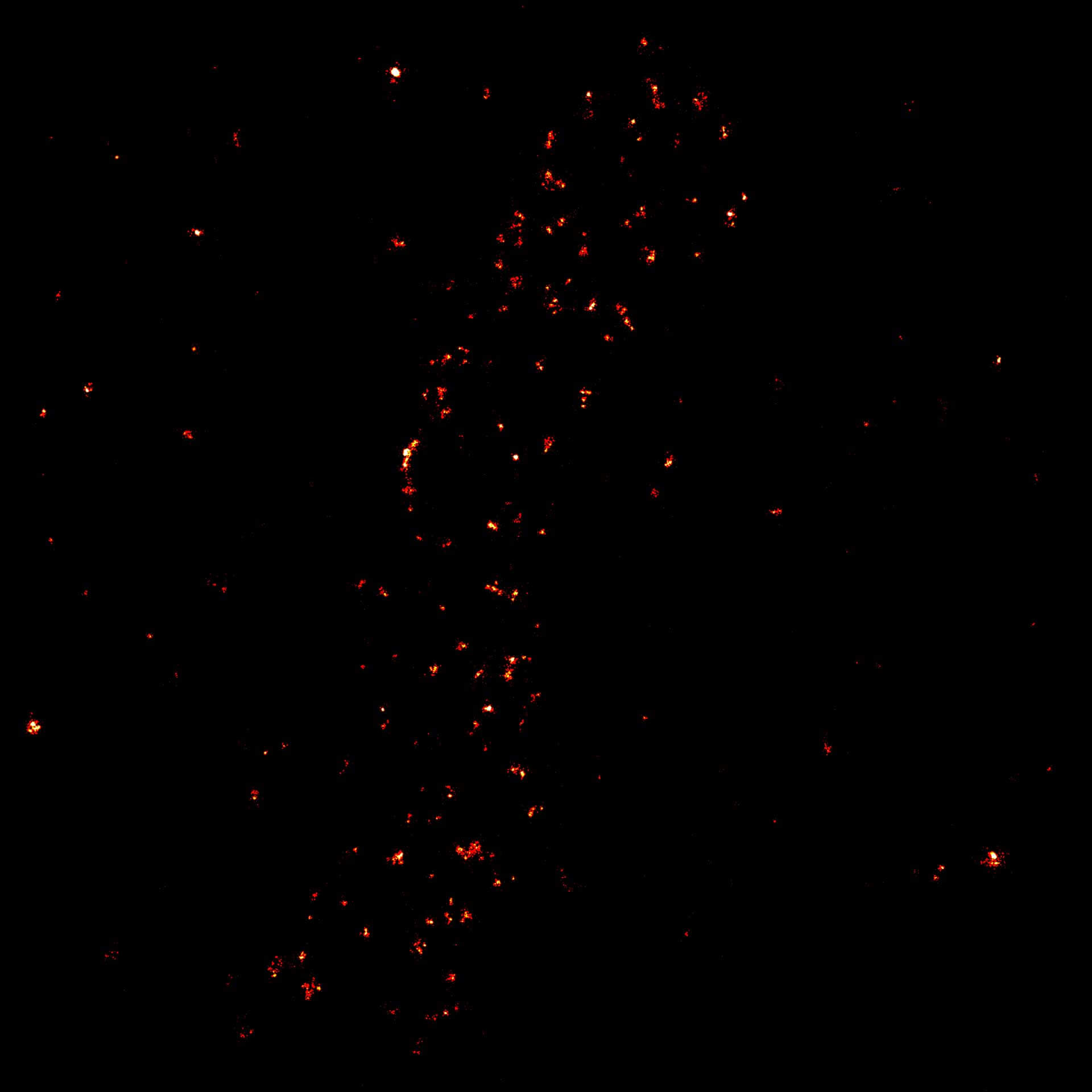
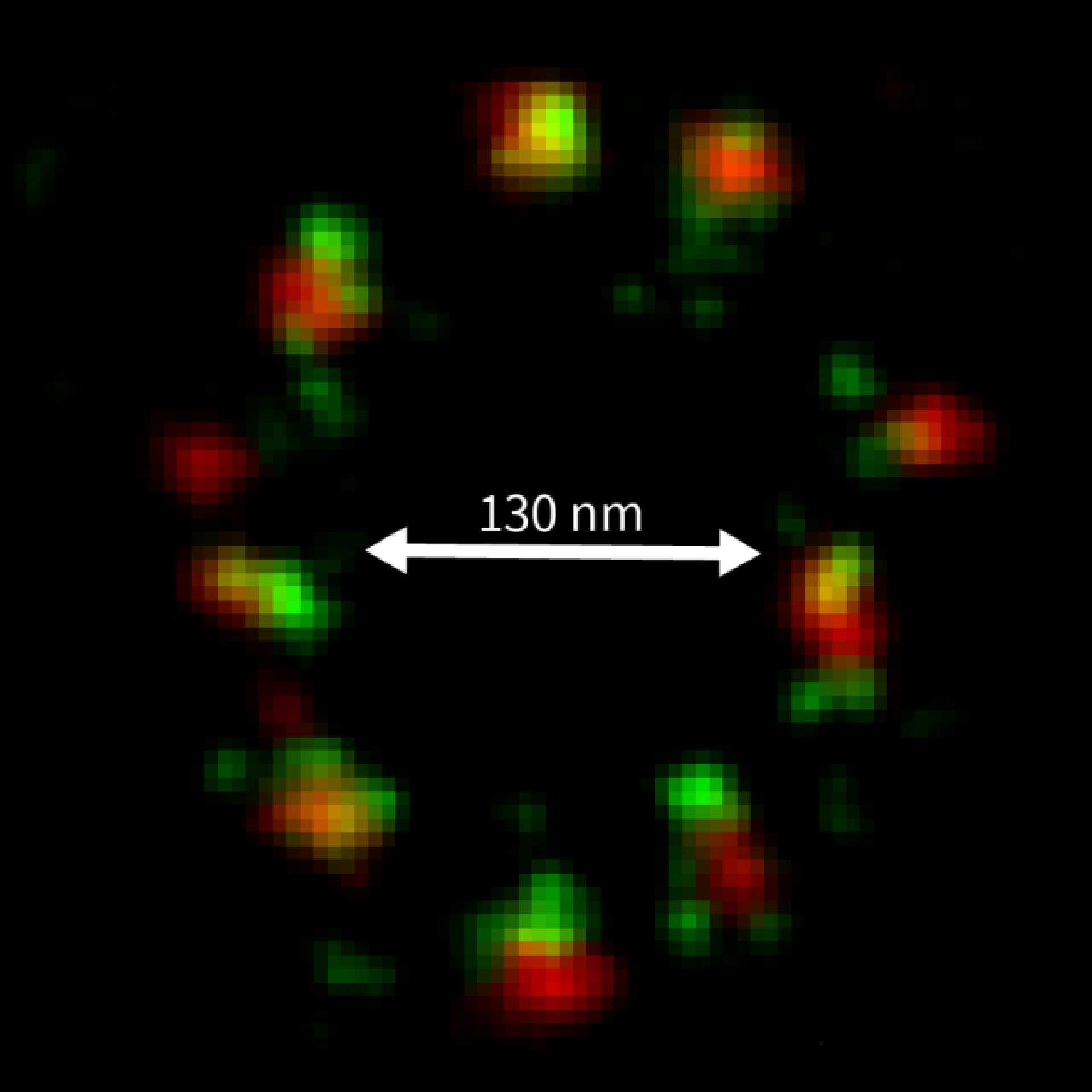
2D MINFLUX imaging of the peroxisomal membrane protein PMP70 labeled with Alexa Fluor 647 in fixed mammalian cells using indirect immunofluorescence

Description
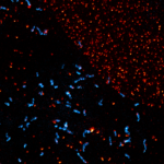
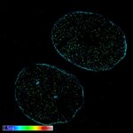
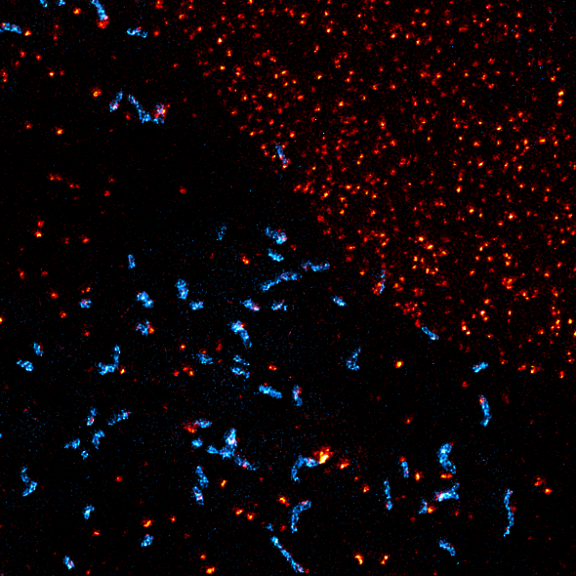
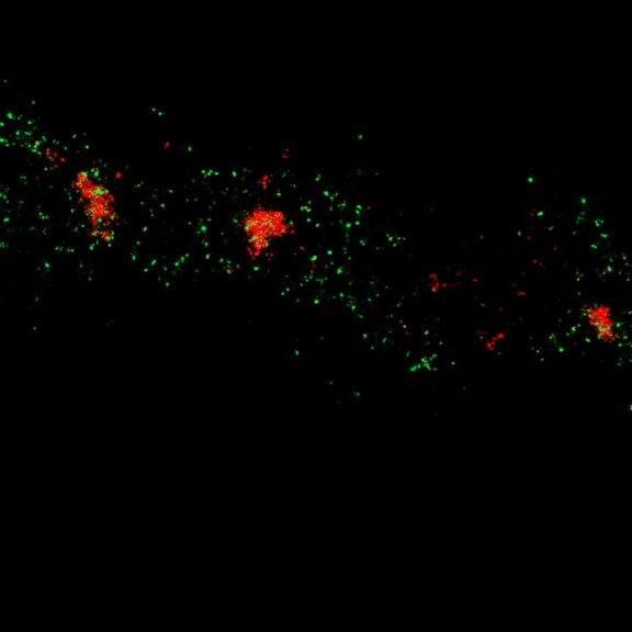
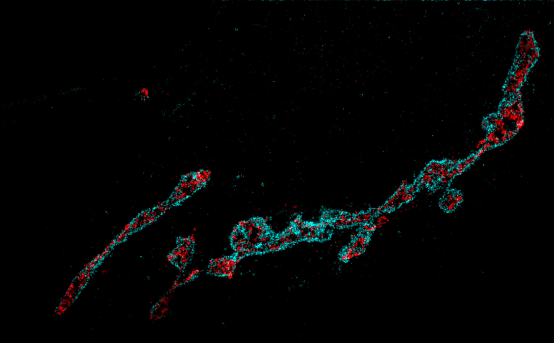
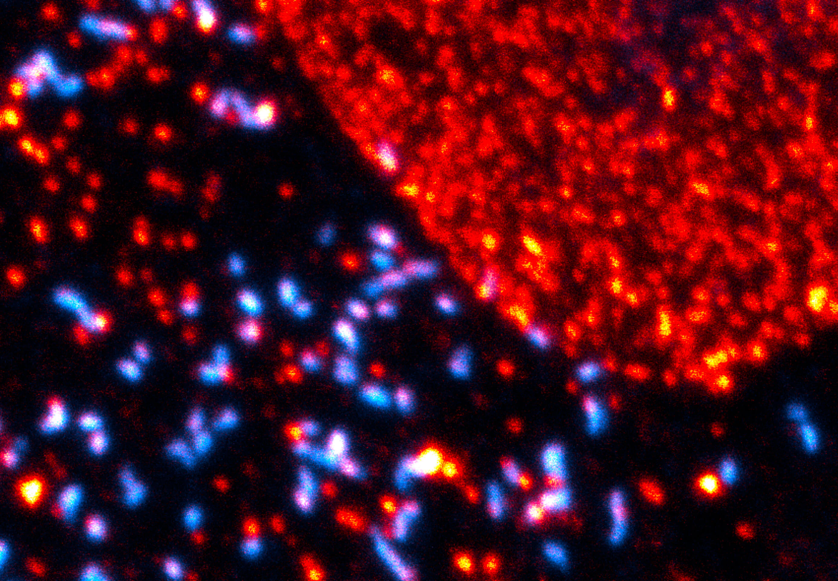
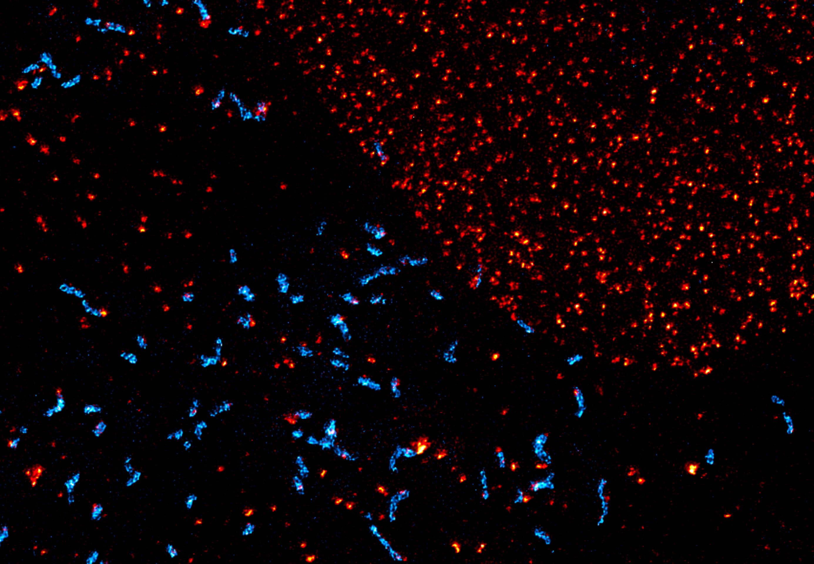
Two-color confocal and MINFLUX images of Tom20 (green) and mitochondrial DNA (red) stained with sCy5 and CF680 in mammalian cells using indirect immunolabeling. The two fluorophores were distinguished by ratiometric detection strategy. Note the dissimilar labeling density of the two imaged structures.
Description
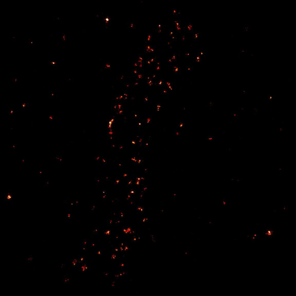
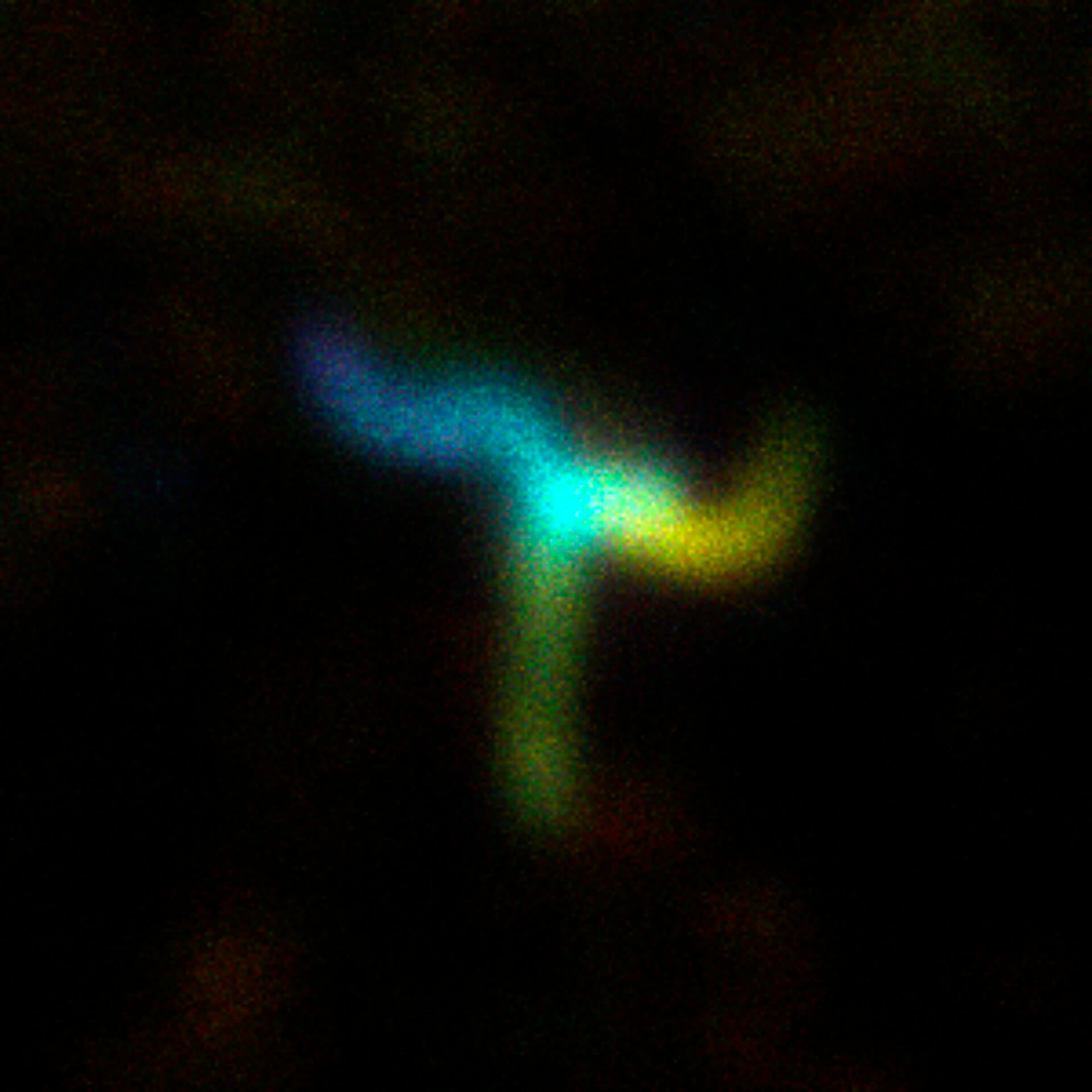
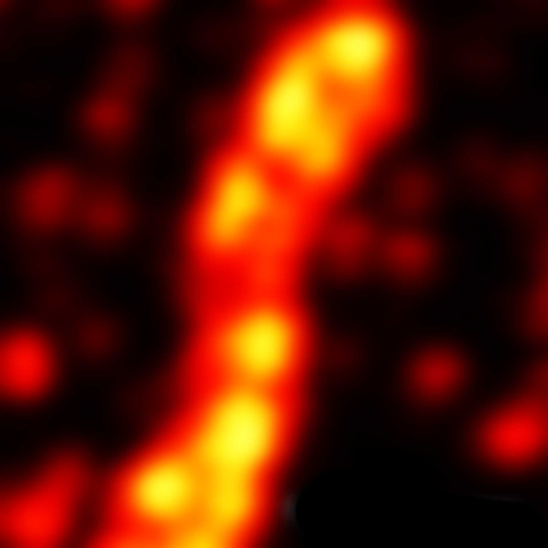
2D MINFLUX image of the mitochondrial import receptor Tom20 labeled with Alexa Fluor 647 in fixed mammalian cells using indirect immunolabeling.
Description
Modules:


Description
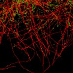
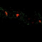
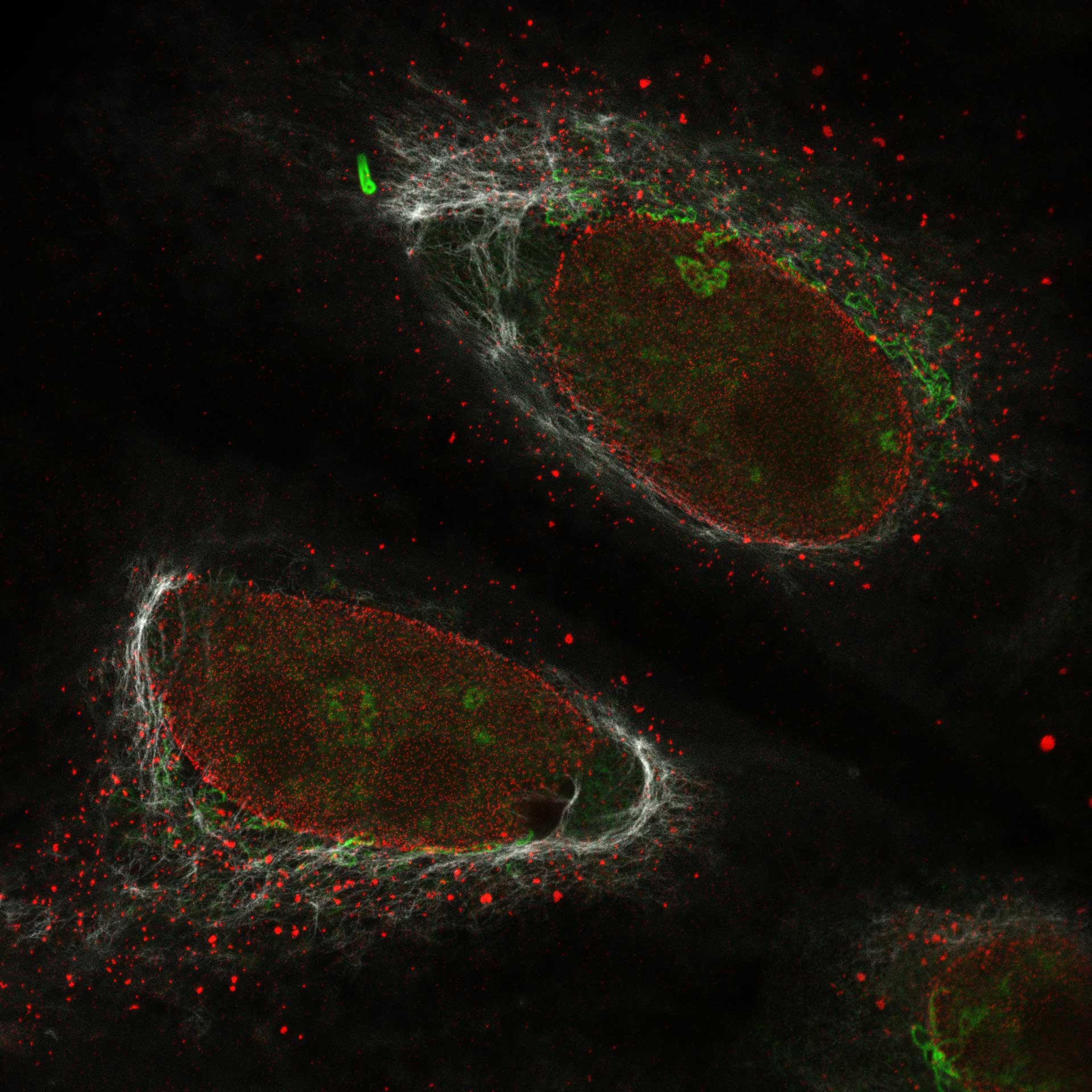
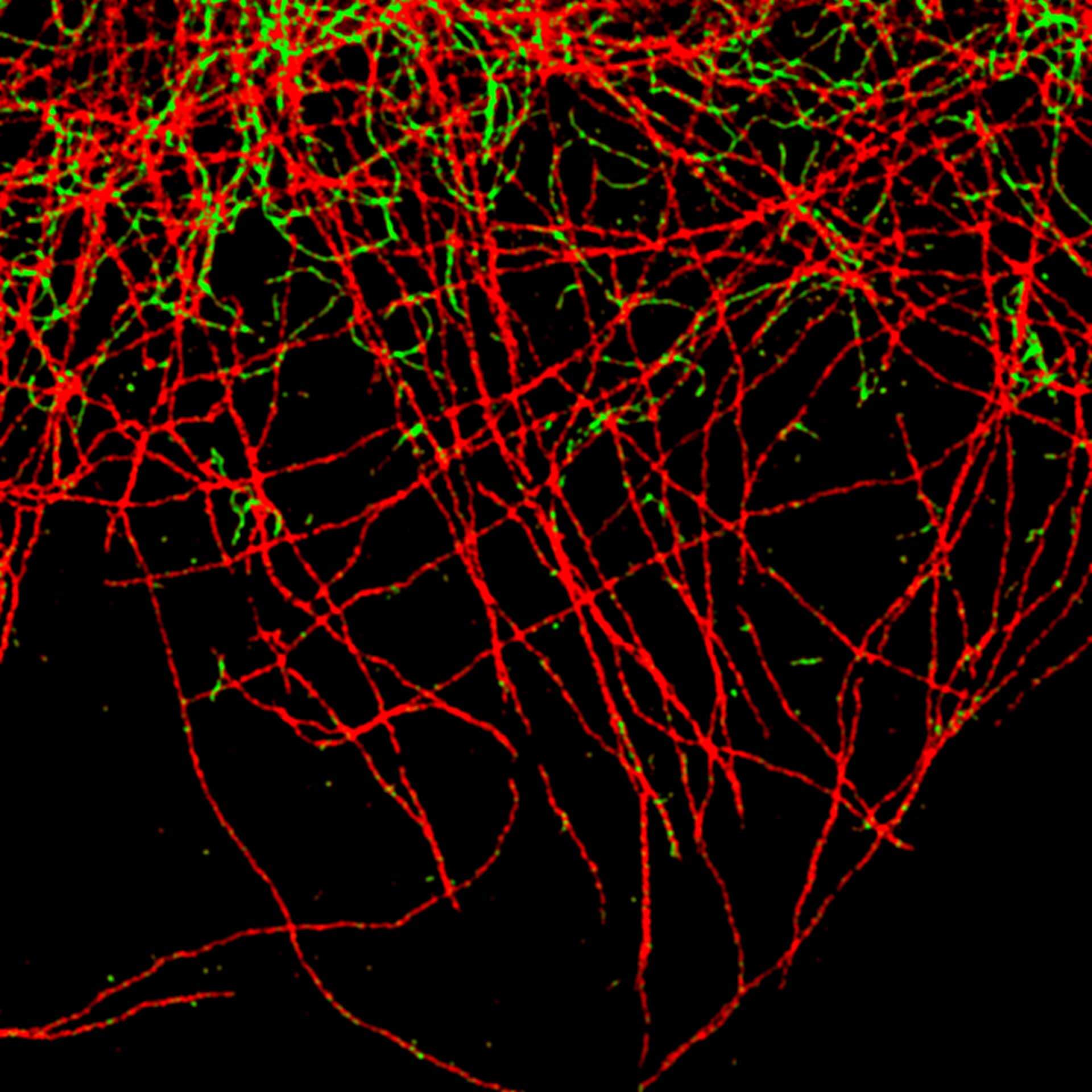
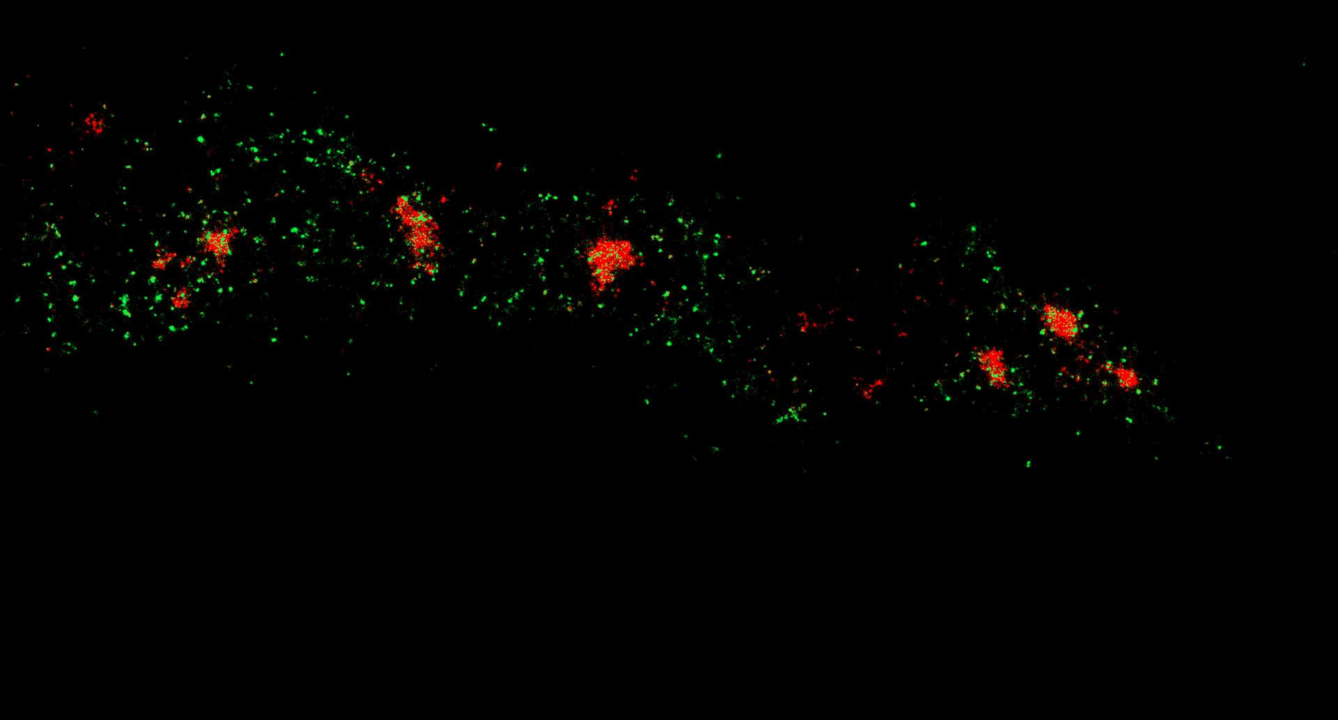
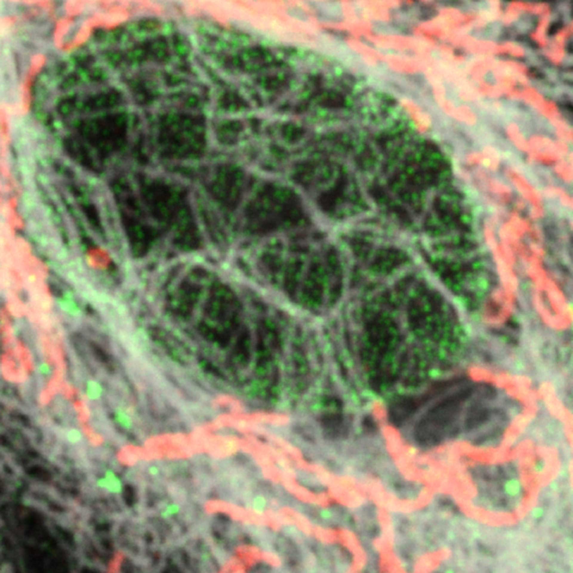
Nuclear pore complex (green), Tom20 (red), and vimentin (white) in cultured mammalian cells.
Modules: