

Sample gallery
Fluorescence imaging, whether at confocal, STED or MINFLUX resolution, guarantees unique insights into the function and structure of life at the molecular level. Besides the scientific information content, some sample portraits provide simply beautiful images. Enjoy browsing our sample gallery.
the fine art of science
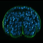
Description
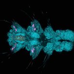
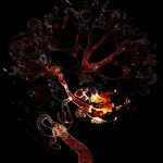
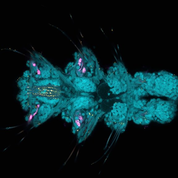
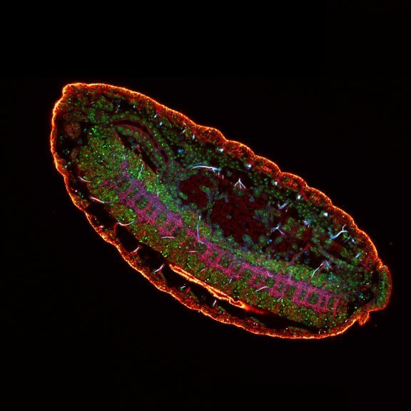
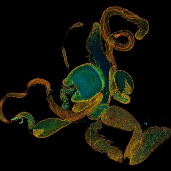
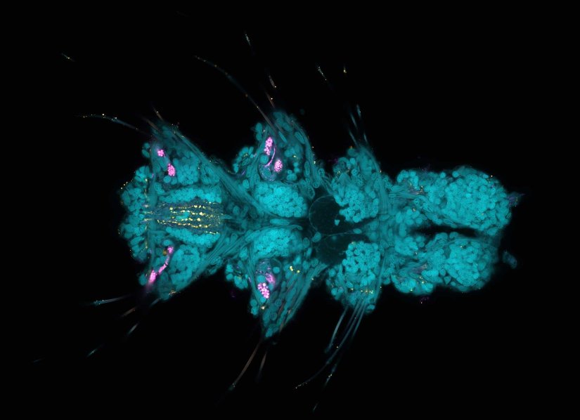
Larva of Platynereis dumerilii acquired in a confocal overview. Nuclei are shown in cyan (DAPI), tubulin in magenta, and serotonin-positive neurons in yellow.
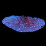
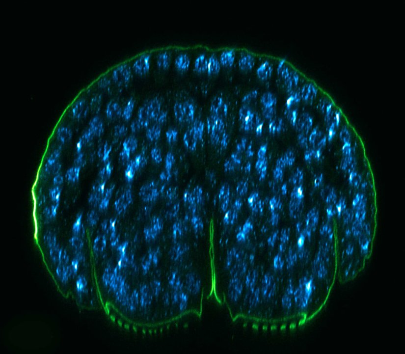
Description
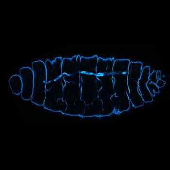
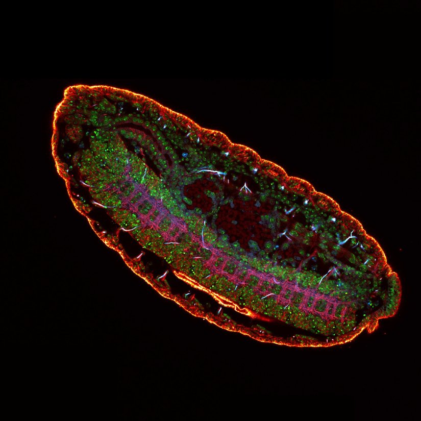
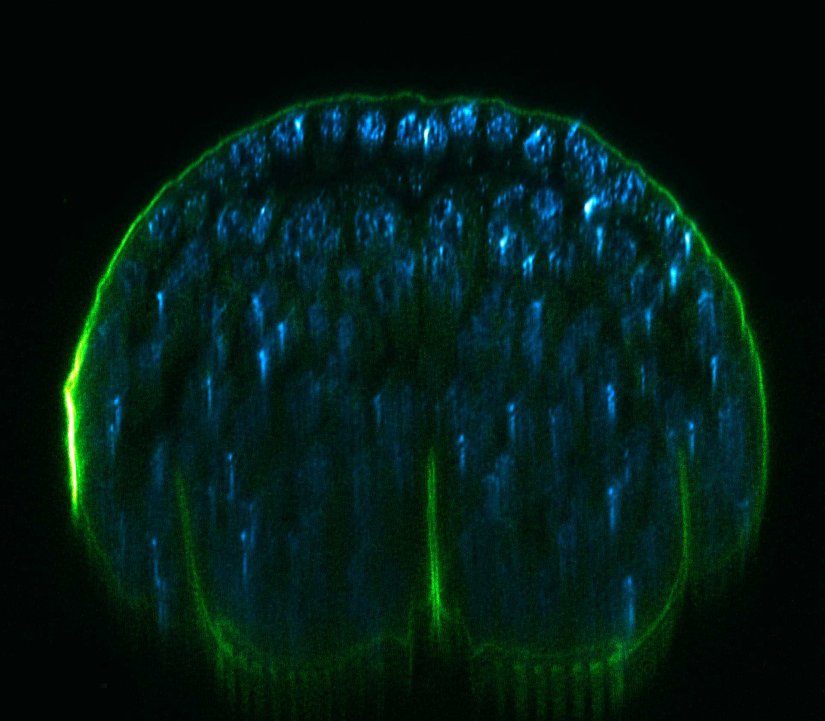
3 color confocal image of a Drosophila embryo stained for chitin (abberior STAR RED, cyan), tubulin (abberior STAR ORANGE, magenta), and DNA (PicoGreen, green).
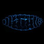
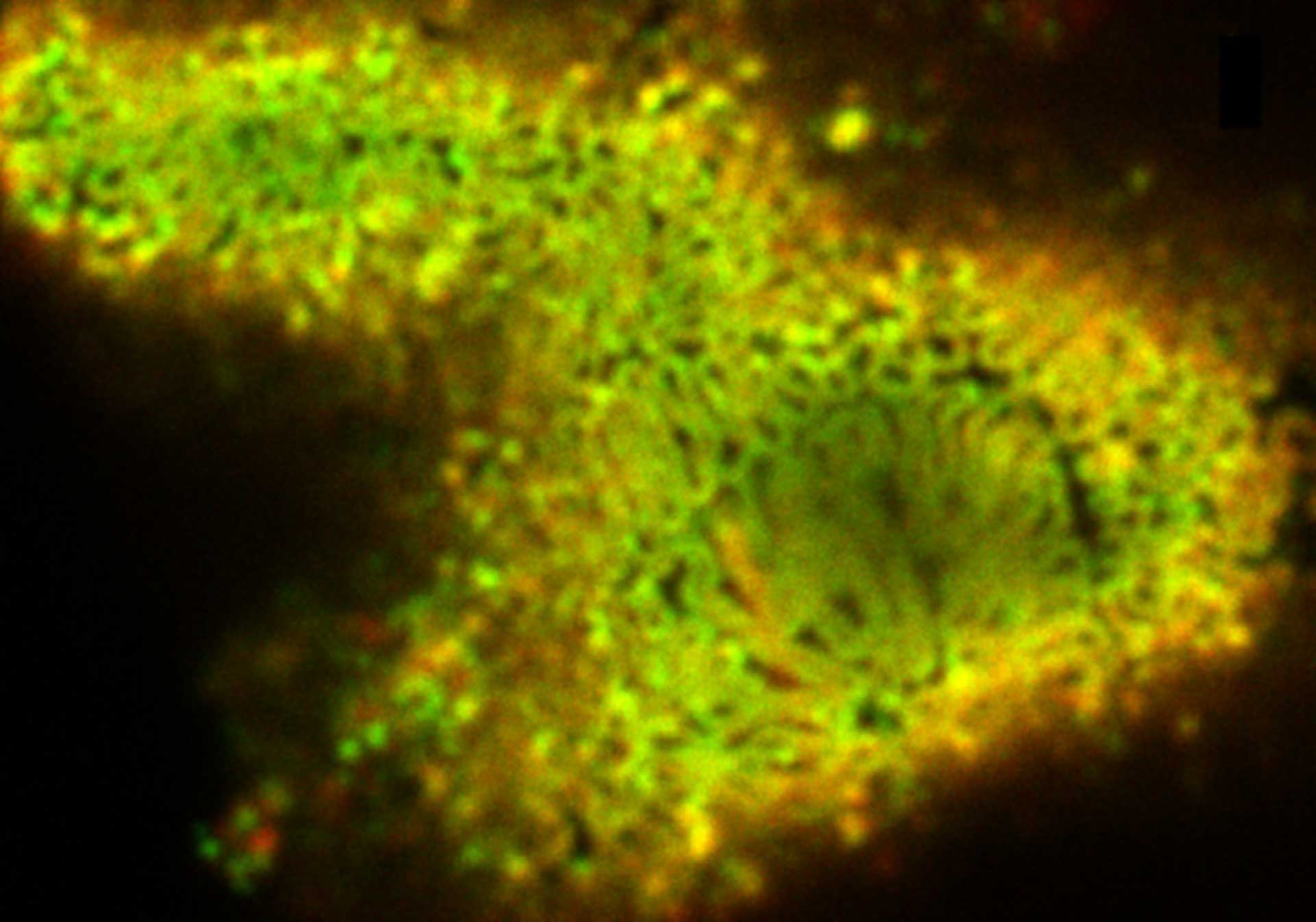
Description
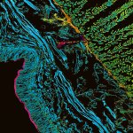
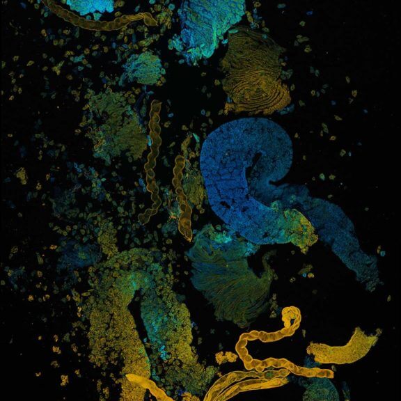
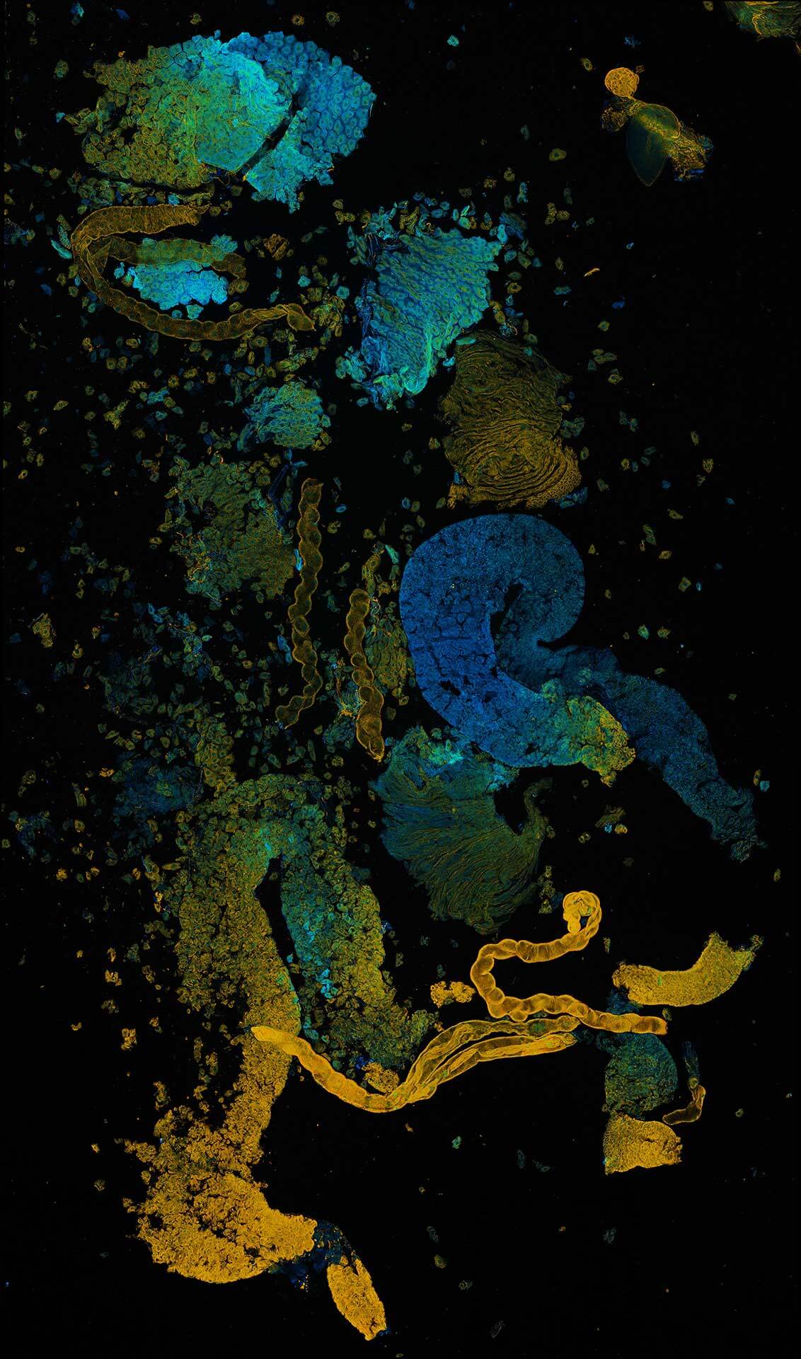
Confocal image of autofluorescence in a cross-section of the earthworm Lumbricus terrestris. TIMEBOW lifetime imaging detects differences in fluorescence lifetime, which depend on the type of autofluorescent molecule and its nano-environment, and visualizes them in distinct colors.
Modules:
Description
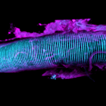
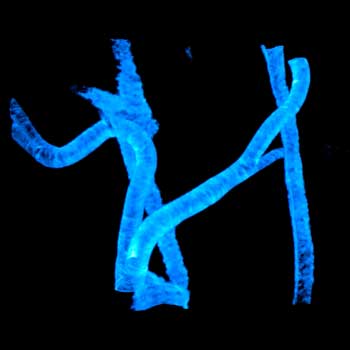
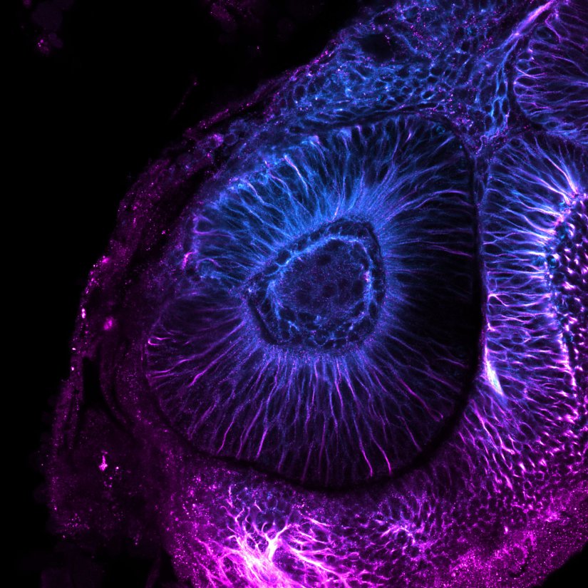
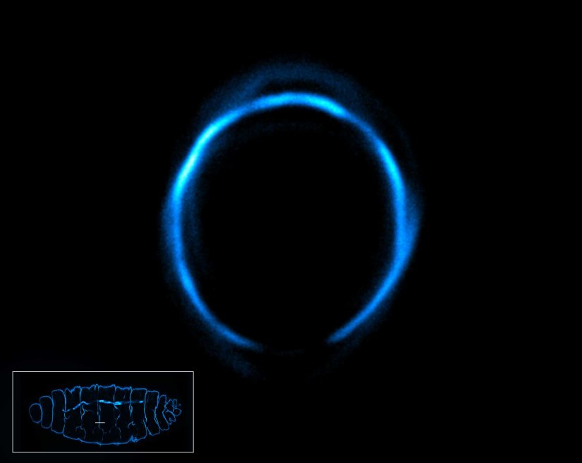
Confocal image of a developing zebrafish eye showing tubulin stained with abberior STAR RED and abberior STAR ORANGE.
Image courtesy of Graziamaria Paradisi, Marco Tartaglia, Antonella Lauri, Ospedale Pediatrico Bambino Gesù, Rome, Italy
Modules:
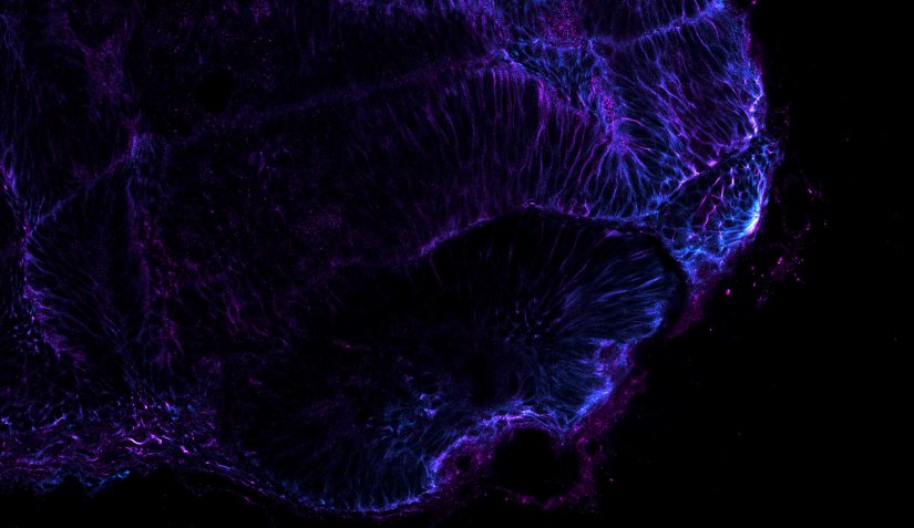
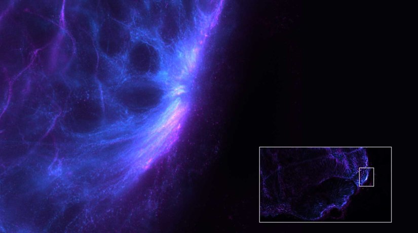
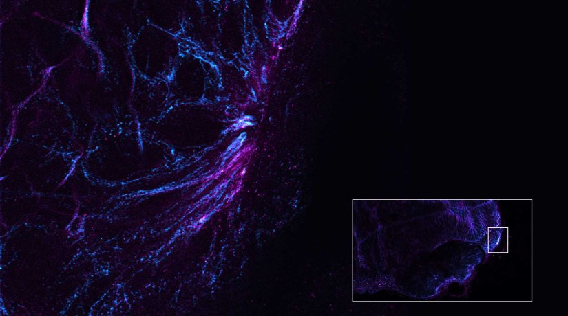
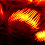
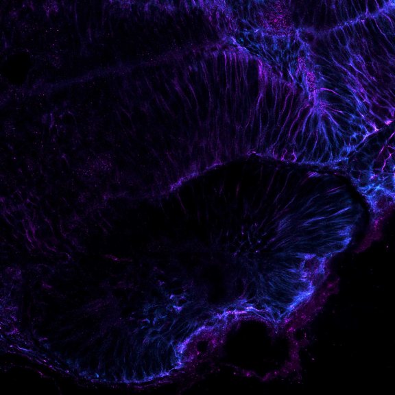
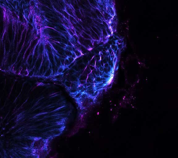
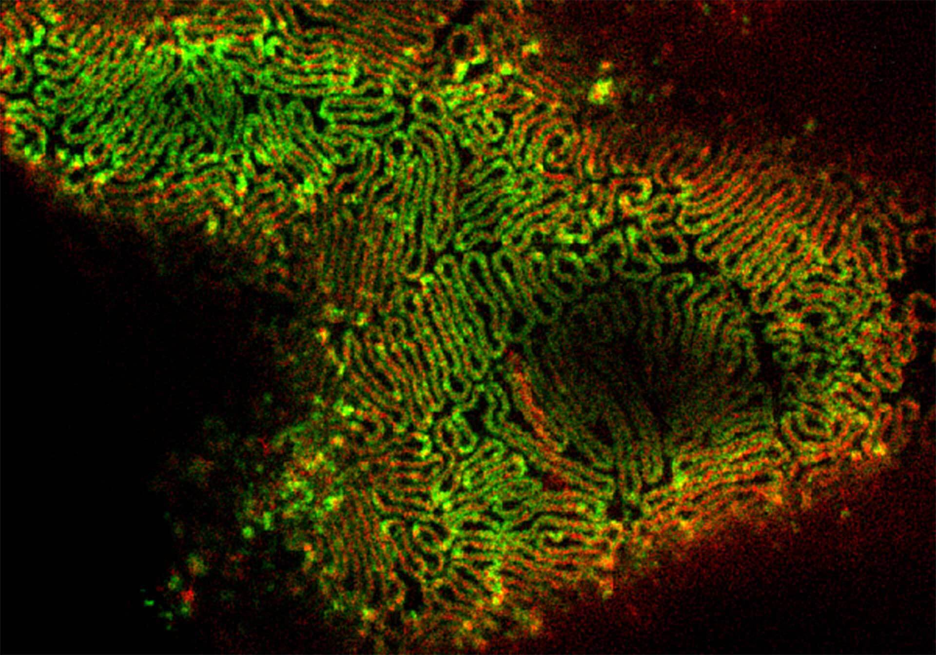
Description
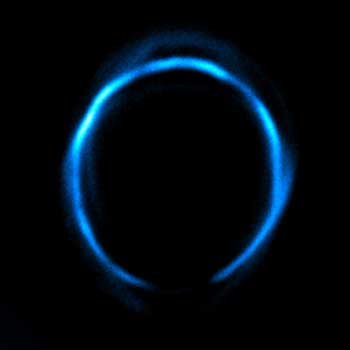
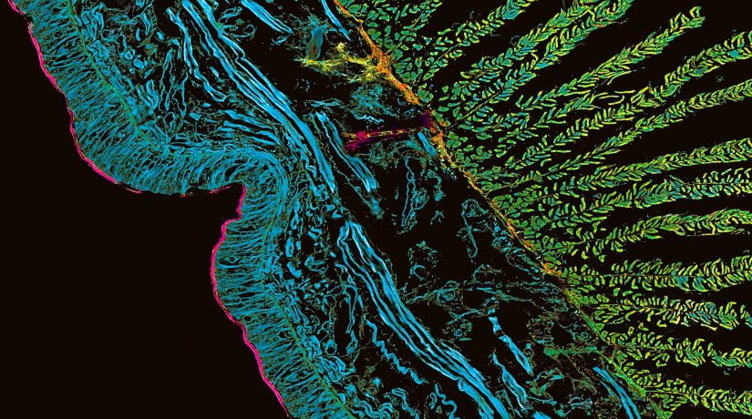
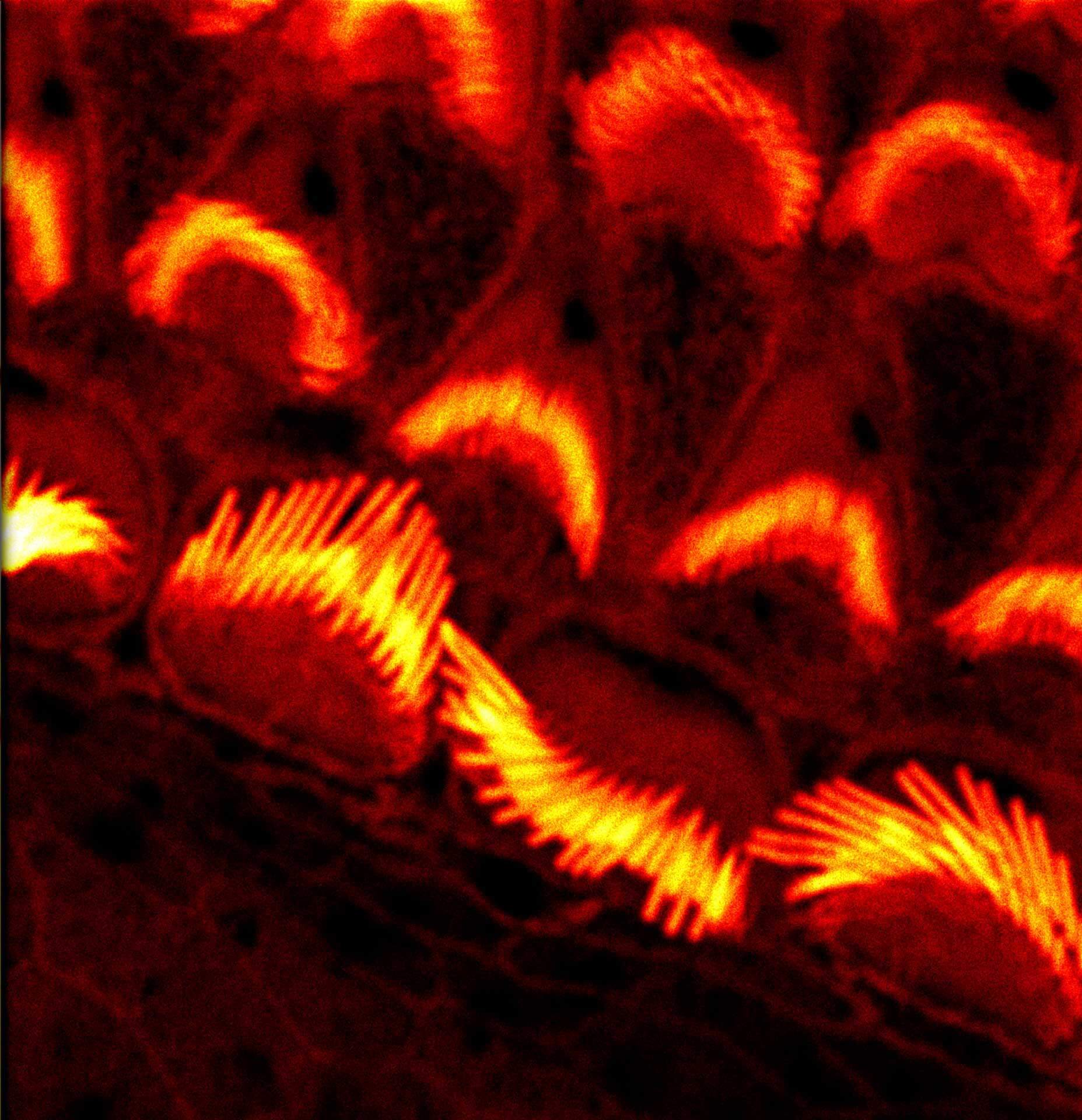
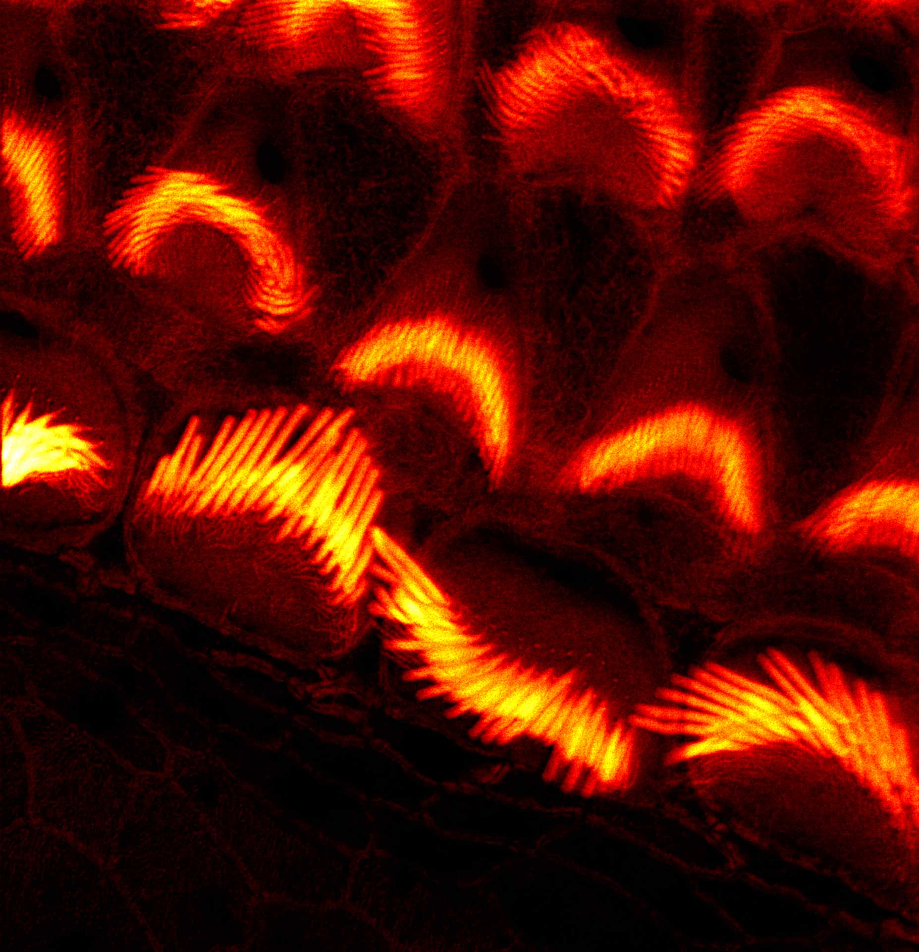
Gain in both signal-to-background ratio and resolution: MATRIX detection dramatically improves a conventional STED image of the zebrafish olfactory epithelium, resulting in a perfect and crystal-clear image revealing even more detail than STED alone can.
Modules:
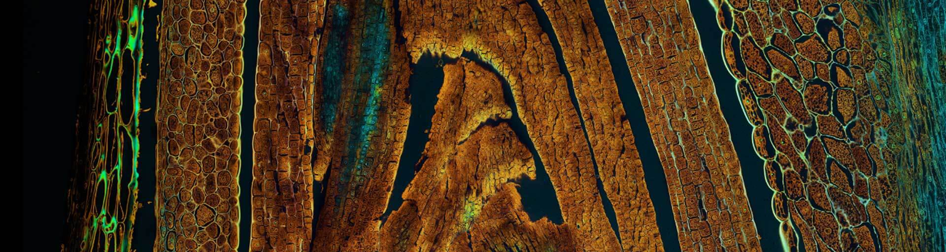
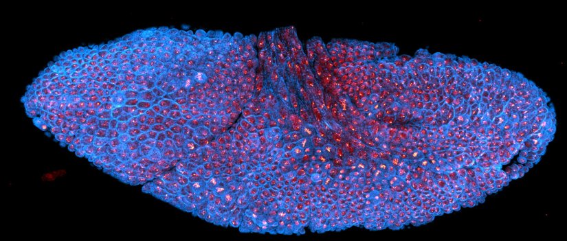
Description
xz section of a stage 17 Drosophila embryo stained for chitin (abberior LIVE 610, green) and DNA (abberior LIVE 550, cyan).
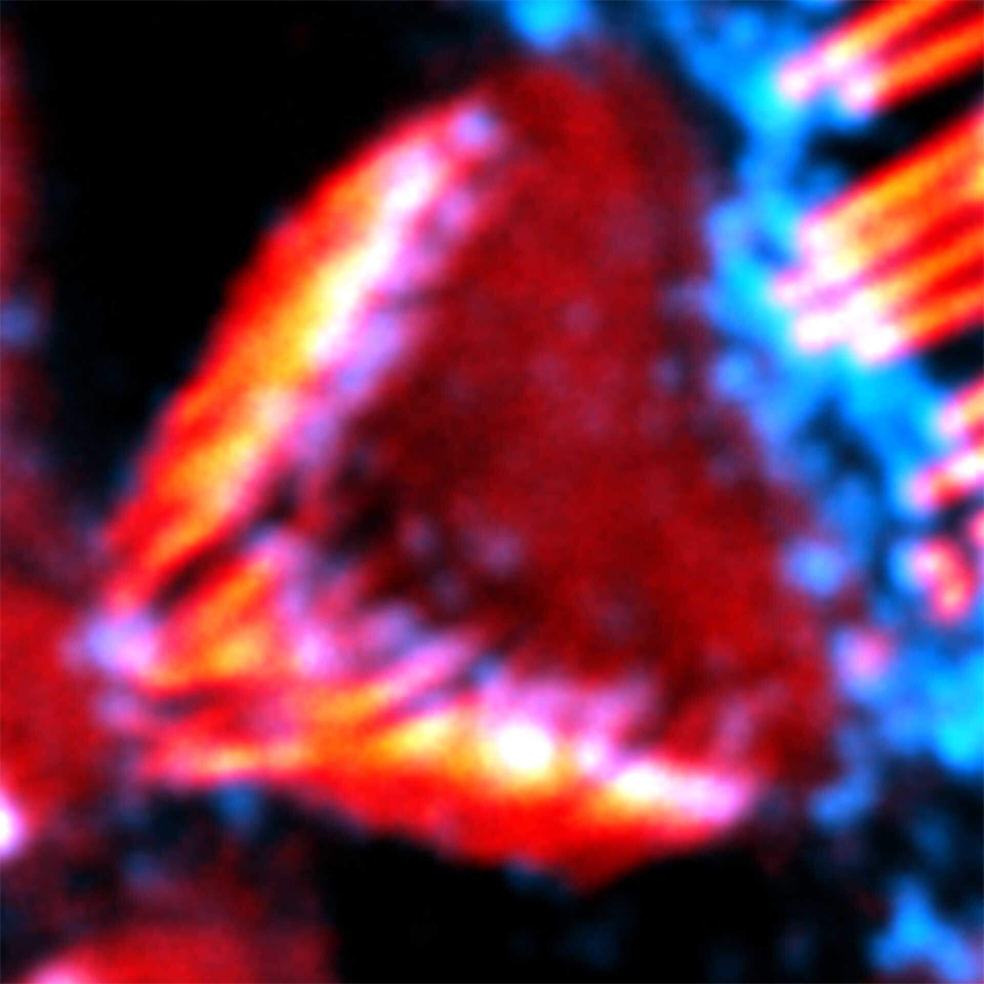
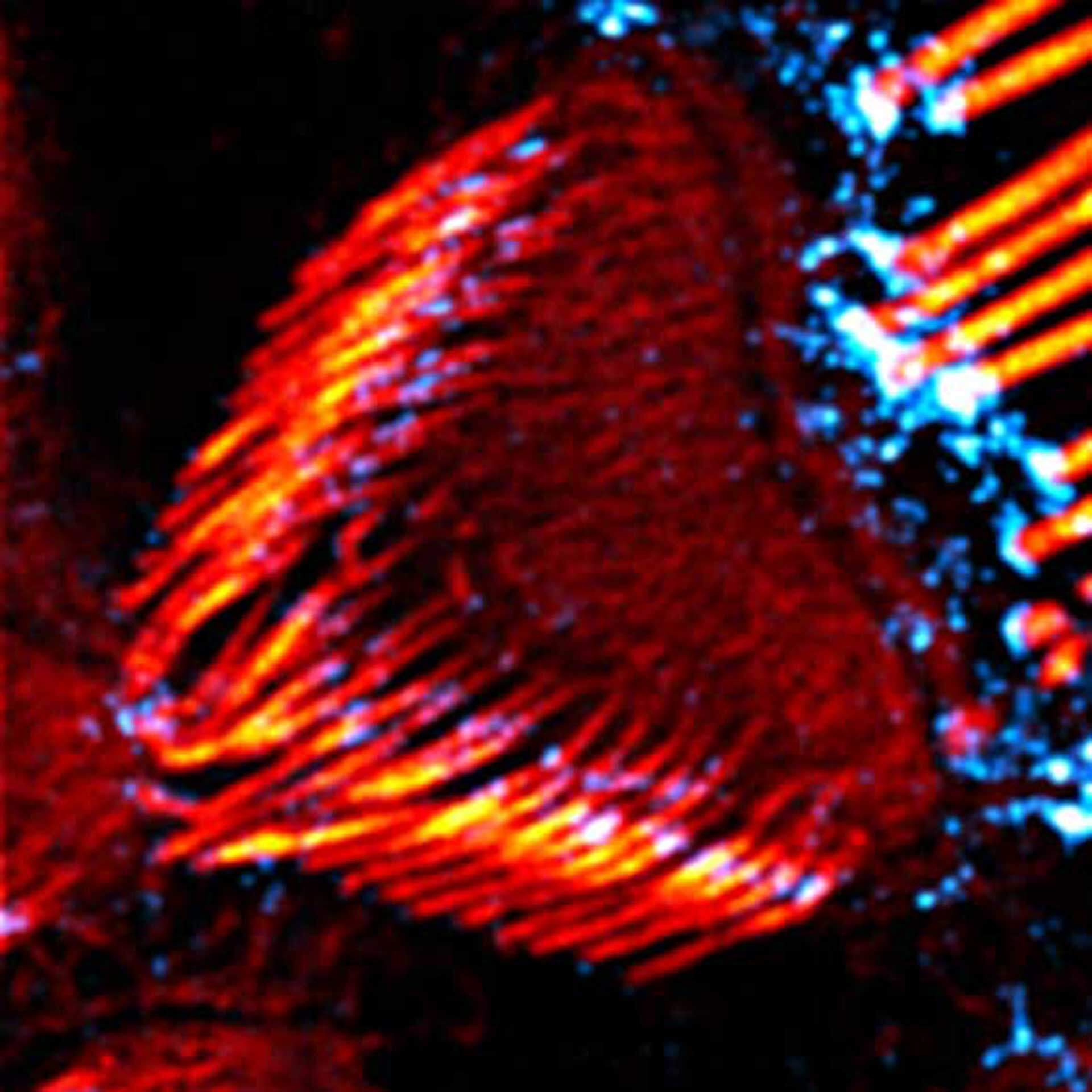
RAYSHAPE preserves resolution and brightness over the whole sample depth of about 200 µm by dynamically redirecting aberrated light to the right places.
In comparison, mechanical optics using a correction collar can only correct a limited z-range of approximately 20 µm
Modules:
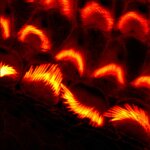
Description
Drosophila stage 12 embryo, imaged with RAYSHAPE, stained for tubulin with abberior STAR RED and for DNA with abberior LIVE 550.
Modules:
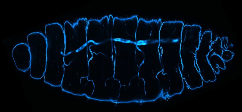
Description
Deep tissue imaging with RAYSHAPE of a stage 17 Drosophila embryo stained for chitin with abberior LIVE 610.
Modules:
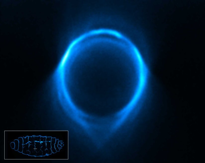
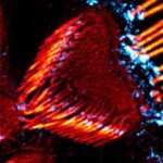
Description
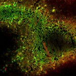
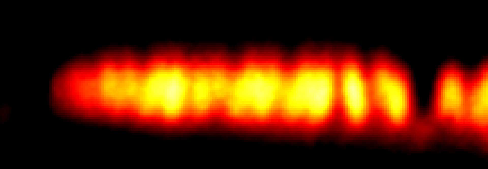
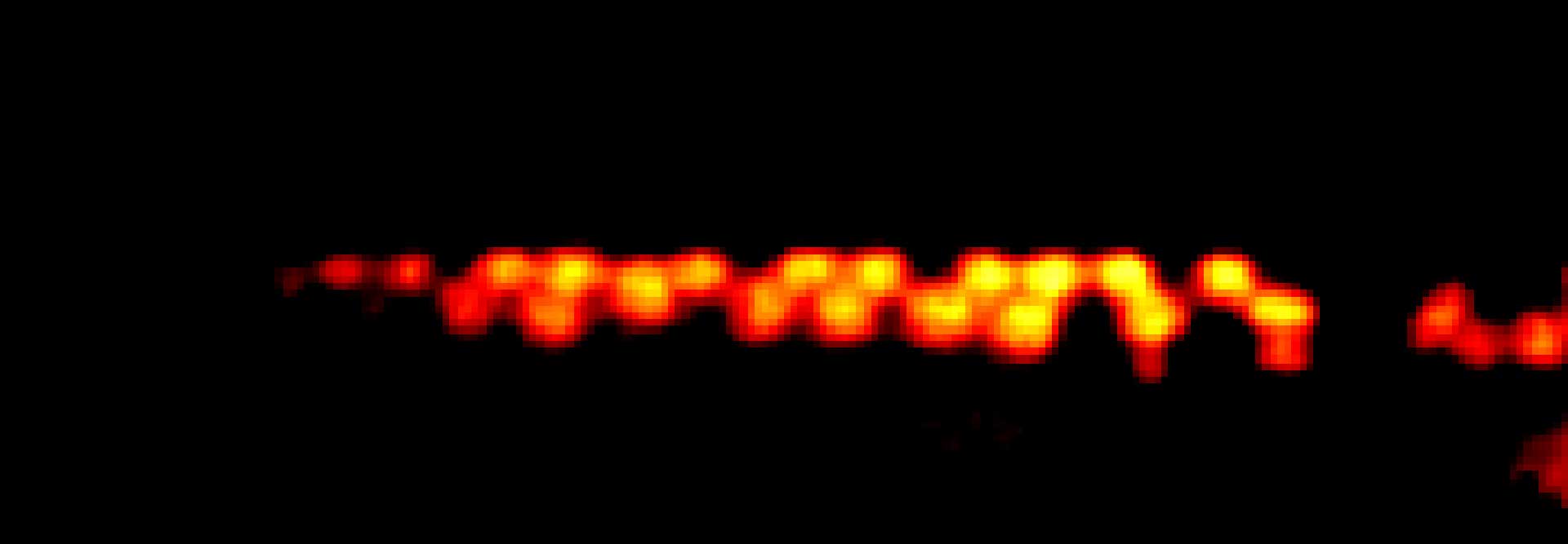
3D STED xz section of a Drosophila embryo trachea, imaged with RAYSHAPE and without abberation correction, depth 15 µm. Chitin was stained with abberior LIVE 610.
Modules:
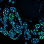
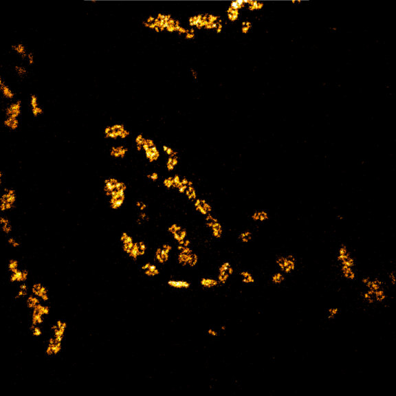
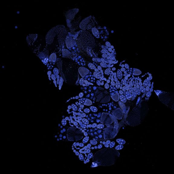
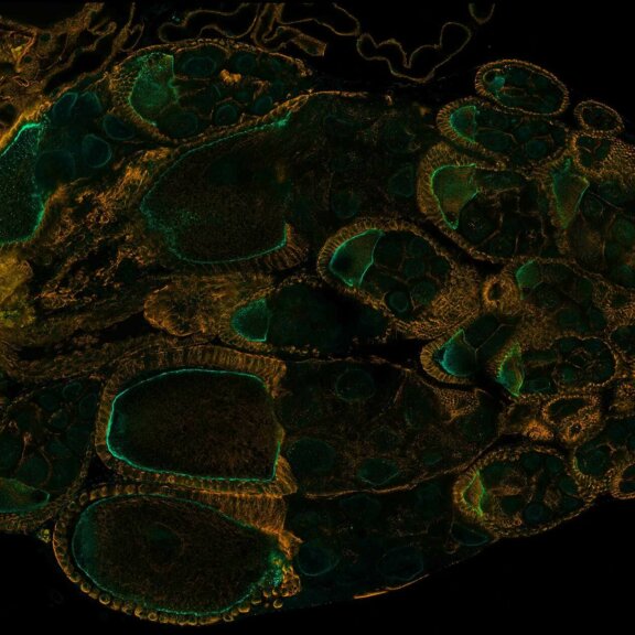
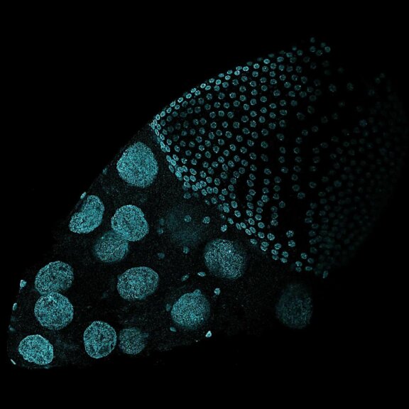
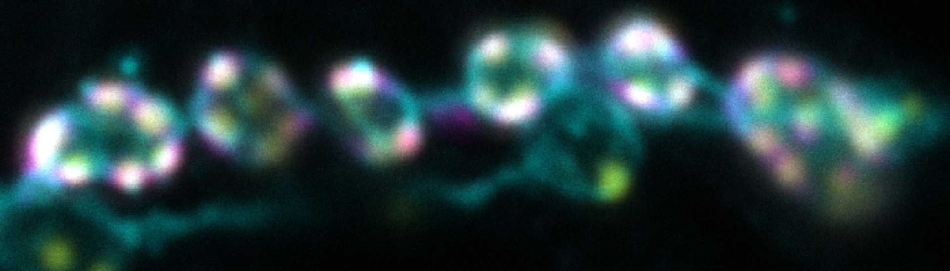
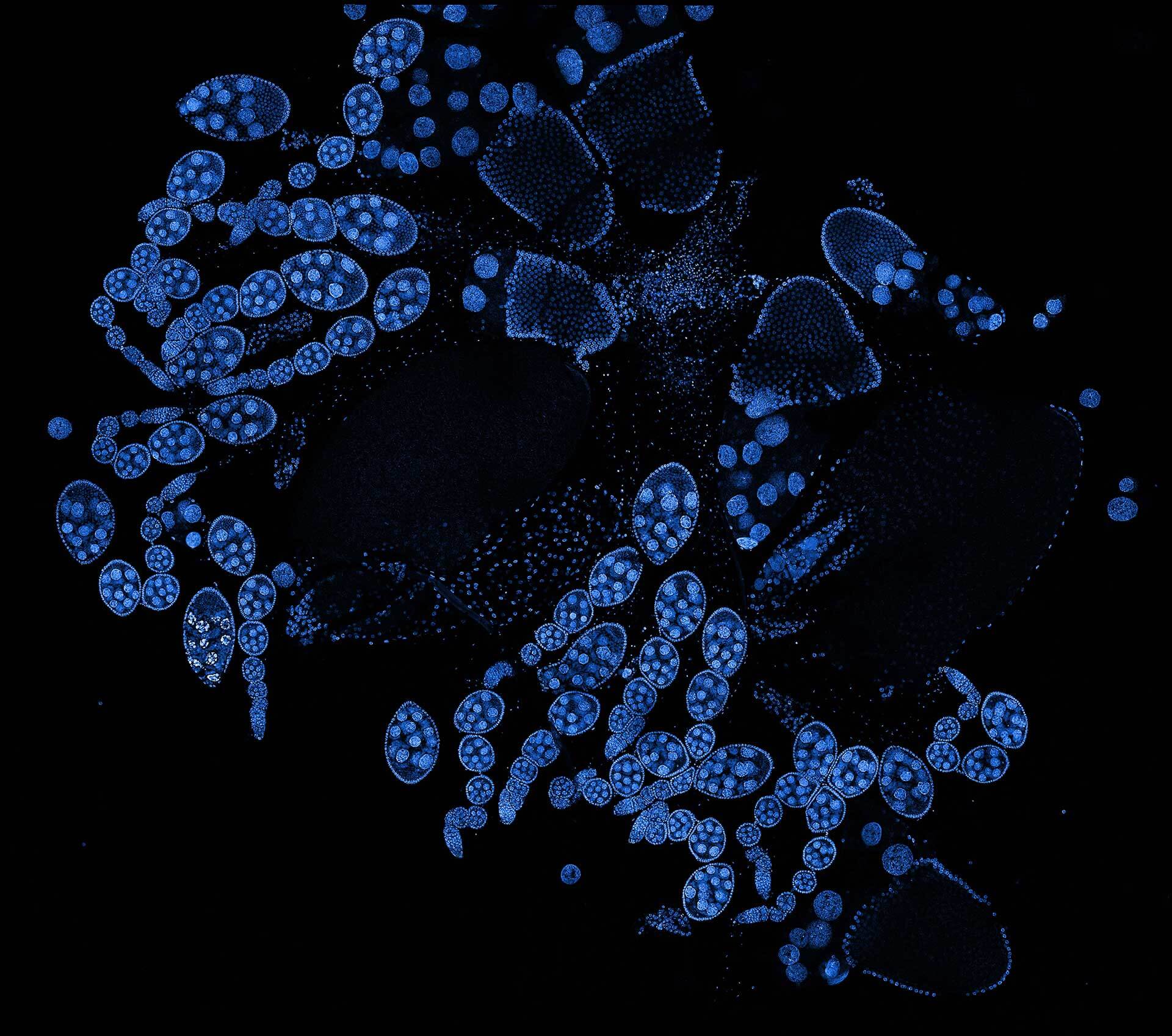
Description
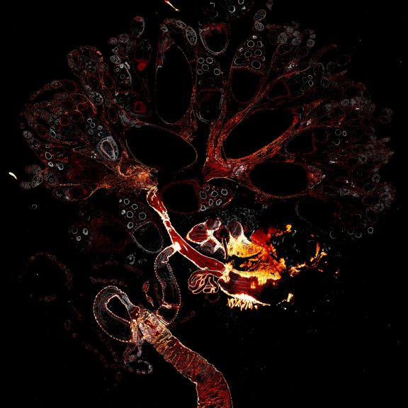
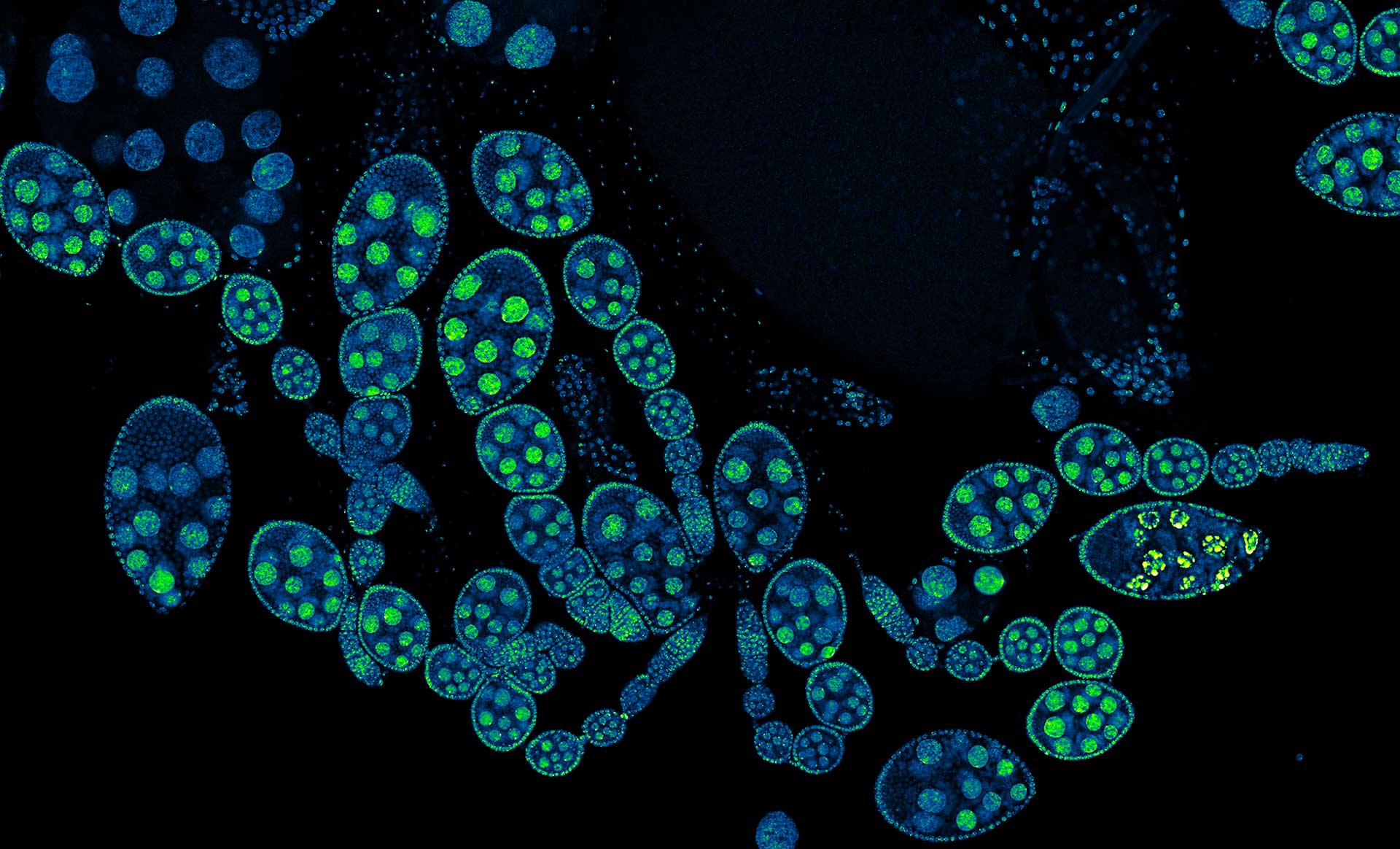
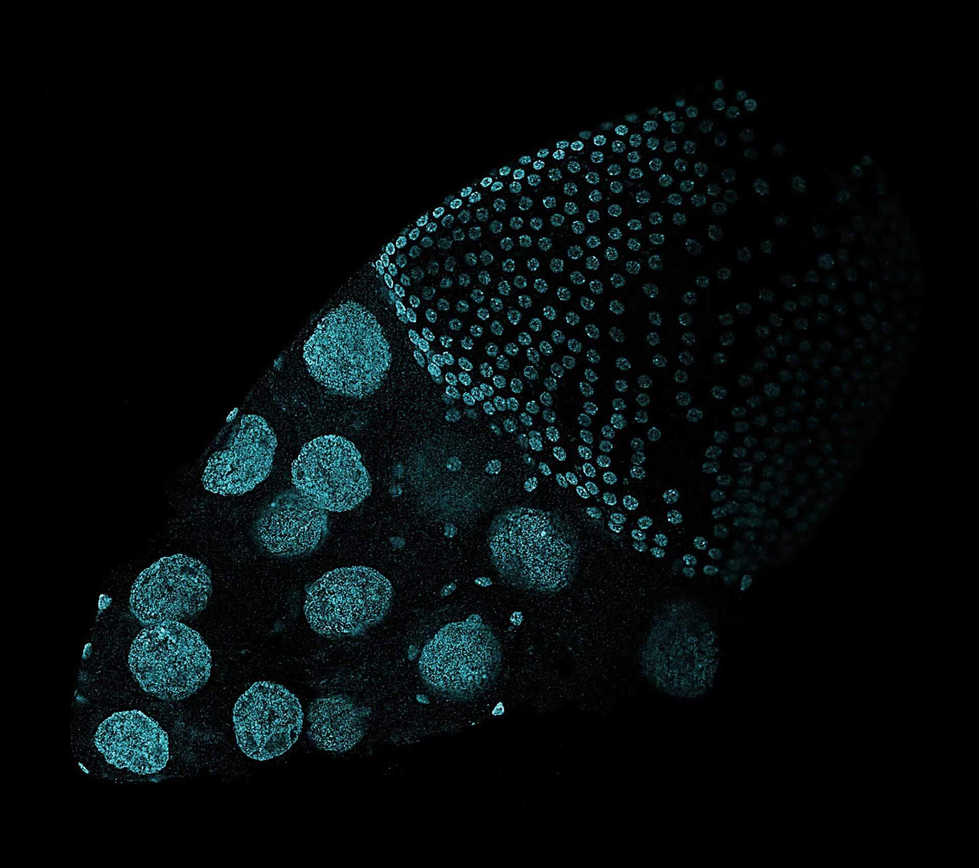
Drosophila ovariole stained with abberior LIVE 560 DNA showing nuclei in different cell types of the egg chamber. Ovaries were dissected from adult female fruit flies and were fixed prior to staining.
Image was acquired with the STEDYCON tiling feature and assembled with the SVI Huygens Stitcher.

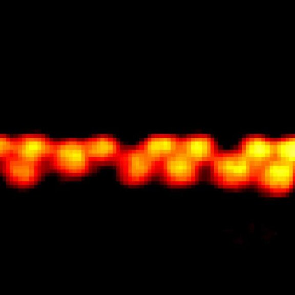
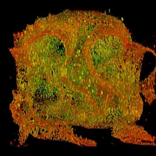
Description
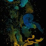
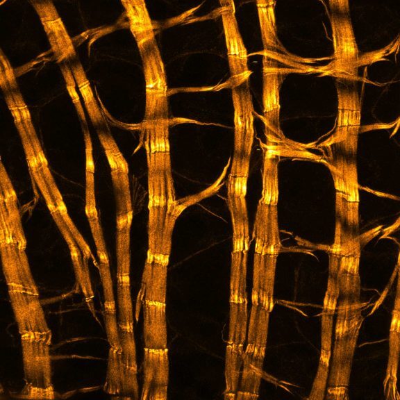
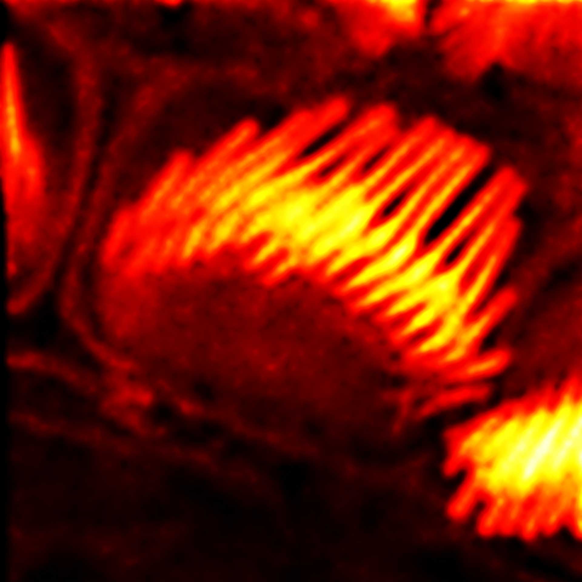
Drosophila spermatid tails were stained with abberior LIVE 610 tubulin. Testis were dissected from adult male fruit flies. Live cell imaging experiment was performed on a INFINITY microscope.

Description
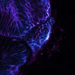
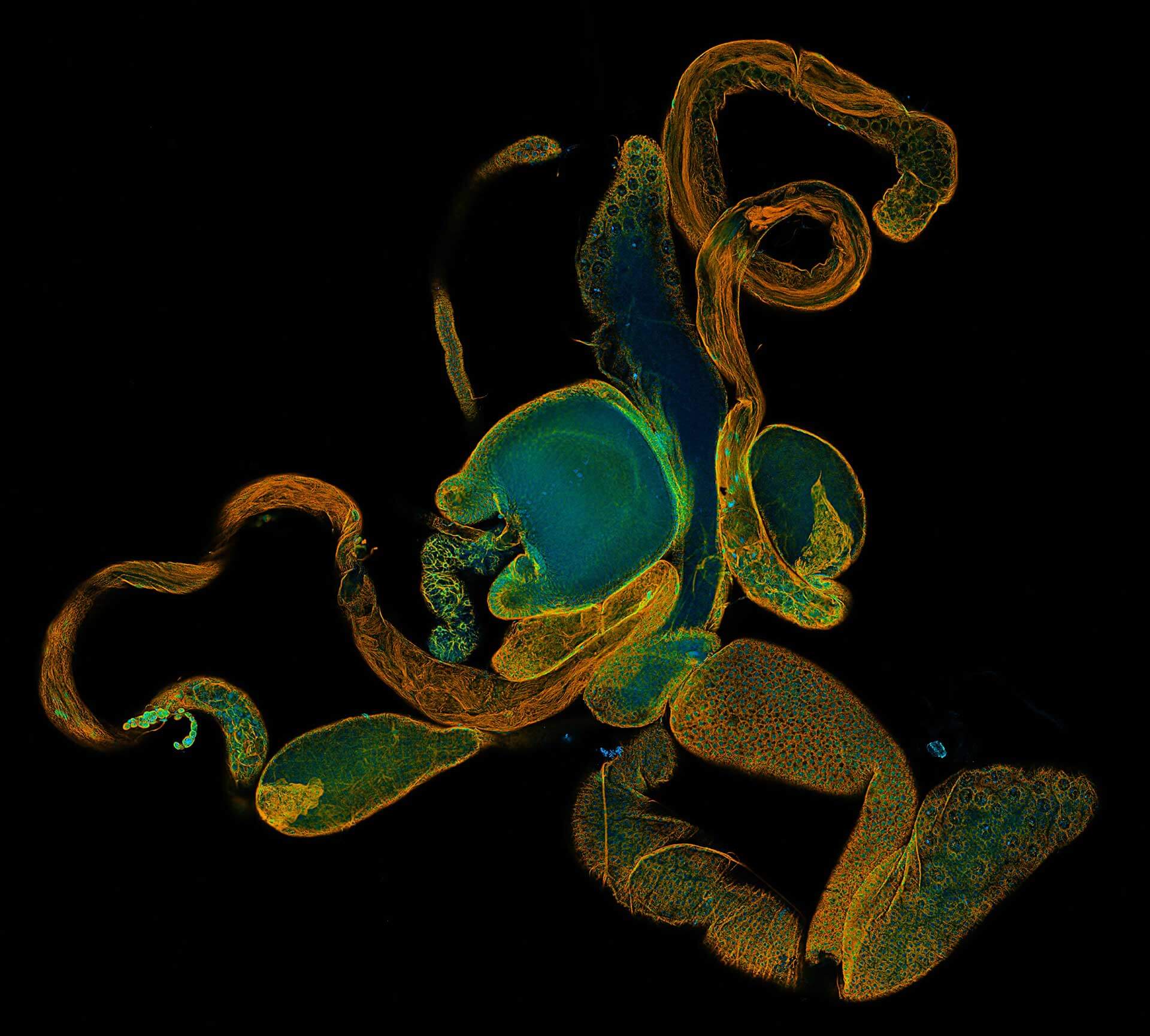
Drosophila male accessory gland stained for F-actin using abberior STAR 580 phalloidin.
Sample was prepared in cooperation with Dr. H. R. Shcherbata at MPl for Biophysical Chemistry, Göttingen, Germany.
Description
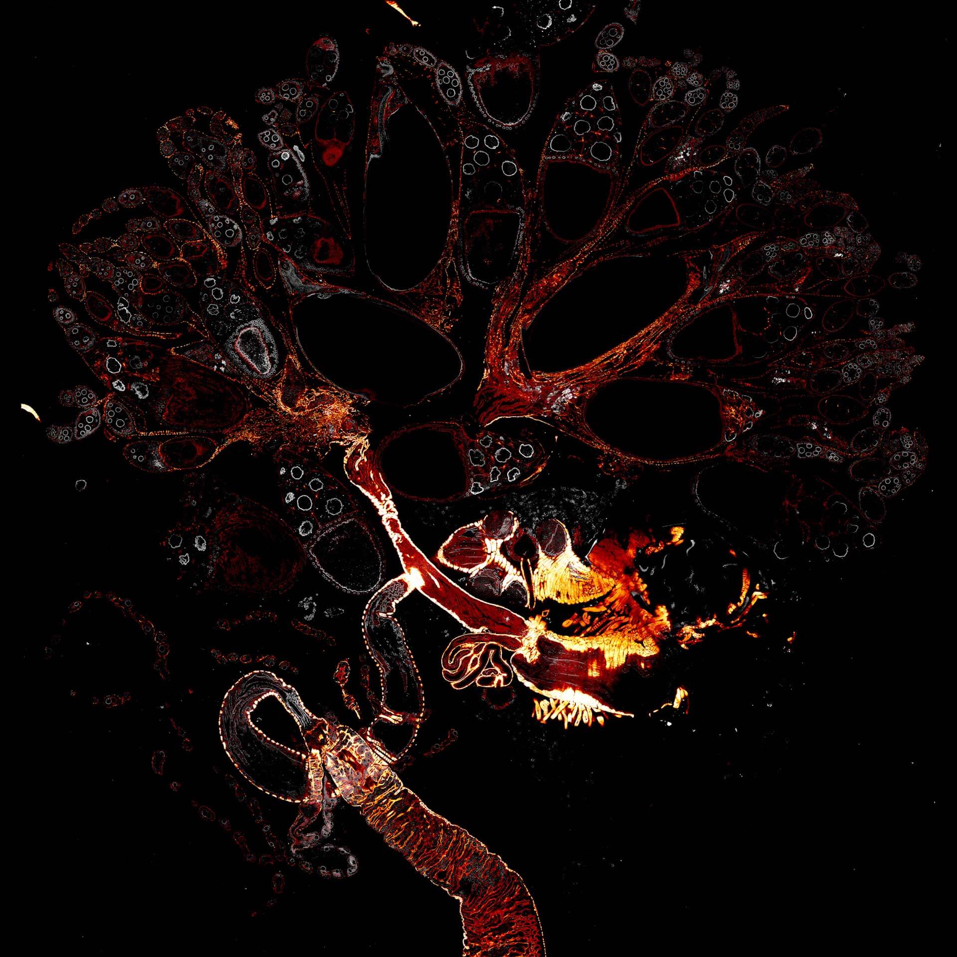
Drosophila female reproductive system stained for F-actin (red) with abberior STAR RED phalloidin. abberior STAR ORANGE is highlighting a nuclear pore protein (gray).
Image was acquired with the STEDYCON tiling feature and assembled with the SVI Huygens Stitcher.

Description
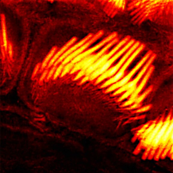
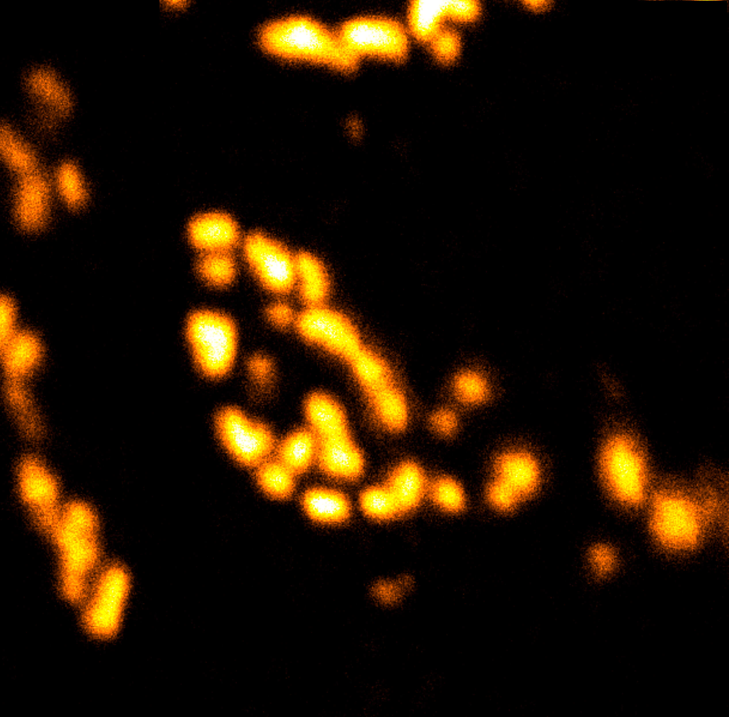
Actin stain of mouse inner ear hair cells using abberior STAR RED phalloidin.
Samples were prepared by Dr. Christian Vogl, InnerEarLab, UMG Göttingen, Germany.
Description
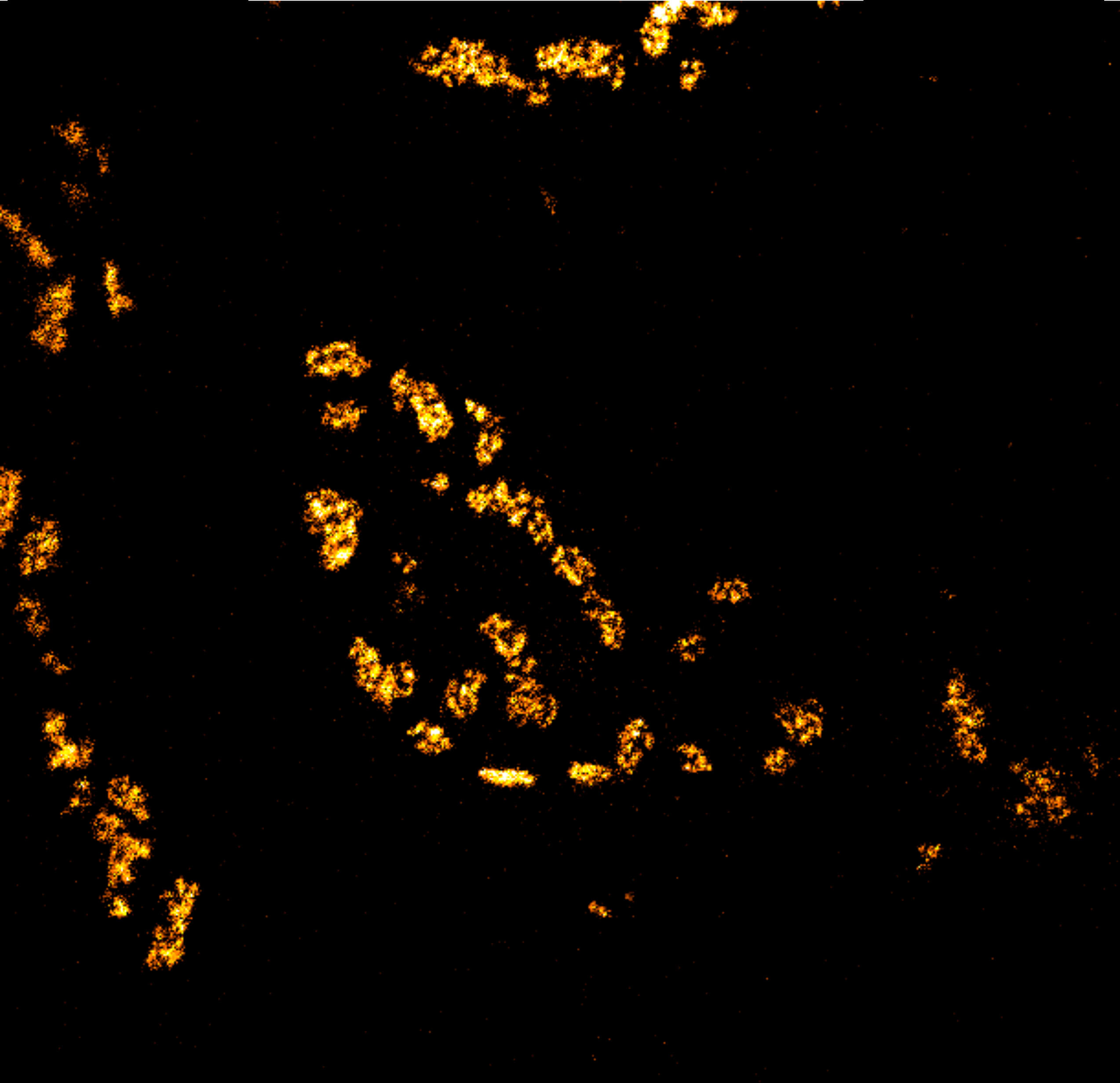
Actin stain of mouse inner ear hair cells using abberior STAR RED phalloidin. Zoom in for full effect.
Samples were prepared by Dr. Christian Vogl, InnerEarLab, UMG Göttingen, Germany.
Modules:
Description
Body wall preparation of a 3rd instar Drosophila melanogaster larva. Synapses at the neuromuscular junction are visualized by immunostaining against Bruchpilot (Brp), an integral component of active zones in Drosophila. Images were acquired on a STEDYCON attached to an Olympus IX81. Images were kindly provided by Dr. Nadine Ehmann, Institute of Physiology, Neurophysiology, University of Würzburg.
Description
Actin stain of mouse inner ear hair cells using abberior STAR RED phalloidin.
Samples were prepared by Dr. Christian Vogl, InnerEarLab, UMG Göttingen, Germany.
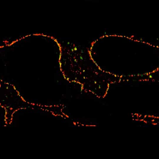
Description
2-color 2D STED image of a cleared adult kidney sample of a rat. Shown is an image of a renal corpuscle showing Nephrin (red, abberior STAR 635P) structures inbetween the Podocin slits (green, AlexaFluor594).
Sample was prepared by D. Unnersjö Jess and H.G. Blom @ KTH Stockholm, Sweden.
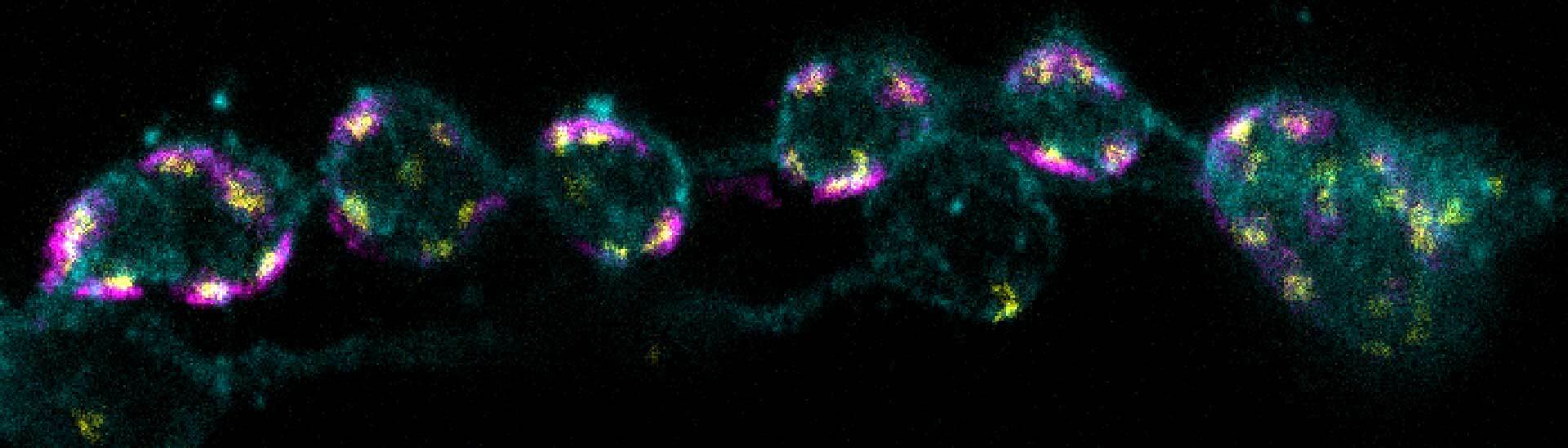
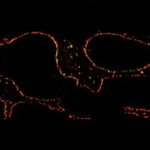
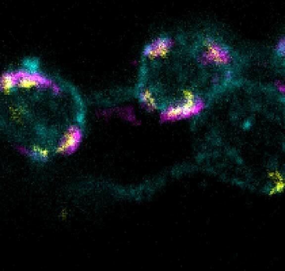
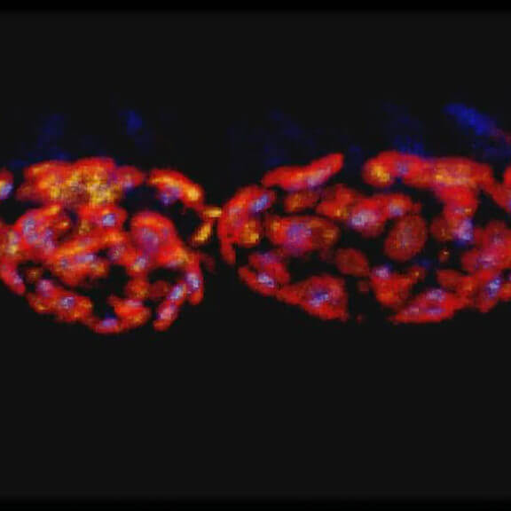
Description
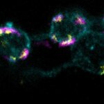
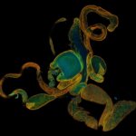
3-color STED imaging: active zones at the Drosophila larval neuromuscular junction immunostained for Bruchpilot and two other proteins.
Two superresolution channels (magenta, yellow) using a 775nm STED laser & one superresolution channel using a 595nm STED laser.
Samples by M. Lenz & M. Landgraf (University of Cambridge, UK).
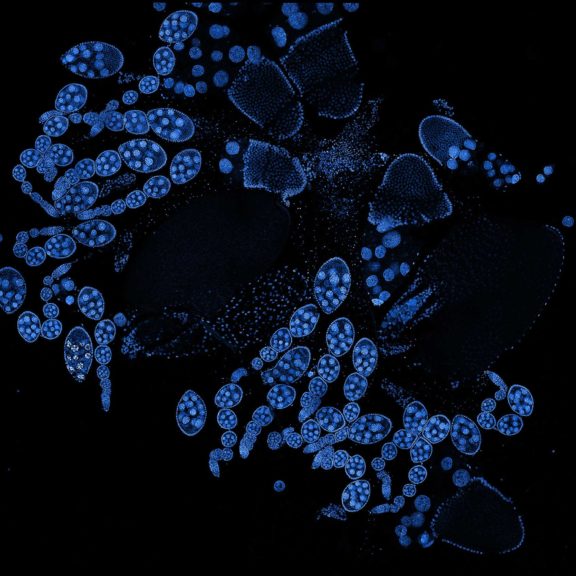
Description
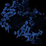
Drosophila ovariole stained with abberior LIVE 560 DNA showing nuclei in different cell types of the egg chamber. Ovaries were dissected from adult female fruit flies and were fixed prior to staining.
Image was acquired with the STEDYCON tiling feature and assembled with the SVI Huygens Stitcher.
Modules:
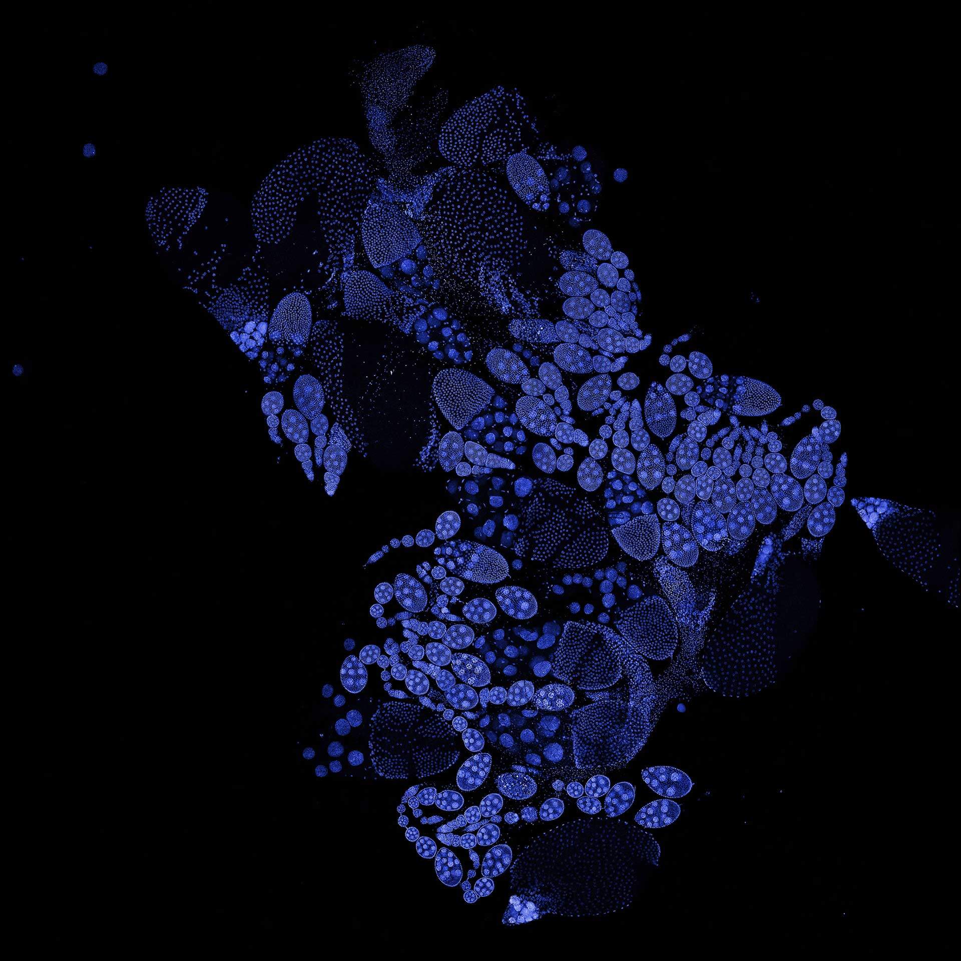
Description
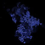
Drosophila ovariole stained with abberior LIVE 560 DNA showing nuclei in different cell types of the egg chamber. Ovaries were dissected from adult female fruit flies and were fixed prior to staining.
Image was acquired with the STEDYCON tiling feature and assembled with the SVI Huygens Stitcher.
Modules:
Description
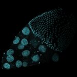
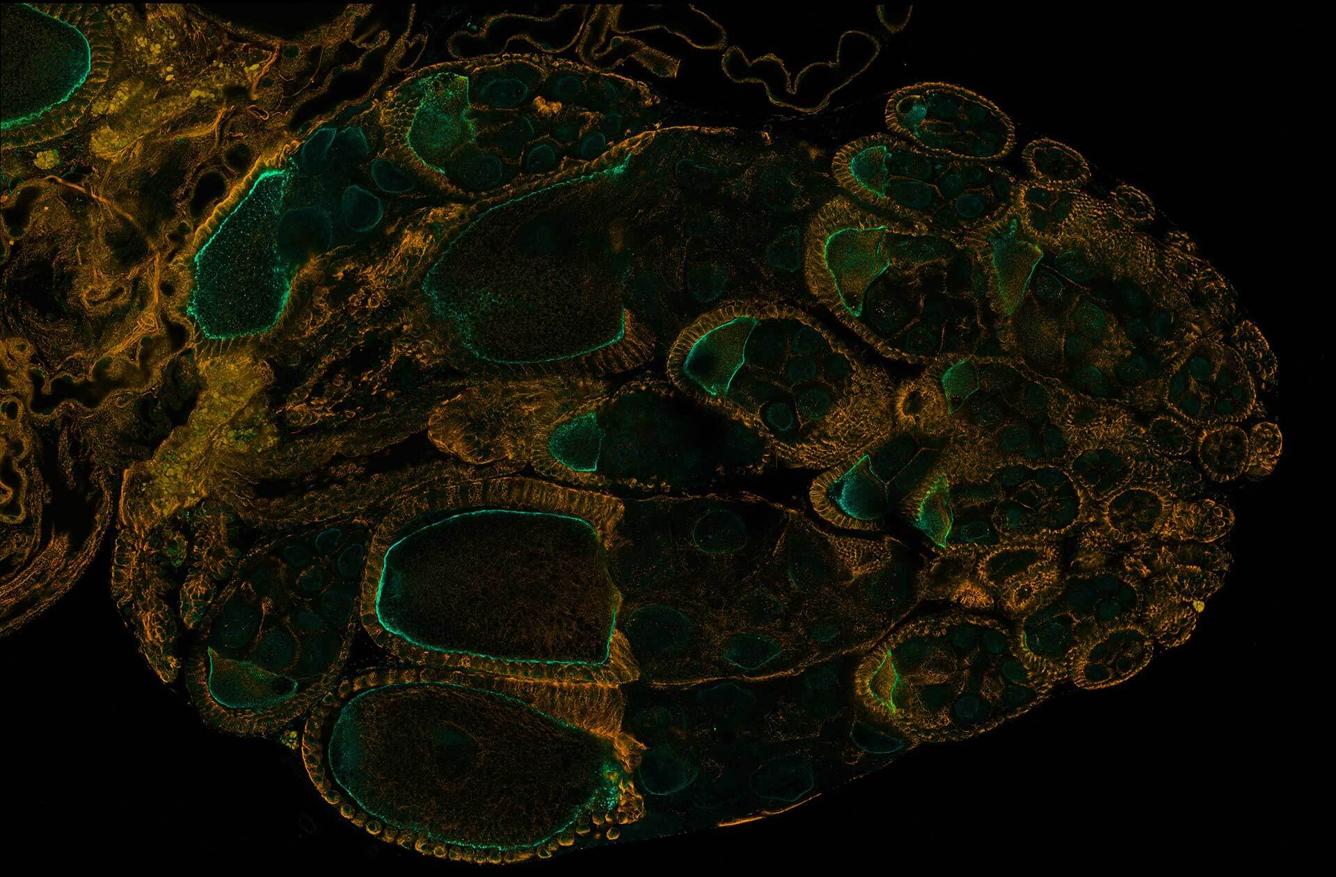
Drosophila ovariole stained with abberior STAR RED phalloidin (actin, red) and nuclear pores in STAR ORANGE (cyan) highlighting different cell types of the egg chamber. Image was acquired with the STEDYCON tiling feature and assembled with the HYGENS stitcher
Description
Drosophila ovariole stained with abberior LIVE 560 DNA, showing the different cell types of the egg chamber. The image was stitched using SVI Huygens.
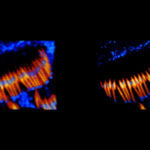
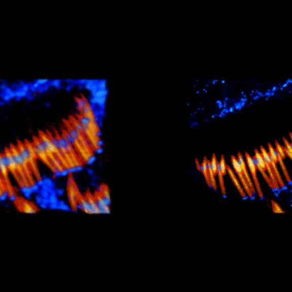
Description
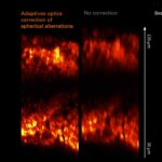
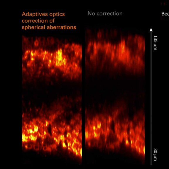
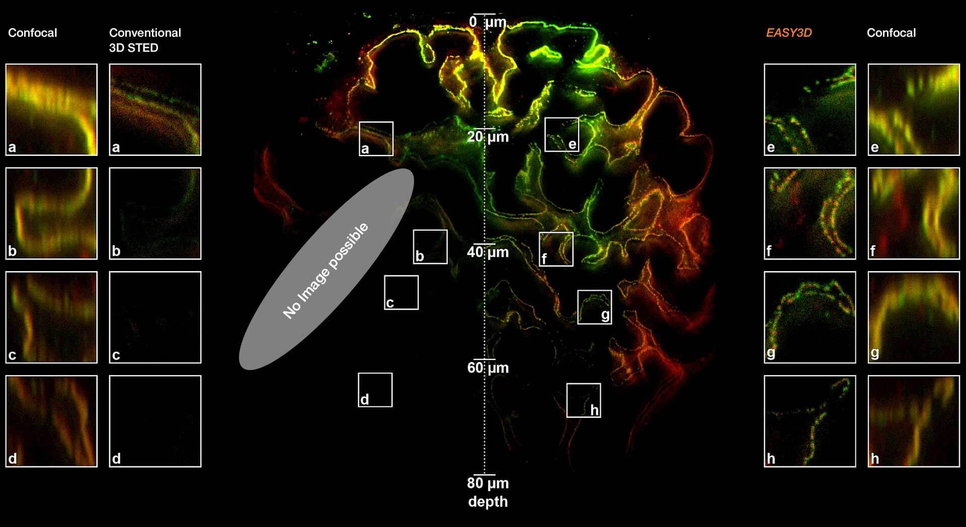
Adaptive optics EASY3D enables deep imaging of cleared adult kidney samples. Comparison of a XZ-slice deep into a renal corpuscle using conventional 3D STED imaging and an EASY3D imaging. EASY3D allowed imaging up to 80 µm into cleared rat kidney tissue using an oil objective. Without adaptive optics, the mismatch between immersion oil and sample leads to complete signal loss. Labels: Nephrin (red, Abberior STAR635P) and Podocin (green, AlexaFluor594).
Sample was prepared by D. Unnersjö Jess and H.G. Blom @ KTH Stockholm, Sweden.
Modules: