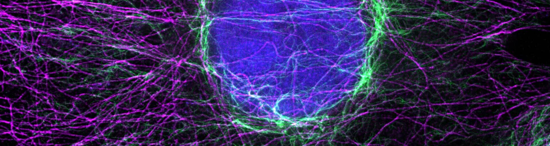
Nanobody labeling protocol

The combination of super bright and highly photostable abberior dyes and polyclonal nanobodies produced in alpacas offer a fantastic solution for high-resolution imaging experiments where small labels, good penetration, and outstanding specificity are necessary.

for easy and proper application of our labels

Introduction

abberior offers a variety of excellent fluorescent dyes with properties optimized for labeling biomolecules, spectroscopic studies, and optical microscopy, particularly superresolution microscopy, and optical nanoscopy.

The combination of super bright and highly photostable abberior dyes and the polyclonal nanobodies produced in alpacas from Jackson ImmunoResearch (AffiniPure-VHH™ Secondaries) offer a fantastic solution for high-resolution imaging experiments where small labels, good penetration, and outstanding specificity are necessary.

Storage

The product contains a vial of nanobody-conjugate in solution and is shipped at room temperature. Upon arrival, the product can be stored at 4 °C for a short period of time. For long-term storage of up to one year add an equal volume of glycerol (ACS grade or better) for a final concentration of 50 %, and store at –20 °C as a liquid. The conjugate should be protected from direct light exposure. Repeated freeze-thaw cycles should be avoided. Therefore, it is recommended to split the abberior secondary nanobody solution into smaller aliquots.

Indirect immunofluorescence staining with abberior secondary nanobodies

The procedure has been successfully tested with our abberior dye conjugates for adherent cells grown on glass coverslips and has yielded consistent results in most instances but may require further optimization for particular model organisms or experimental conditions.

Required reagents; not provided

  • Phosphate-buffered saline pH 7.4 (PBS)
  • Fixative depending on the primary antibody
  • 0.1% – 0.5% Triton X-100 in PBS pH 7.4 (permeabilization buffer)
  • 1% – 3% Bovine Serum Albumin + 0.1% Tween20 in PBS pH 7.4 (blocking buffer, PBT)
  • Primary antibody
  • Mounting Medium
  • Glass coverslips with a glass thickness of ~170 µm (No. 1.5 or No. 1.5H)

    Note: We do not recommend using plastic coverslips because frequently only suboptimal imaging results are achieved. If possible, coverslips with grids, gratings, or similar should be avoided, as these structures can interfere with imaging causing aberrations that degrade image quality.
  • Humid chamber
  • Fluorescence microscope with suitable excitation light source and detection filter

Protocol for cultured cells

All steps are carried out at room temperature and in a petri dish unless stated otherwise.

  1. Incubate cells with a fixative suitable for the primary antibody in use for 5 min to 15 min.

    Note: When using Methanol as a fixative, step 2 can be skipped.
  2. Replace the medium with permeabilization buffer and incubate the sample for 5 min to 15 min.
  3. Wash cells 3 x 5 min in PBS.
  4. Replace the medium with a blocking buffer and incubate the sample for 30 min to 60 min.
  5. Dilute the primary antibody to the final staining concentration in the blocking buffer.

    Note: The staining concentration depends on the primary antibody used.
  6. Take the coverslips out of the petri dish; remove the excess blocking buffer by placing the cover slip edge onto a piece of tissue paper. Transfer the coverslips into a humid chamber, cells facing upwards. Add the primary antibody onto the coverslips and incubate for 1 h in the humid chamber.
  7. Remove excess primary antibodies by placing the cover slip edge onto a piece of tissue paper. Wash the cells in PBS (3 x 5 min) using a fresh petri dish.
  8. Replace the medium with a blocking buffer and incubate the sample for 30 min to 60 min.
  9. Dilute the secondary nanobody to the final staining concentration in PBS (pH 7.4).

    Note: For most applications, a dilution of 1:200 to 1:800 of a 1 mg/ml stock solution is sufficient. However, staining protocols may vary with cell type and application.

    Note: To dilute the nanobodies, BSA or other serum should be avoided, as the nanobodies can bind to free IgG antibodies within the serum and thus prevent optimal staining.

    Optional: At this step abberior phalloidin can be added to the staining solution (1 Unit/ml).
  10. Take the coverslips out of the petri dish. Remove excess blocking buffer by placing the cover slip edge onto a piece of tissue paper. Transfer the coverslips into a humid chamber, cells facing upwards. Add the secondary nanobody onto the coverslips and incubate for 1 h in the humid chamber under the exclusion of light.
  11. Remove excess secondary nanobody by placing the cover slip edge onto a piece of tissue paper. Wash the cells in PBS (3 x 5 min) using a fresh petri dish.
  12. Take the cover slip out of the washing solution, remove excess PBS by placing the cover slip edge onto a piece of tissue paper, and mount the coverslip with a suited mounting medium.

Abbreviations

PBS Phosphate-buffered saline
PFA Paraformaldehyde
PBT 1% – 3% Bovine Serum Albumin + 0.1% Tween20 in PBS
min Minute
h Hour