


abberior DNA-PAINT, FLASH kits
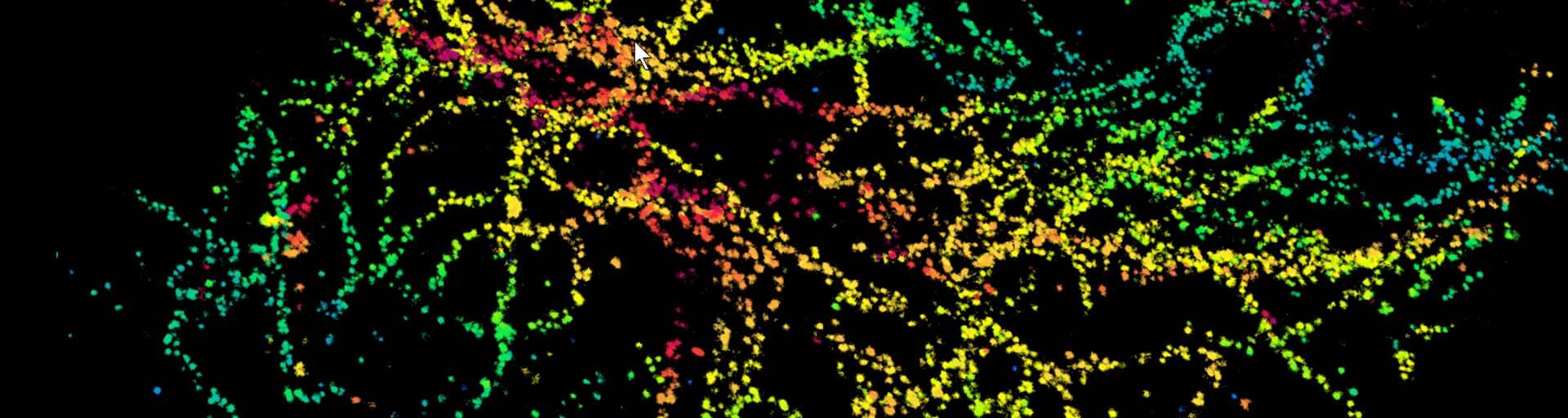
The newly developed in-house abberior FLASH DNA-PAINT kits combine the unmatched localization precision of DNA-PAINT with an adapter-mediated multiplexing strategy. Designed for MINFLUX and SMLM. All FLASH kits can be adapted and expanded upon your specific requirements.
for superresolution microscopy
Introduction
The following protocol has been validated with abberior DNA-PAINT, FLASH dye conjugate. Depending on the model organism or target of interest, further optimization steps may be necessary. The following sections provide step-by-step instructions for: indirect immune FLASH-DNA-PAINT staining using anti-mouse, anti-rabbit, and anti-GFP secondary nanobodies (Indirect Immunostaining Kits), as well as direct immune FLASH-DNA-PAINT staining with a primary anti-GFP nanobody (Direct IF – GFP Kit).
The protocol is designed for adherent cells cultured on glass coverslips.
Storage
The product is shipped at room temperature. Upon arrival, store buffers at 4 °C and sdAb, Imager, and Adapter at −20 °C.
Immunofluorescence staining with abberior DNA-PAINT, FLASH for MINFLUX microscopy
The procedure below has been validated with abberior DNA-PAINT, FLASH dye conjugates, though further optimization may be needed for specific model organisms or targets. The following protocols describe indirect immune FLASH-DNA-PAINT staining using anti-mouse and anti-rabbit secondary nanobodies, as well as direct immune DNA-PAINT, FLASH staining using an anti-GFP nanobody, for adherent cells grown on glass coverslips.
Reagents, provided for indirect immunostaining kits
Secondary single domain antibodies (sdABs)
- FLASH sdAB Anti-rabbit + Docking Site A1, 100 µl (5 µM) (to be combined with adapter A1, imager IMG1) or FLASH sdAB Anti-mouse + Docking Site A2, 100 µl (5 µM) (to be combined with adapter A2, imager IMG2)
- Concentration: 5 µM Protein, 5 µM DNA (1 DNA strand per protein)
- Volume: 100 µL
- Storage: -20 °C
- Storage buffer: PBS, 50 % glycerol
- Recommended dilution: 1:200 – 1:500 (for optimal results the dilution needs to be optimized (depending on the target accessibility and expression level)
Adapter
- abberior DNA PAINT, FLASH adapter A1, 600 µl (20 µM) or abberior DNA PAINT, FLASH adapter anti-mouse A2, 600 µl (20 µM)
- Concentration: 20 µM in TE buffer (10 mM Tris, 1 mM EDTA, pH 8)
- Volume: 600 µL
- Storage: -20 °C (1 µM imager solutions are stable for multiple freeze-and-thaw cycles). Optional: Prepare 50 µL aliquots and store them at -20 °C. Working aliquots can be stored at 4 °C for short-term or -20 °C for long term.
Imager
- abberior DNA PAINT, FLASH imager IMG1, 300 µL (1 µM) or abberior DNA PAINT, FLASH imager IMG2, 300 µL (1 µM) carrying the respective fluorophore (580/660)
- Concentration: 1 µM in TE buffer (10 mM Tris, 1 mM EDTA, pH 8)
- Volume: 300 µL
- Storage: -20 °C (1 µM imager solutions are stable for multiple freeze-and-thaw cycles). Optional: Prepare 50 µL aliquots and store them at -20 °C. Working aliquots can be stored at 4 °C for short-term or -20 °C for long term.
Note: Further dilutions should be prepared fresh before use. Low imager concentrations are not stable in plastic tubes.
Buffers
- abberior Antibody incubation buffer, 50 ml, stored at 2 – 8 °C
Note: Blocking agents (BSA, serum, DNA etc.) are not provided and have to be added in the desired amounts before use. - abberior Washing buffer (10×), 50 ml, store at 2 – 8 °C (to be diluted 1:10 in water before use)
- abberior Imaging buffer, 50 ml, store at 2 – 8 °C
Reagents, provided for the direct IF/GFP kit
Single domain antibodies (sdABs)
- FLASH sdAB Anti-GFP + Docking Site A3, 100 µl (5 µM)
- Concentration: 5 µM clone 1H1, 5 µM DNA (1 DNA strand per protein)
- Volume: 100 µl
- Storage buffer: PBS, 50 % glycerol
- Storage: -20 °C
- Recommended dilution: 1:200 – 1:500 (for optimal results the dilution needs to be optimized depending on the target accessibility and expression level)
Adapter
- abberior DNA PAINT, FLASH adapter A3, 600 µl (20 µM)
- Concentration: 20 µM in TE buffer (10 mM Tris, 1 mM EDTA, pH 8)
- Volume: 600 µl each
- Storage: -20 °C (1 µM imager solutions are stable for multiple freeze-and-thaw cycles). Optional: Prepare 50 µl aliquots and store them at -20 °C. Working aliquots can be stored at 4 °C for short-term or -20 °C for long term.
Imager
- abberior DNA PAINT, FLASH imager IMG3, 300 µL (1 µM) carrying the respective fluorophore (580/660)
- Concentration: 1 µM in TE buffer (10 mM Tris, 1 mM EDTA, pH 8)
- Volume: 300 µl each
- Storage: -20 °C (1 µM imager solutions are stable for multiple freeze-and-thaw cycles). Optional: Prepare 50 µl aliquots and store them at -20 °C. Working aliquots can be stored at 4 °C for short-term or -20 °C for long term
Note: Further dilutions should be prepared fresh before use. Low imager concentrations are not stable in plastic tubes.
Buffers
- abberior Antibody incubation buffer, 50 ml, 30 ml, store at 2 – 8 °C
Note: For longer-term storage, we recommend to store aliquots at -20 °C. - abberior Washing buffer (10x), 50 ml (10×), 20 ml, store at 2 – 8 °C (to be diluted 1:10 in water before use)
- abberior Imaging buffer, 50 ml buffer, 50 ml, store at 2 – 8 °C
Required reagents (not provided)
- Fixative depending on cell-line, primary antibody or GFP construct (e.g.: 2 % – 8 % Formaldehyde in PBS (PFA), Methanol (abs.), or 4 % PFA + 0.02 % Glutaraldehyde in PBS)
- 1x Phosphate-buffered saline pH 7.4 (PBS)
- 1x PBS supplemented with 10 mM MgCl2 (Magnesium chloride)
- BBI 150 nm gold colloid solution (SKU:EM.GC150), or Nanopartz Gold Nanoparticles 150 nm (A11-150-CIT-DIH-1-10)
- Two-component silicon glue
Staining procedure for cultured cells
- Step 1 for indirect immunostaining kits
Prepare the sample using a protocol optimized for your target and primary antibody staining. Wash with washing buffer (1×).
- Step 1 for the direct IF/GFP kit::
Perform the fixation of the sample using a protocol optimized for your GFP-tagged protein target. - Before the first use, add your blocking agents of choice to the abberior Antibody incubation buffer in the desired concentrations (Recommendation: 2% BSA).
- Block cells in abberior Antibody incubation buffer for ~30 minutes.
- Dilute sdABs in Antibody incubation buffer.
Note: For most applications, a dilution of 1:200 of the sdAbs is sufficient. However, staining protocols may vary with cell type and application. - Incubate for 1 h at room temperature
- Wash three times with washing buffer (1×).
- Incubate the sample for 5 min with undiluted gold particle solution.
- Wash three times with abberior Washing buffer (1×).
- Incubate in abberior Washing buffer supplemented with 10 mM MgCl2 for 5 minutes.
- Wash once with abberior Imaging buffer.
- Before imaging: Add imager strands and adapters diluted in imaging buffer. We recommend starting concentrations of 500 pM (1:2000) for imagers and 20 nM (1:1000) for adapters. However, the optimum imager concentration strongly depends on the target and labeling density. Thus, the imager concentration should be adjusted such that distinct single-molecule blinking events can be observed.
- Samples can be mounted on cavity slides and sealed with two-component silicon glue for easy imaging and storage. Samples should be stored at 4 °C.
Abbreviations
MINFLUX Minimal fluorescence photon fluxes
GFP Green fluorescent protein
sdABs Single domain antibodies, nanobodies
PBS Phosphate-buffered saline
PFA Paraformaldehyde
MgCl2 Magnesium chloride
min Minute
h Hour