


FLEXPOSURE
FLEXPOSURE adaptive illumination dramatically reduces the light dose on the sample by up to two orders of magnitude through decreasing or shutting down laser irradiation, whenever it is clear that there is no structure present at the current pixel. This substantially reduces photobleaching. Often, FLEXPOSURE makes all the difference between a crisp image and no superresolution image at all!
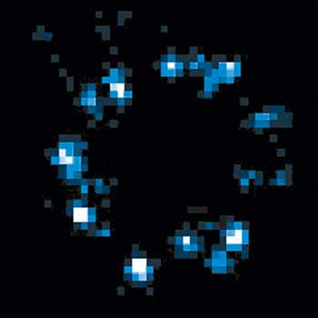
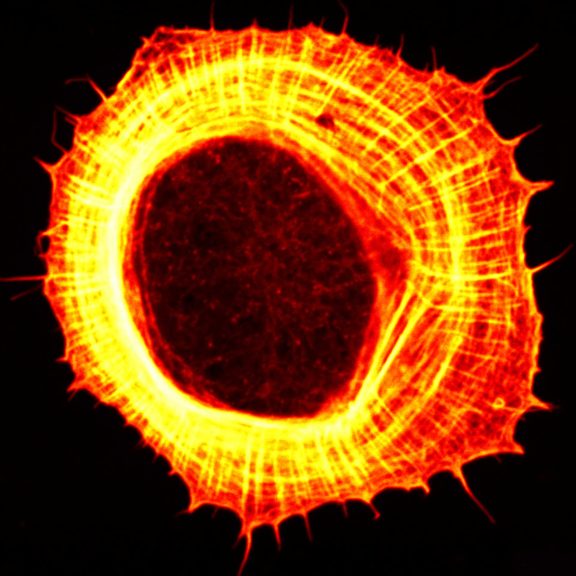
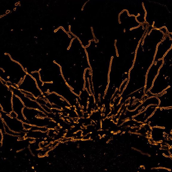
100% resolution, 4% light dose
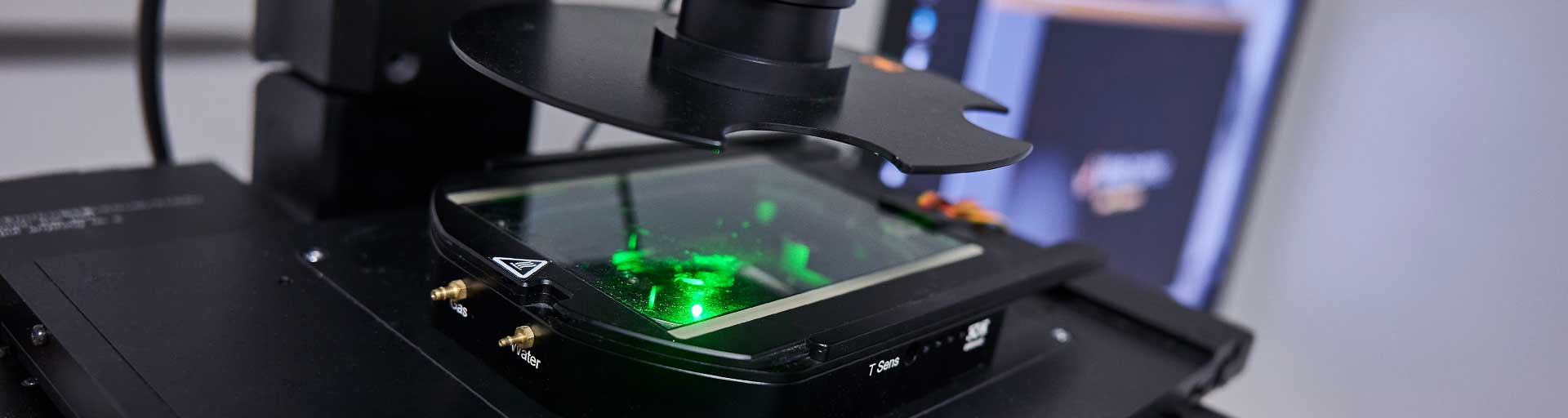
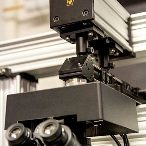
MINFLUX Module
Single-digit nanometer resolution with the MINFLUX module for our MIRAVA POLYSCOPE
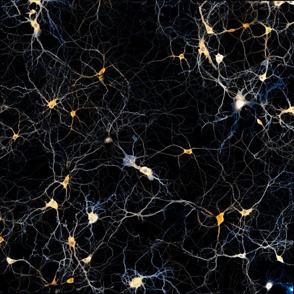
MATRIX Detector
Many eyes see more than one. The MATRIX detector drastically improves signal-to-background ratio, resolution, and dynamic range.
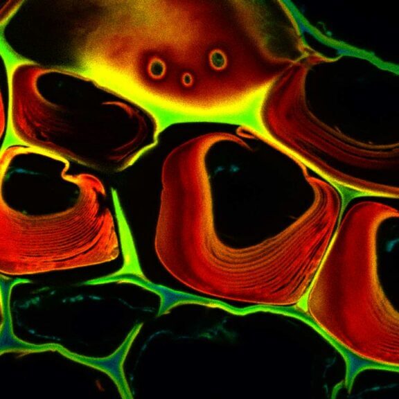
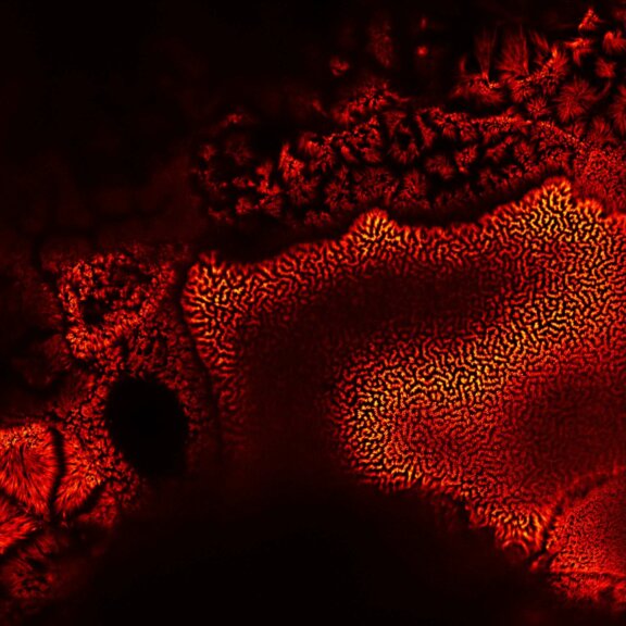
TIMEBOW Imaging
TIMEBOW lifetime imaging for stunning results at confocal and STED super-resolution.
FLEXPOSURE Illumination
Brings down the light dose on your sample and lables dramatically. Key ingredient for volume and live-cell superresolution.
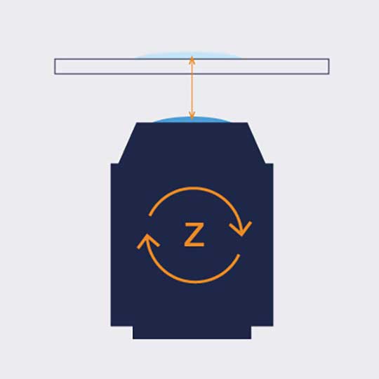
RAYSHAPE Mirror
Dynamic aberration correction with a deformable mirror over about 200 µm z-range. 140 digital actuators adjust the mirror surface within milliseconds.

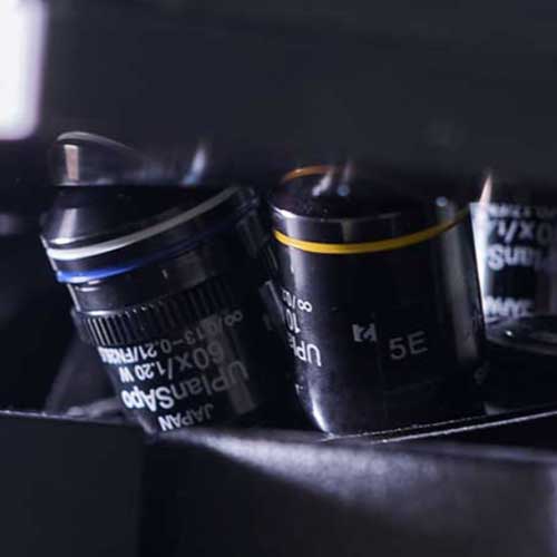
Custom Solutions
We offer solutions for even the most challenging applications. Everything that can be done, we will do.


Be gentle to the sample
and turn the lasers only on when needed
Fluorescence requires light. Light inevitably bleaches fluorophores. What a dilemma.
That’s why photobleaching is an eternal nuisance in fluorescence microscopy, exacerbated by phototoxic effects in live-cell imaging.
But eternal?… Absolutely not! abberior has the solution: FLEXPOSURE! It protects the sample by bringing down the light dose by up to two orders of magnitude, without compromising on contrast and resolution.
FLEXPOSURE does so by shining light only where it has an effect, and nowhere else. Instead of illuminating everything at full intensity, FLEXPOSURE strongly reduces or completely shuts down the lasers where there is no signal to be detected. Dark or out-of-focus areas experience almost no irradiation, while regions containing in-focus
fluorescent markers are imaged with 100% signal and resolution. This reduces photobleaching and phototoxic effects to the absolute minimum.
Adaptive illumination with FLEXPOSURE thus facilitates imaging over volumes, many frames, or long-term measurements where conventional imaging has long bleached the sample.

Adaptive illumination
the intelligent way of live-cell imaging
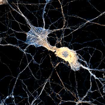
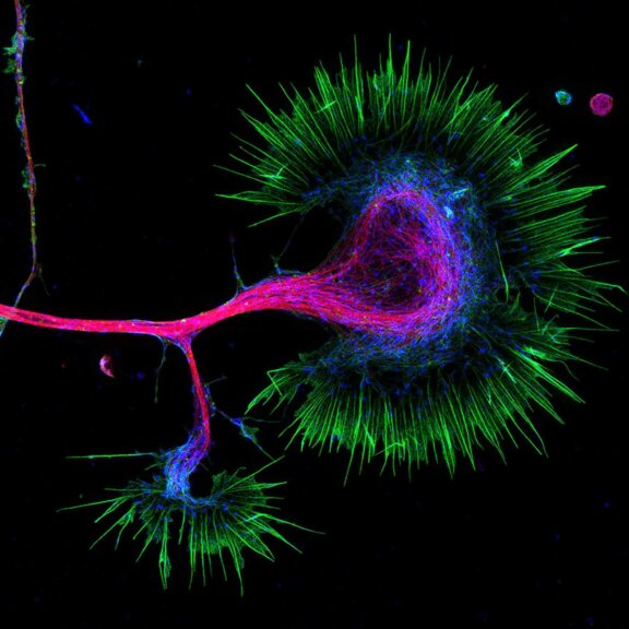
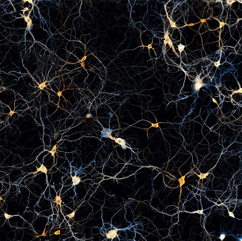
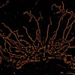
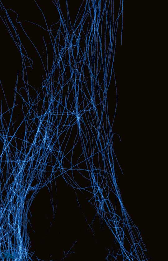
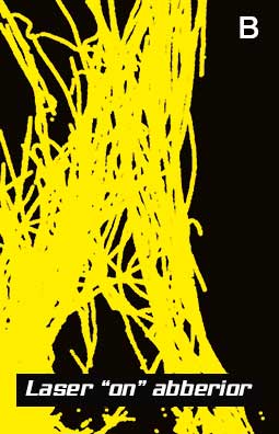
Adaptive illumination versus full laser power. For acquiring the image of the delicate tubulin filaments on the left, a basic microscope pointlessly illuminates the whole image area (A).
Based on low-light probing directly before imaging each pixel (this is no pre-recorded mask!), abberior’s FLEXPOSURE dynamically shuts down lasers where no structure is detected (B).
This intelligent protection scheme is particularly important for live-cell imaging.
- for volume and live-cell imaging with dramatically reduced light dose
FLEXPOSURE adaptive illumination

Why is TRUESHARP better at dealing with noise and background?
Learn how TRUESHARP images boosting deals with noise and background, why noise is random and how we analyze and identify signal and background separately.

How can TRUESHARP improve your everyday imaging experience?
See how TRUESHARP will strongly enhances resolution, sharpness, and brilliance of your images and why abberior’s TRUESHARP, MATRIX, and TIMEBOW technology allows you to get as close as possible to…
Microscope Systems
Fluorescence microscopy is an indispensable tool and for your science, you need the finest technology combined with hassle-free operation from abberior.
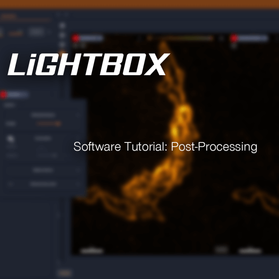
How does LiGHTBOX post-processing work? – Software tutorial
Unlock the full potential of your microscopy data with efficient post-processing in the LiGHTBOX software, using TRUESHARP deconvolution, MATRIX array detection, TIMEBOW lifetime imaging and more!