

Sample gallery
Fluorescence imaging, whether at confocal, STED or MINFLUX resolution, guarantees unique insights into the function and structure of life at the molecular level. Besides the scientific information content, some sample portraits provide simply beautiful images. Enjoy browsing our sample gallery.
the fine art of science
Description
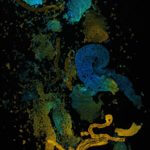
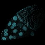
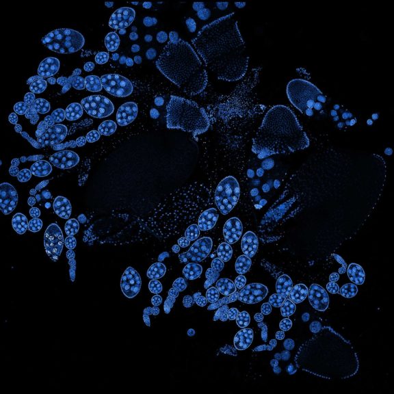
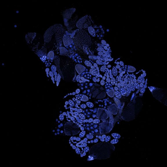
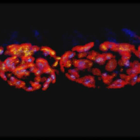
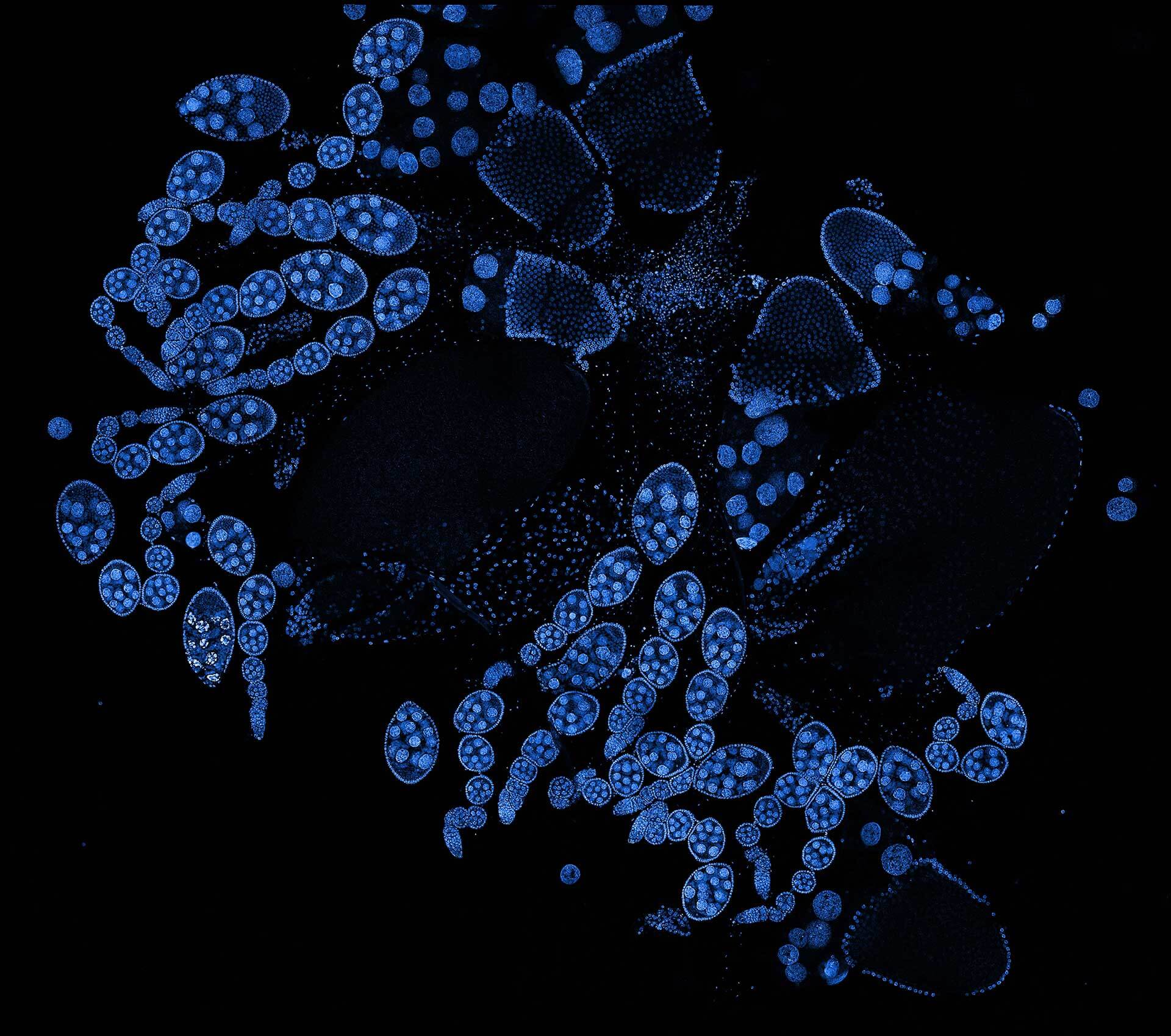
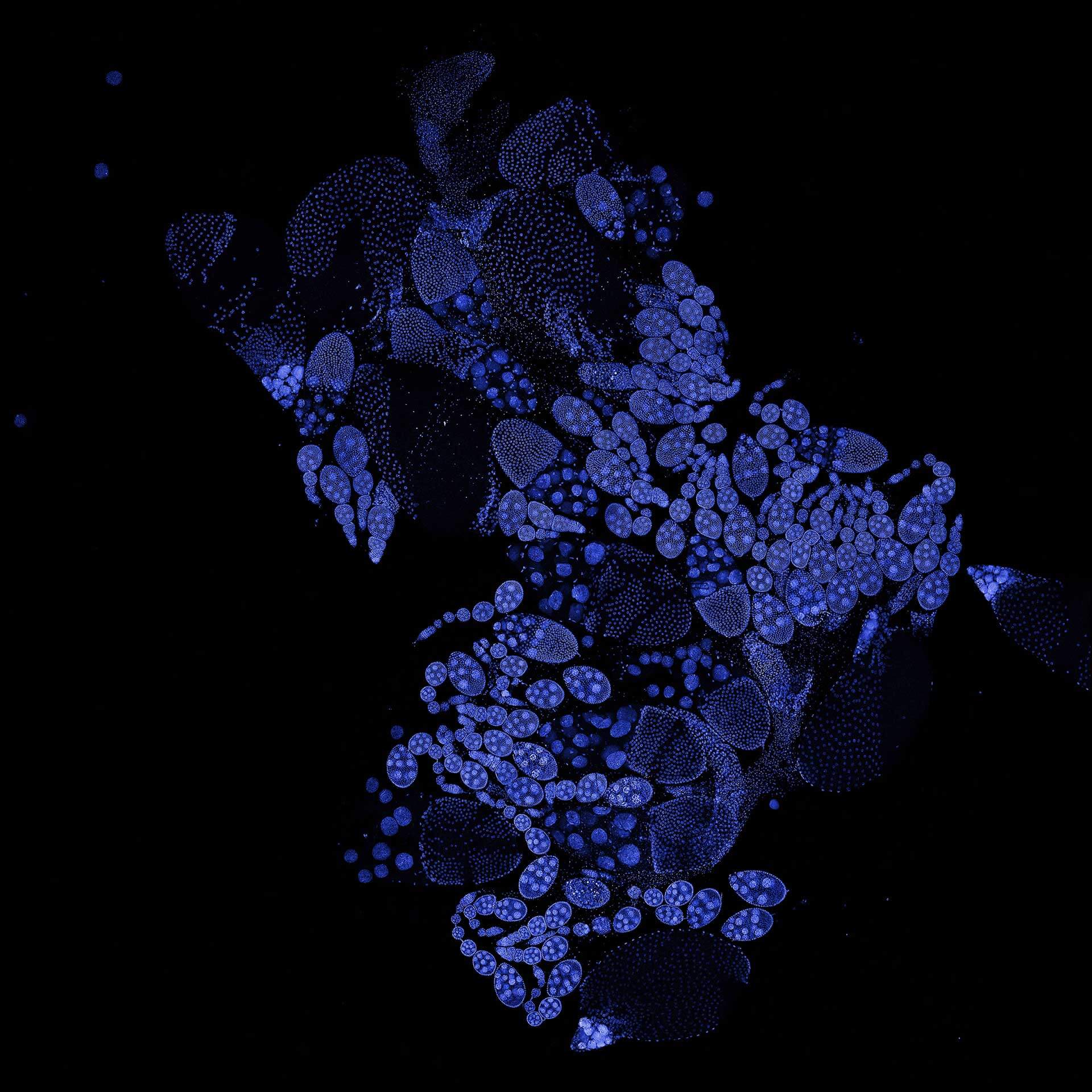
Drosophila ovariole stained with abberior LIVE 560 DNA showing nuclei in different cell types of the egg chamber. Ovaries were dissected from adult female fruit flies and were fixed prior to staining.
Image was acquired with the STEDYCON tiling feature and assembled with the SVI Huygens Stitcher.
Modules:
Description
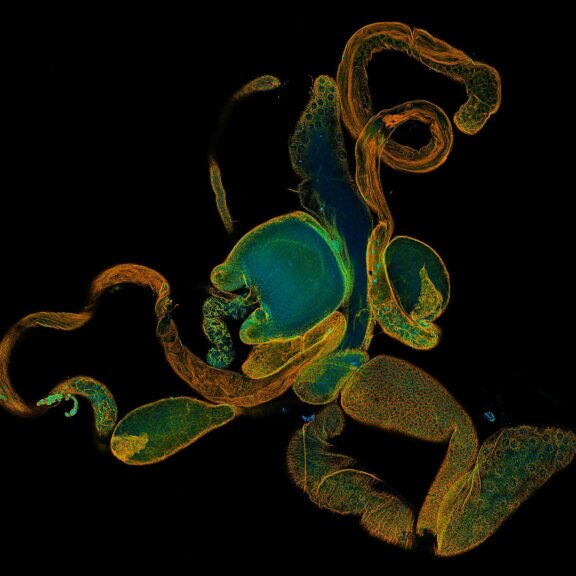
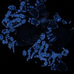
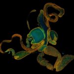
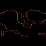
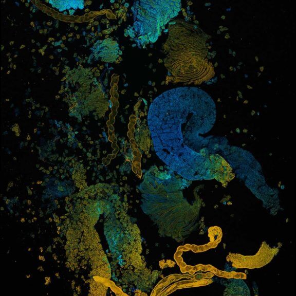
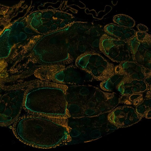
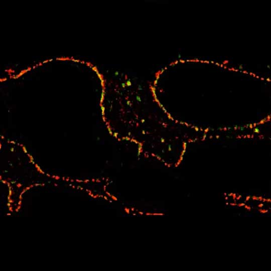
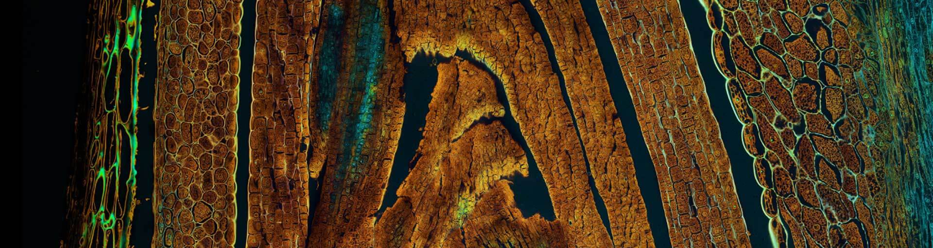
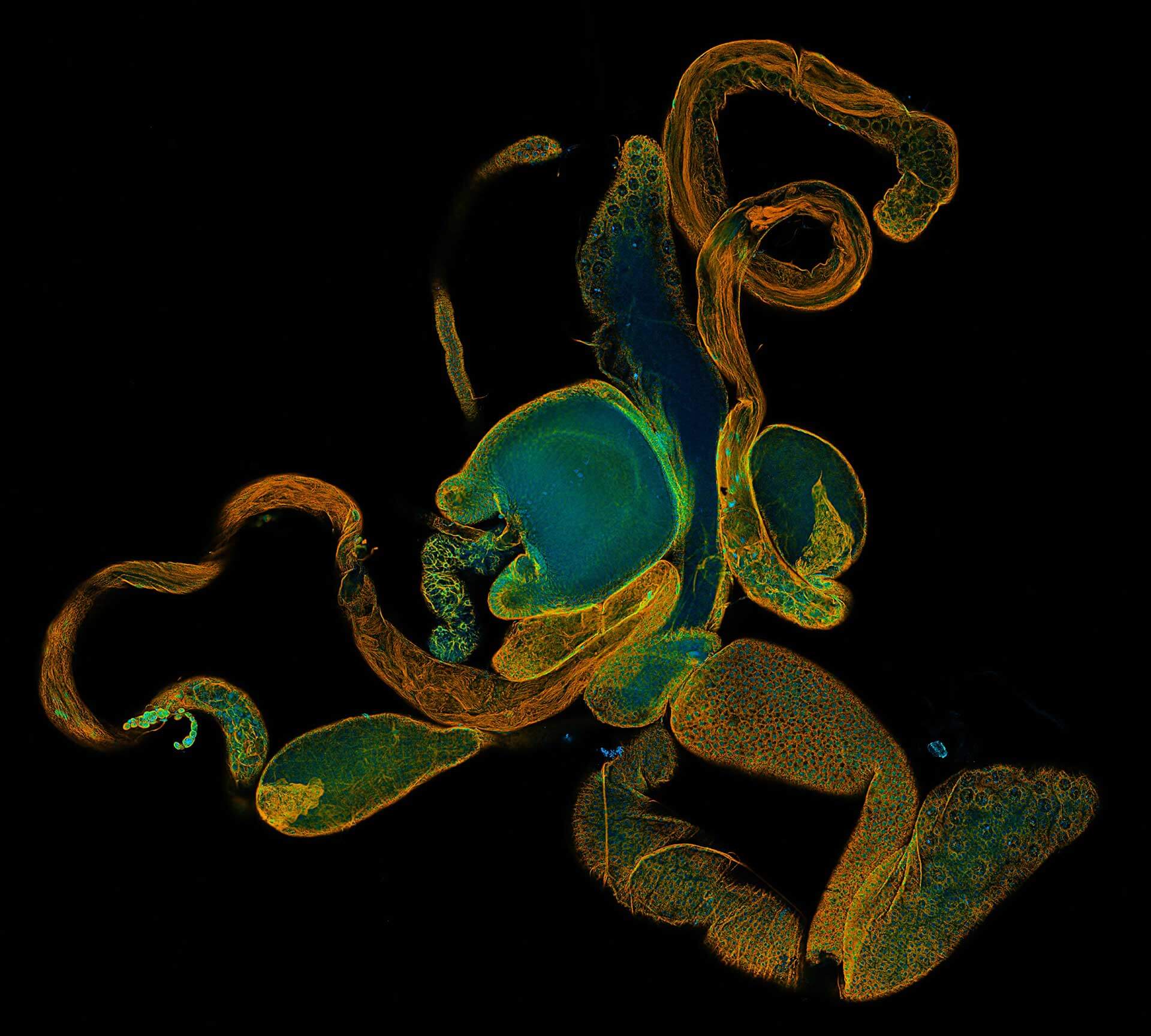
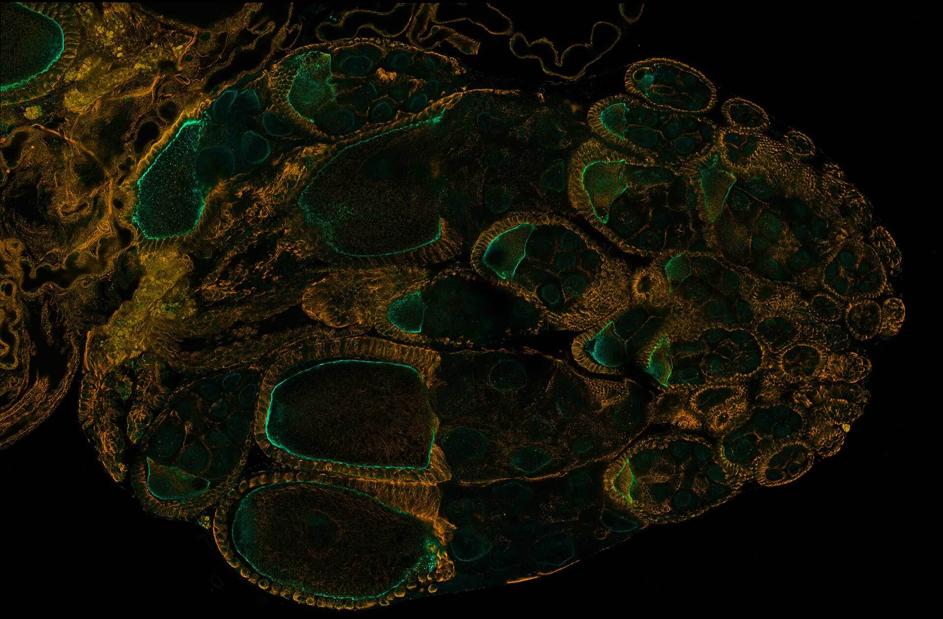
Drosophila ovariole stained with abberior STAR RED phalloidin (actin, red) and nuclear pores in STAR ORANGE (cyan) highlighting different cell types of the egg chamber. Image was acquired with the STEDYCON tiling feature and assembled with the HYGENS stitcher
Description
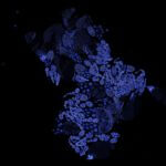
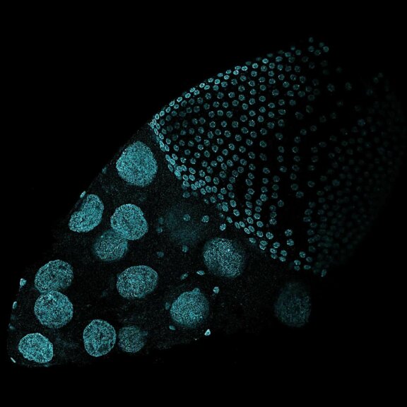
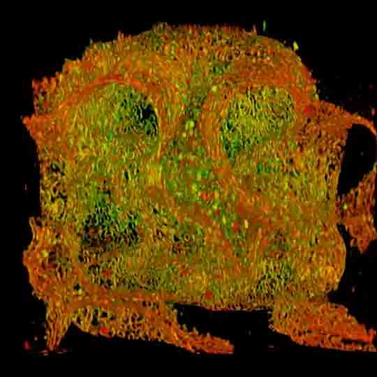
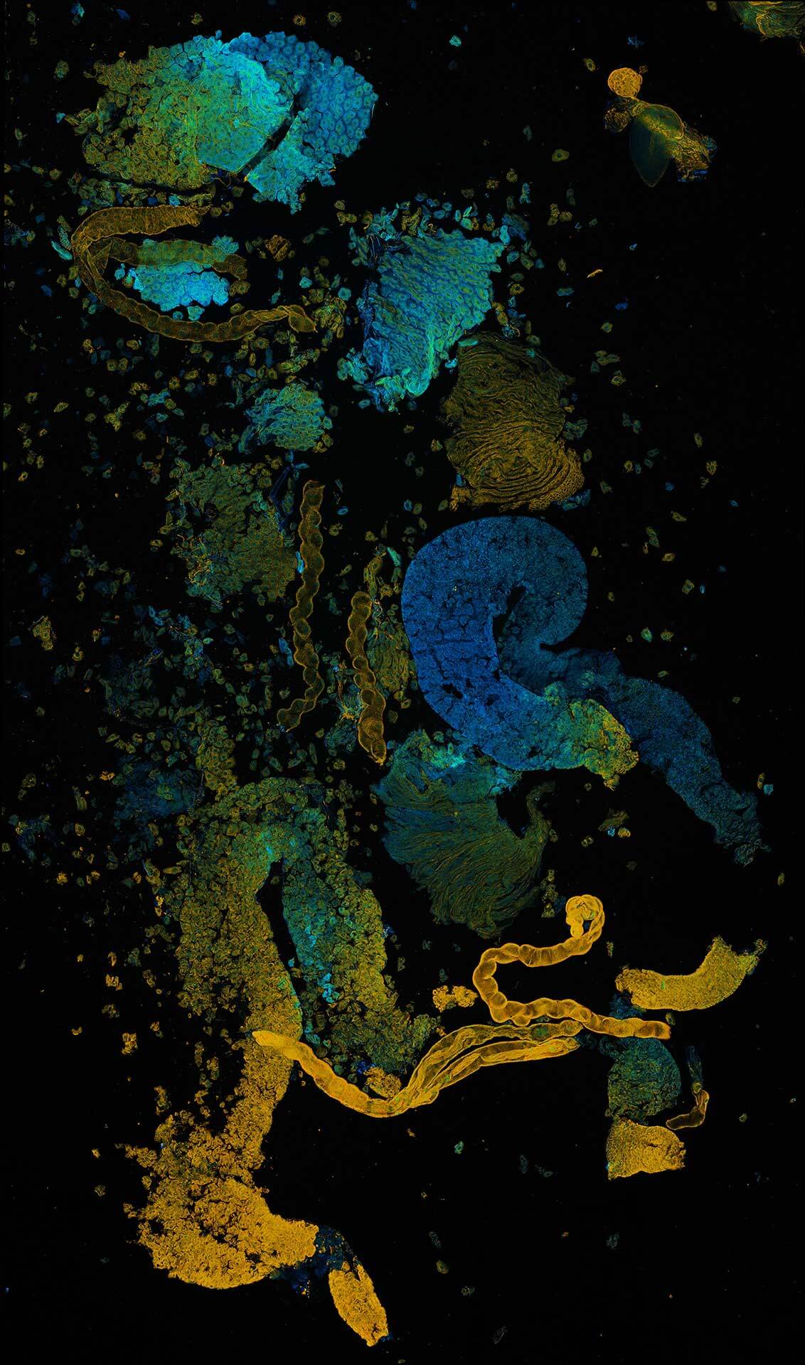
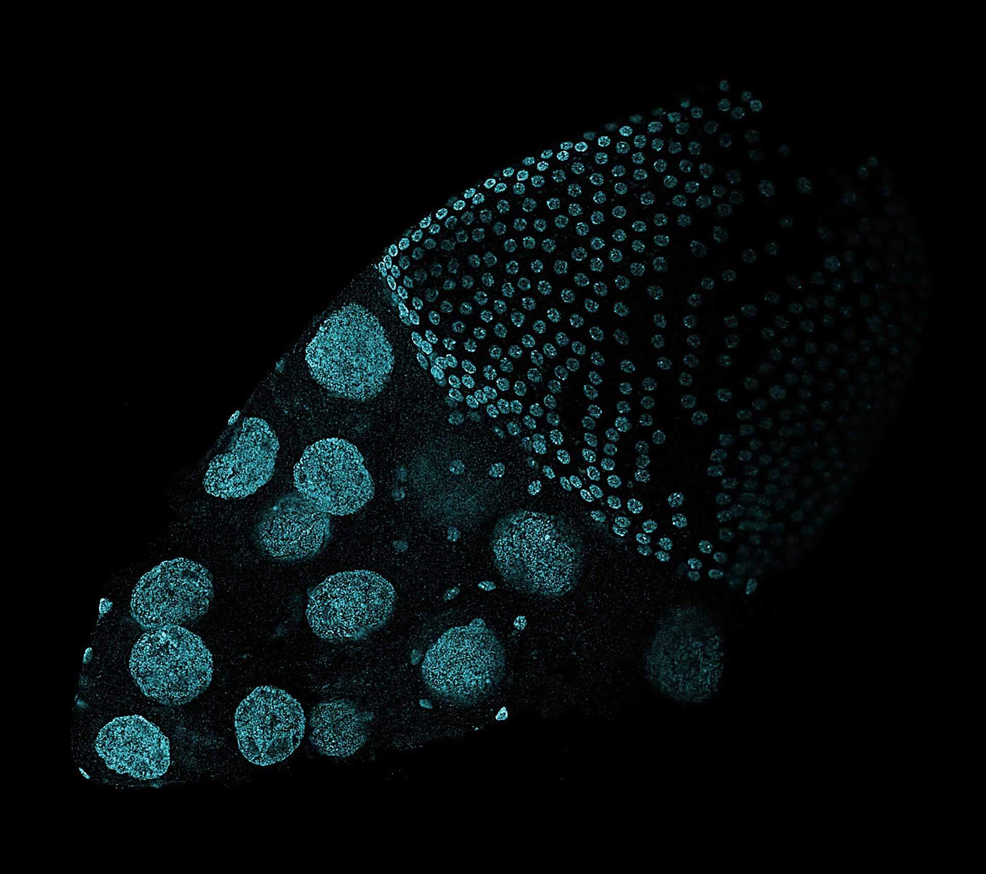
Drosophila ovariole stained with abberior LIVE 560 DNA, showing the different cell types of the egg chamber. The image was stitched using SVI Huygens.
Description
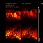
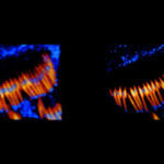
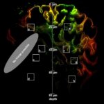
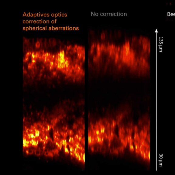
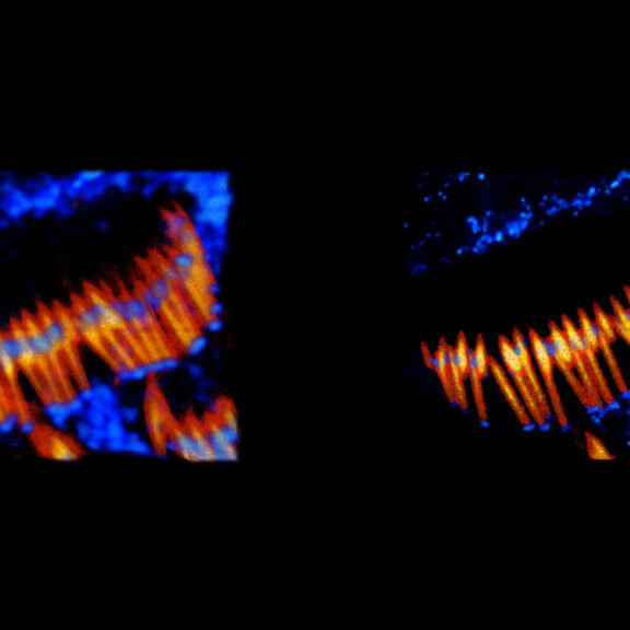
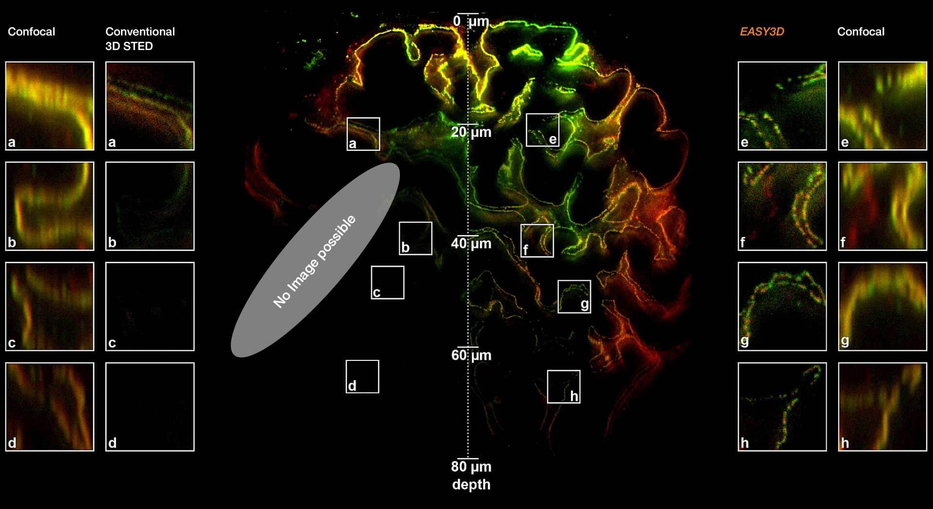
Adaptive optics EASY3D enables deep imaging of cleared adult kidney samples. Comparison of a XZ-slice deep into a renal corpuscle using conventional 3D STED imaging and an EASY3D imaging. EASY3D allowed imaging up to 80 µm into cleared rat kidney tissue using an oil objective. Without adaptive optics, the mismatch between immersion oil and sample leads to complete signal loss. Labels: Nephrin (red, Abberior STAR635P) and Podocin (green, AlexaFluor594).
Sample was prepared by D. Unnersjö Jess and H.G. Blom @ KTH Stockholm, Sweden.
Modules: